Nano based Drug Delivery System for Cancer Therapy: A Next Generation Theranostics
Cancer is considered as one of the foremost cause of illness and death with very complex pathophysiology even though prominent advancement has been made on innovative tumor treatments. Therapeutic properties and the global survival rate are still disappointing for the patients with cancer. There is a shortfall in the capabilities of these cancer therapies, some novel strategies are developed to provide better treatment therapies to improve their quality of life and also aids in reducing the number of deaths. Amongst the cardinal phases towards ensuring ideal cancer management is early diagnosis and targeted drug delivery of anti-tumor to decrease its toxicities. Recently the progress of nanotechnology as novel therapeutics, have advanced and trialed to overwhelm numerous limitations of previously available drug delivery systems for cancer treatment. Nanobased therapeutics has provided the chance to directly contact the tumorous cells selectively with improved drug localization, cellular application as well as providing targeted drug delivery eluding the interaction with the healthy cells. In this review, we summarize about various novel nanomaterials as anti-tumour drug delivery carriers for cancer treatment; also provide insight into the superlative necessities of nanotechnology in cancer therapy and its challenges in targeted drug delivery.
Introduction
Well-organized research in the field of cancer has showed that a group of diverse problems, together with the rapid spreading and uncontainable growth of abnormal cells (tumor cells), results in the development of malignant cancers [1, 2]. Tumor cell growth is commonly considered as the impact of genetic mutations [3]. Presently, the second foremost cause of death all over the world after cardiovascular disease is cancer. Not only this, there is a constant rise in the severity of cancer among the individuals [4]. Now cancer is extensively recognized as a universal problem that lacks a global resolution [5]. According to estimation in 2018, about 18.1 million cancer cases and around 9.6 million casualties were produced by cancer [6]. As per the estimation of Global Cancer Observatory (GCO) organization in 2030, approximately 30 million individuals will die from these malignant cancers [7]. Furthermore, the high death rate of cancer and the financial burden imposed on cancer affected patient families are massive. So, efforts on the development of novel strategies in cancer diagnosis and treatment are of great importance. A diversity of methods are existence for treating the cancers; however, some of them are unsuccessful due to the adverse side effects as well as their poor bioavailability and water solubility [8], which leads to reduce the pharmacological activity of antitumor drugs on the cancerous tissues [9]. The existing cancer treatments produced outstanding contributions in extending the endurance period of malignant patients and also aids in the improvement of patient’s lifetime [10].
Despite the substantial developments in health science and technology, cancer stands as an ailment with very limited treatment methods. Metastasis of tumor contributes a lot to the mortality and infirmity, and the precise mechanisms involved in the ailment remain to be illustrated clearly [11, 12]. In recent years, there is an increasing demand for the enhancement of new strategies for targeted treatment of cancer to address the limitations of prevailing treatment methods [13, 14]. Nanoparticle-based drug delivery systems (NDDSs) are being broadly employed in the early, diagnosis, imaging as well as used in the cancer therapy owing to their high cancer-targeting efficiency, controlled release property and also due to their low toxicity profile [15].
Nanoscience generally deals with few nanometres (nm) to several hundred nm size range, possess greater surface area along with adaptable optical, magnetic, electronic, and biological properties than the macroparticles [16]. It has been the field of concern for evolving the targeted drug delivery systems as it very beneficial in overcoming the restrictions of traditional therapeutics [17, 18]. It is considered as a promising approach in both the tumor diagnosis as well as treatment of malignancy at molecular level. Extensive researches are investigated to improve the nano-based tumor therapeutics with very less toxicity profile than the existing traditional therapy [16]. Nowadays, nanomaterials are designed to assist as therapeutic mediators to permeate the biological barriers to facilitate the molecular interactions also helps to identify the changes at molecular levels.
Present novel Drug delivery system (NDDS) for cancer therapy, which are being marketed, in addition under research consisting polymeric micelles, polymeric nanoparticles (PNPs), liposomes, dendrimers, nanocapsules, nanospheres, and nanotubes [19, 20]. With the rapid development of nanotechnology, in this review we have summarized the basic principles of the targeted drug delivery of nanotherapeutics, current progress, its challenges, nanomaterials in targeting tumor cells, Tumor Microenvironment (TME) and its application in cancer diagnosis its treatment with focus on their benefits and limitations as well as describes the path of future research.
Nanotechnology in Cancer Diagnosis
Cancer diagnostic strategies are very essential at early detection and targeted to inhibit the carcinogenic cell proliferation. Prominent early diagnostic tools for tumors are Magnetic Resonance Imaging (MRI), Positron Emission Tomography (PET), Computed Tomography (CT) and Ultrasound [21]. These imaging tools have limitation due to their insufficient provision of appropriate clinical evidence about different types of cancer and its various stages. Henceforth it makes very difficult to acquire a full estimation of the ailment state and to provide optimum therapy [22]. In the recent research, nano-based tools and techniques aids in the imaging of tumor cells at the cellular and molecular levels to explore the TME [23].
Near Infrared (NIR) Quantum Dots
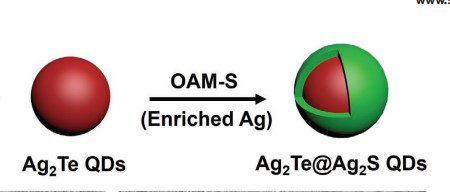
NIR quantum dots are based on the in vivo fluorescence imaging which can carry out concurrent, non-invasive imaging with high resolution to precisely acquire the biological figures in vivo and play a substantial role in the initial diagnosis of tumours [24]. Quantum dots emits fluorescence in the near-infrared spectrum around 700-1000 nm are used for imaging different cancers like pancreatic cancer, liver cancer, colorectal cancer and lymphoma [25]. However, this conventional fluorescence imaging technique (near-infrared (NIR)-I windows) are rigorously obstructed by the auto fluorescence, strong tissue scattering and absorption. The emergence of second near-infrared (NIR) window (NIR-II, 900-1700 nm) with higher tissue penetration depth, higher spatial and temporal resolution significantly overwhelms the imaging limitations of NIR-I and also possesses high brightness and good photo stability to aid cancer imaging [24]. Apart from this, in vivo fluorescence imaging with the silver-rich Ag2Te quantum dots (QDs) comprising a sulfur source (Ag2Te@Ag2S QDs), shows better spatial resolution images over a wide infrared range with enormous penetration depth [26]. Hence, NIR-QDs are considered as one of the promising fluorescent biomarkers in the arena of in vivo fluorescence imaging. The Ag2Te quantum dots [26] are depicted in Figure 1.
Nanoshells
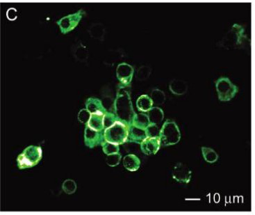
Nanoshells size ranging approximately between 10 and 300 nm composed of dielectric cores usually made of silicon is surrounded with a thin metal (gold) shell [27, 28]. Nanoshells work by altering plasma-mediated electrical energy into light energy, also it modifies the optically over UV-infrared emission/absorption arrays. Nanoshells are free from the heavy metal toxicity [29] although it is not used widely due to their bulky sizes. Wu, et al. Demonstrated the imaging with 20-nm thin gold nano shells loaded into gel phantoms via optical tomography [30]. Apart from this, gold nanoshells are fused with the immunoparticles to target the tumour molecules specifically. By using the in vitro culturing technique, Loo, et al. Targeted HER-2 with the HER- 2 antibody-conjugated nanoshells which were formulated accordingly to scatter light by using optical coherence tomography [31]. These nanoshells are also fabricated as carriers for anticancer agents as they were theranostic in nature. Sengupta, et al. Revealed the high anticancer activity from the novel nanoshells enclosed with an antiangiogenic factor (combrestatin and doxorubicin) while associating only with combrestatin or doxorubicin and further they confirmed the progressive discharge of these agents, which endorsed vascular interruption within the tumour microenvironment [32]. Schematic diagram for the encapsulation of anticancer drug in nanoshell [32] is shown in Figure 2.
Colloidal Gold Nanoparticles
Colloidal gold nanoparticles (AuNPs) are good contrasting agent due to its small size, high atomic number and good biocompatibility. Various researches on AuNPs demonstrated that when the energy surpasses 80kev, the mass attenuation rate of gold becomes very high than the other metal nanoparticles [33]. Rand, et al. Formulated AuNPs with liver tumor cells and found that with X-ray imaging, the liver tumor cells in the gold nanocomposite cluster were stronger than the liver tumor cells alone [34]. In the colon cancer, Paciotti, et al. Demonstrated that systemically conveyed AuNPs with tumor necrosis factor (TNF) amassed in tumor cells and TNF-conjugated AuNPs had better theranostic effect compared with that of inherent TNF alone [35]. Chen, et al. Revealed about the gold nanocages, a different variety of AuNPs aids in the detection of erythroblastic oncogene B (erbB2) and epidermal growth factor receptor 2 (EGFR2) by in vitro assay techniques which has a prospective solicitation in photo thermal imaging [36] shown in Figure 3 [36].
Nanotechnology in Cancer Therapy
On the heels of the advancements of nanotechnology with specially modified nanomaterials and surface modification of the anti-tumour drug with nanoparticles as carrier can provide controlled drug release as well as aids in the targeted drug delivery to improve the stability and bioavailability of anti-tumour drugs [37].
Properties of Nanomaterials
Nanomaterials used in biomedical application as therapeutic drugs and devices are in the particle size around 1–100 nm [38]. As the materials proportion reduces into nanoscale which develops some exclusive properties like optical, electrical and magnetic properties, thus making the nanomaterials are more potent from traditionally used macromolecules. In addition, these nanomaterials have enhanced electrical conductivity, high surface-to-volume ratio, super-paramagnetic behaviour, and exceptional fluorescent characteristics. In the medical arena, noticeable features of nanomaterials are it aids in transportation of therapeutic drugs with targeted release property and also used to increase the permeability and biocompatibility [39]. These specific properties of nanoparticles make it as a reliable material which can be utilized especially as cancer therapeutics. These nanomaterials can amass with biomolecules, which can improve the specificity of biochemical drug complex in targeted therapy, thus enhancing the efficiency of nano-based treatment and also reducing its toxicity in the healthy cells [40].
Progress of Nanotechnology in Targeted Drug Delivery
Nanomaterials have been made as a recent advancement with the aim of targeted drug delivery for accurate directing to the explicit tumor cells, and it is accomplished by both passive targeting as well as active targeting. In passive targeting, Enhanced Permeability and Retention (EPR) effect is being used. On the other hand, active targeting is accomplished by coupling with the peptides, antibodies and small molecules. In comparison with the free drugs, targeted drug delivery assists in reducing the toxicity level in normal cells, increase the half-life, solubility, loading capacity and also protects drugs from degradation [38, 41]. Over subtle design and modification, nano-based drugs provide improved specificity, less cytotoxicity to normal tissue, high bioavailability, longer half-life period, larger loading capacity, and controlled drug release forms, overwhelming the disadvantages of orthodox chemical therapy. Last few years, great progress has been made in the cancer pathophysiology and Nano Science Technology and Industry (NSTI) produced abundant nanomaterials for the treatment of cancer.
Nanomaterials for Cancer Therapy
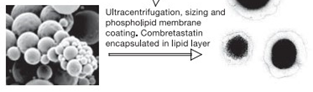
The fundamental arrangement of nanomaterials is very complex in nature, with a surface coating, shell layer and the central portion (core) which is generally known as NP. These NPs have deep tissue penetration property to increase the EPR effect. Besides, the surface characteristics it impacts on the bioavailability and half-life by crossing epithelial fenestration effectively [42]. The advancement progress on nano-based anti-tumour drug carriers [43] is being depicted in Figure 4.
Nano Carriers For Cancer Therapy
Based upon the general shape of NPs, it is being categorized into zero dimension, One-D, two-D and three-D [44]. It predominantly comprises of nanobased polymeric substances, inorganic nanoparticles and various nano drug carriers. Various nanomaterials used in anti-tumour drugs delivery as carriers are represented in Table 1.
| S.No | Nanomaterials | Anticancer drug | Outcomes | Reference |
|---|---|---|---|---|
| 1 | Chitosan | Chlorambucil | Decreased the abnormal toxicity and improved the uptake of tumor cells. | [45] |
| 2 | Silica nano particles | Fluconazole | Enhanced the targeted delivery, permeability and decreased side effects | [46] |
| 3 | Zinc Oxide nanoparticles | Doxorubicin | Increased blood concentration and anti-tumor efficacy. | [47] |
| 4 | Poly-Lactic Acid nanoparticles (PLA) | Verapamil and Doxorubicin | Decreased drug resistance and improved anti-cancer effect. | [48] |
| 5 | Poly (Lactic-co-Glycolic Acid) nano particles (PLGA) | Curcumin | Enhanced the drug targeting. | [49] |
| 6 | Polyethylene glycol (PEG) nano particles | Doxorubicin | Enhanced the therapeutic effect. | [50] |
| 7 | Liposomes | Bortezomib | Decreased side effects and improved anti-tumor effect. | [51] |
| 8 | Gold (Au) nanorods | Platinum and Doxorubicin | Targeted delivery and response activity. | [52] |
| 9 | Carbon nanotubes | Doxorubicin and propidium iodide | Progresses the drug loading and stability. | [53] |
| 10 | Quantum dots | Doxorubicin | Progresses the drug activity and anti-tumor ability. | [54] |
Table 1: Nanomaterials used as carrier for the delivery of anti-tumor drugs.
Nano-Polymers
Nano-polymer carriers have very good biodegradability, bioavailability and biocompatibility. It is the utmost significant and extensively studied nanomaterial in the therapeutic field [55]. At present, nano-polymer carriers are primarily classified into natural and synthetic, which are used specifically based on the anti-tumor drugs. Natural nano-polymer carriers basically include agarose, collagen, chitosan and hyaluronic acid-based polymers. In contrast, manmade nano-polymeric carriers are poly anhydrides, PEG (polyethylene glycol), PGA (Polyglutamic acid), PCL (poly-ϵ-caprolactone), PLA (polylactic acid), and PLGA (poly D, L-lactide-co-glocolide), etc. Saneja, et al. Demonstrated the co-polymerization of glycolic acid and lactic acid to form Polylactic-co-glycolic acid (PLGA) possesses better biocompatibility with EPR effect [56]. Furthermore Zhou, et al. Investigated about the polymeric micelles by polymer self-assembly into nano-aggregates for the characterisation
![Figure 5: Further, Opaxio [paclitaxel poliglumex] is an important polymer nano-drug which has been arrived to the preclinical stage of research [60].](/fulltextimages/9641/fig_5.jpeg)
as they comprises of amphiphilic copolymers [57]. Then according to the study of Cagel, et al. Nano polymers with the hydrophilic section enhances the stability and reduces the drug uptake in the reticuloendothelial system, thus delaying their circulation time period while the hydrophobic core facilitates the impenetrable anticancer drugs to be absorbed and carried efficiently [58]. In the midst of different synthetic nano-polymers PEG possesses very less toxicity and it has been the emphasis of research in the recent period of time. PEG polymer is obtained by the polymerization of ethylene oxide. Its key features are governable polymerization degree, its steady structure [59], and also it can evade the recognition of human immune system. Clinically approved polymer nanomedicine formulations for cancer treatment is paclitaxel as Paclical® in Russia and Genexol-PM® in Korea are shown in Figure 5. Further, Opaxio [paclitaxel poliglumex] is an important polymer nano-drug which has been arrived to the preclinical stage of research [60].
Inorganic Nanoparticles
Recently the use of inorganic nanoparticles has been increased gradually especially in tumour imaging and treatment [61]. Inorganic NPs used in the tumour imaging and treatment includes metals like iron, gold, silver and zinc nanoparticles, metal oxides like iron and titanium oxide NPs, carbon nanotubes, carbon dots and semiconductors [62, 63]. Owing to their excellent physical and chemical properties like good stability, optical, magnetic responsiveness, these nanoparticles are considered more appropriate for the treatment of cancer than the conventional carbon-based nanocarriers [64]. Chen, et al. [65] Fused the Ferric oxide (Fe3O4) NPs on carbon nanotubes to afford a double- targeted drug delivery based on the multifunctional nanoplatform showed highest drug-loading capacity [at pH 8.5] for tumor cell targeted visual imaging and magnetic- targeted drug delivery by coating transferrin on the surface of modified nanotube. Though, the toxicity profile of the inorganic nanoparticles limits its clinical applications [66]. Basoglu, et al_. and Mandriota, et al. Demonstrated a study on the magnetic NPs (MNPs) in order to enhance the biocompatibility and stability by coating various organic polymers which showed high efficiency in gene therapy and chemotherapy for the treatment of malignancy [67, 68]. Further Hoopes, et al._ achieved the thermal ablation of malignant tumor by magnetic hyperthermia using MNPs and the results highlighted that this technique can be a better alternative for tumour treatment [69].
Nano-Liposomes
Nano-Liposomes are mostly similar to the structure of biofilms. It possesses a lipophilic tail and hydrophilic head, which helps to encapsulate the drugs in aqueous environment to form multilayer vesicles [70]. It is considered as a promising nano-carrier of drugs because of its high biocompatibility, controlled drug release, strong drug loading capacity, high safety, and low toxicity profiles [71]. Wu, et al. [72] formulated Luteolin (LUT) in the nano-liposomes with the encapsulation effectiveness up to 90% which aids to increase the LUT water solubility, bioavailability and `anti-tumor activity. Further in vitro studies illustrated that liposome-LUT inhibits the tumor cell evolution by persuading apoptosis of cancerous cells and showed superior anti-tumor activity on mouse colon cancer cell CT26, when compared with LUT without nano- liposome. Pastorino, et al_. Modified the doxorubicin drug by loading Anti-disialoganglioside (Anti-GD2) antibodies which ultimately aids to improve the targeting of drugs to Human Neuroblastoma [73]. Zuccari, et al. [51] fused Bortezomib in the nanoliposomes by encapsulating the asparagine-glycine- arginine (NGR) peptides possess high therapeutic efficacy as well as very less toxic side effects when compared to the administration of bortezomib alone. Han, et al. [74] revealed that multiple paclitaxel liposomes have very high anti-tumor efficiency and improved bioavailability compared to free paclitaxel. According to O’Brien et al.,_ liposomal doxorubicin showed a comparably high efficiency in treating the breast cancer with reduced cardiotoxicity [75]. Moreover, liposome-based Nanocarriers presented an alternative for the combination of drugs, which can increase the therapeutic activity and also it can inverse the drug resistance [76].
Nano-Gene Carriers
Nano-gene carriers in the gene therapeutics have a substantial part in treating malignancy [77]. These carriers used in the gene therapy are the key success in gene therapy [78]. Presently, the most frequently used gene vectors are viral and non-viral vectors. In gene therapy, viral vectors are limited in application due to its complication and higher cost for the preparation [79]. In contrast, non-viral vectors are potential substitute for viral vectors, because they are very simple to formulate, higher transportability, lower toxicity, and almost all of them are nano-vectors including liposomes, peptides, and polymers [80, 81, 82]. Wang, et al. formulated a multi-functional cancer therapeutic plasmid namely Cas9-sgPlk-1 as a carrier transport by the electrostatic interaction by lipid encapsulation of gold nanoparticles. It provided a well-organized targeted gene editing in the in vitro and in vivo experiments [83]. Then Kim, et al. demonstrated the macrophage derived exosomes encapsulated with paclitaxel by sonication, incubation, and electroporation [84]. By using the sonication approach, exosomes exhibited very high paclitaxel loading efficiency with higher drug release in vivo [84]. In a recent study performed by Lou, et al. Performed miR-122 expression plasmid on the Adipose tissue-derived Mesenchymal Stem Cells (AMSC) aided in the treatment of hepatocellular carcinoma cells [85]. In another study demonstrated by Yuan, et al. Proved that treating the triple-negative breast cancer (TNBC) cell line Human Mammary Carcinoma (MDA- MB-231) with exosomes secreted by human umbilical cord mesenchymal stem cells overexpressing miR-148b- 3p (HUCMSC-miR-148b-3p) showed an inhibitory effect on the proliferation of MDA-MB-231 and also it highlights the prospective of miR-148b-3p comprising exosomes for the treatment of breast cancer [86]. According to the study of Gong, et al. Exosomes as endogenous nanocarriers has very beneficial potential in co-delivery of doxorubicin and hydrophobically modified miR-159 for triple-negative breast cancer therapy [87].
Targeted Delivery of Nano-Drug Carrier
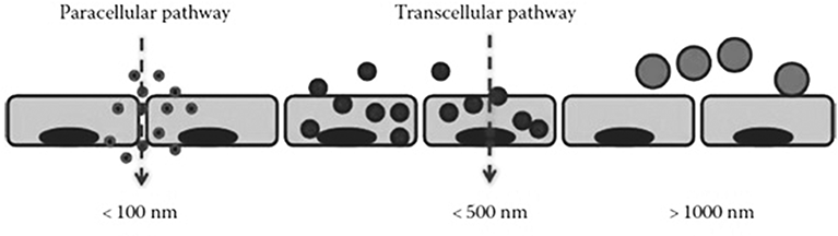
Although traditional chemotherapy drugs can kill tumor cells with high efficiency, but due to lack of its specificity they have toxic and side effects on normal tissues. This nanotechnology provides a new opportunity to overcome the above issues especially in tumour targeted therapy [88]. Generally, two major pathways are involved in the entry of anti-tumour drugs to the targeted site which are depicted in Figure 6 [89]. At present, anti-tumour drugs in the form of nanoparticles can be transport the drugs for its targeted delivery through three ways: passive transport, active transport and physical and chemical transport. These transport techniques can identify cancerous tissues more accurately in complex organisms and release the drugs at cancerous tissues and reduce toxic effects on normal cells.
Passive Targeted Transport
Passive targeting, mainly through EPR effect, enables the drug by macrophages as a foreign body immediately after entering into the human body, so as to reduce non- specific binding with non-target sites and reach the targeted sites for selective binding [90]. Drug carriers, such as liposomes, mainly transport the drugs through passive targeting [91]. Mitra, et al_. [92] embedded Adriamycin glucan complex as long-circulating nanoparticles. This embedded formulation enriched the drug targeting into the tumor site of mice by EPR effect, achieved the high efficiency and low toxicity of drugs. At present, many passive targeting nanoparticle formulations like Marqibo, Myocet and Doxil showed promising therapeutic effects in clinical trials [93]. Carmeliet and coworkers demonstrated the induction of neovascularization by high propagation of cancer cells in the vascular barrier which leads to the deterioration of tumor vessels. In the meantime, the reduced lymphatic drainage concomitant with malignancy intensifies the retention of NPs, which allows the nano-carriers to discharge the drugs in the cancer cells [94]. Carita, et al. demonstrated that nanocarriers loaded with antitumor drugs have better penetrability into the malignant cells rather than into the healthy cells [95]. On the other hand, Cirpanli, et al. Revealed the encapsulation efficiency of camptothecin in nano particulates by using amphiphilic cyclodextrins, polycaprolactone or poly (lactide-co-glycolide) with an intention to retain the bioactive lactone system and also to avoid the camptothecin drug from hydrolysis to the inactive carboxylate form [96]. Gaur, et al._ formulated a nanoparticle by conjugating 20(S)-camptothecin and cyclodextrins polymer (CRLX101) which aids in the high biocompatibility and also to control the release of drug constituents over a period of time in tumor cells [97].
Active Targeted Transport
The limitation of passive targeting is lower specificity to tumour site, whereas active targeting has higher targeting. It is found that some antigens or receptors are over-expressed on the surface of tumor cells [98]. Where normal cells express the various receptors like folate [99], prostate- specific membrane antigen [100], biotin [101], transferrin [102], peptide [103] and carbonic anhydrase IX [104] them normally. Active targeting is based on the specific recognition between receptor and ligand or the covalent modification of targeting groups on the surface. Mackiewicz, et al_, Designed multifunctional poly (ethylene glycol)-block-poly (lactic acid)) nanoparticles modified by folic acid and fluorescent probes. The study results demonstrated that the formulated nanoparticle can be used for both the cell imaging as well as targeted delivery of anti-tumor drugs at the same time [105]. Generally, transferrin receptors will be overexpressed in the cancer cells than the normal cells, Amreddy, et al. fused transferrin with NPs to transport antitumor drugs for the cancer therapy. When compared to the unmodified NPs, transferrin-conjugated NPs have exhibited higher cellular uptake efficacy and also enhanced the intracellular delivery of antitumor drugs [106]. Moreover, Soe, et al. indicated about the transferrin-conjugated polymeric NPs which plays a substantial role in overwhelming drug- resistant chemotherapy [107]. Epidermal Growth Factor Receptor (EGFR) is one of the associates of ErbB family of tyrosine kinase receptors which is basically overexpressed in various kinds of cancer, as it is involved cancer cell growth and proliferation is being widely employed as a target for malignancy [108]. Furthermore, Alexis et al. Incorporated altered ligands to target EGFR-overexpressed tumor cells to improve the targeted drug delivery [109]. In addition, Balasubramanian, et al._ Conjugated two different kinds of altered cancer-specific ligands curcumin and 5-fluorouracil- loaded, folate and transferrin into a distinct polymeric magnetic nanoformulation for active targeting, showed enhanced target specificity with high efficiency [110].
Physical and Chemical Targeted Transport
The microenvironment of tumor cells is different from that of normal cells. Based on the unique physical and chemical environment of tumor site, researchers have developed a series of nano-drug carriers with stimulus response, which can achieve targeted release of drugs by controlling exogenous stimulus (change of temperature, magnetic field, light or electric pulse) or endogenous stimulus (change of pH value or redox), thereby improving drug efficacy and reducing side effects [111, 112, 113, 114].
Temperature responsive nanocarriers: Corato et al_. enhanced the magnetic heating in the dense magnetic cores by encapsulating the magnetite NPs with a diameter around 9 nm [115]. A nanocomposite of magnetite with porphyrin was prepared and studied for its magnetism with the photo-responsiveness [116]. Shah, et al._ [117] wrapped photosensitizer tetrakis (hydroxymethyl) phosphonium chloride and anticancer drug doxorubicin in hydrophobic lipid bilayer membrane. Further, wrapped the magnetic nanoparticles in hydrophilic inner capsule which realizing the simultaneous magnetocaloric therapy, photodynamic therapy and chemotherapy. Experimental results showed that this combined therapy can almost eliminate completely cancer cells, and produce remarkable therapeutic effect.
pH-responsive nanocarriers: Tumour cells/tissues has lower pH value than the normal tissues. The pH value of normal tissue is about 7.4, while the pH value of tumor extracellular microenvironment is about 6.5~7.2 [118]. Deng et al_._ [119] found that the amino protonation caused by chitosan swelling would lead to release the tumour necrosis factor-α (TNF- α) in local acidic environment in treating the tumour tissues.
Photo-responsive nano-carrier: The photo-responsive nano-carrier can respond to specific wavelength light to achieve targeted drug delivery [120]. You, et al_._ designed and synthesized multifunctional doxorubicin hollow gold nanoparticles, which accelerated the release of drugs under the irradiation of near infrared light [121]. Compared with traditional chemotherapy methods, the NIR technology increased the anti-cancer activity and reduced the systemic toxicity. The study results highlighted that the NIR technology has a broad prospect in treating the tumour tissues [121].
Redox responsive nanocarriers: Concentration of Glutathione (GSH) in tumour cells was about several hundred times higher than that in the extracellular cells [122]. Based on this principle, Wang et al_. [123] developed camptothecin (CPT) conjugated core cross-linked micelles. This conjugation can break down into disulfide bonds by oxidation-reduction, thus, destroying the micelle structure and releasing CPT rapidly. _In vitro cytotoxicity study showed that the anti-cancer activity of redox-responsive core cross-linked micelles was significantly higher than that of nonresponsive micelles.
Magnetic-responsive nanocarriers: Widder, et al_._ [124] proposed the targeted therapy of magnetic drugs during 1970. The research on magnetic targeted drug delivery system (MTDS) has become an important consideration in the current research especially in tumour diagnosis and treatment. Magnetic nanoparticles were fixed by external magnetic field, and then heated by alternating magnetic field to kill tumour cells [125]. Core-shell nanoparticles [126], magnetic liposomes [127] and nano porous metal capsules [128] are used as magnetic responsive nanocarriers in MTDS.
Nanomaterials for Cancer Treatment
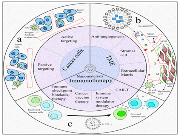
Up to now, quite a few conventional approaches are being applied to tackle the various types of cancer. Furthermore, despite the variations in working platforms and mechanisms, maximum of the researchers assumes two foremost targets. They are tumor cells and Tumor Micro Environment (TME) which consists of the immune system associated to the tumour. The schematic system [129] is being diagrammatically represented in Figure 7.
Strategies in Targeting Tumor Cells
Directly targeting the tumour cells is an ordinary method to eradicate cancer. With active targeting and EPR, improved nano-carriers like NPs, carbon nanomaterials (CNMs), and dendrimers can release the therapeutic antitumor drugs or biomaterials into the tumour cells [130]. In these platforms, antibodies targeting the exact overexpressed antigens on tumor cell surfaces are being used widely. After the endocytosis by tumor cells, the nucleic acid constituents induce cell apoptosis or the encapsulated therapeutic drugs produces cytotoxicity. Advancement has been made in nano- DDS of nucleic acids and exosomes [131, 132], liposomes, PNPs [133]; dendrimers [134] are immensely investigated in the cancer therapy.
Strategies in Targeting TME
The next important targeting strategy is TME, which contains cancerous cells. In accordance with various researches, angiogenesis is enormously very active almost in all the cancers due to the abnormal cell growth. Based on this distinctive characteristic, Sengupta prepared a NP system unambiguously targeting only the abnormal tumor cell angiogenesis along with combretastatin. This formulation was further encapsulated into the PLGA core with doxorubicin (DOX). Therefore, the DOX was proficiently utilized in cancer after a short break of cancerous vessels persuaded by combretastatin, and an enhanced overall therapeutic activity was attained [32]. Further abnormal vasculature and extracellular matrix (ECM) has been investigated in the tumor treatment. ECM also plays a major role and acts as a controlling scaffold in tumor cell proliferation, passage, incursion as well as angiogenesis [135]. Some foremost constituents contributing to these carcinogenic properties are collagen and other enzymes like Hyaluronidase (HA). Collagen is the chief structural protein of the ECM, which forms movement paths for the abnormal cells, whereas HA subsidizes to very high interstitial fluid pressure (IFP) and averting drug diffusion as well as penetration [136, 137]. Enzymes, like matrix metalloproteinases (MMPs), can control TME by deploying the action of non-ECM constituents, together with the various receptors, growth factors and cytokines [138]. In nanocarrier strategy, ECM is considered as one of the major parameters especially when combined with the traditional organic drugs. ECM hyaluronic acid produces a good therapeutic property against the metastatic pancreatic cancer, especially in patients with high expression of HA [139]. Numerous efforts are made to improve the penetration capability in tumors by nano carriers with hyaluronidase (HAase) for the antitumor drugs. It is not a complex method but has been considered as a very effective approach which exhibits improved anti-tumour efficacy [140].
Cancer Immunotherapy
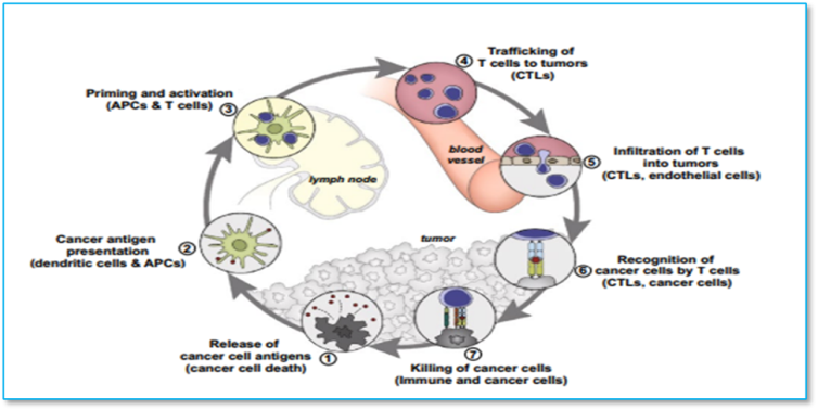
The immune system plays a vital role in cancer formation and progression. There are several approaches in immunotherapy and it is theoretically possible that each step in the cancer immunity cycle might be the potential therapeutic target with various methods including immune checkpoint blockade therapy, chimeric antigen receptor (CAR)-T cell therapy, cancer vaccine therapy and immune system modulator therapy [141]. In cancer immunotherapies, natural or synthetic molecules are used extensively to improve the function of immune system and exert anti-tumor effect. The most significant immune check points which are studied includes immune check point inhibitors (ICIs), targeting Programmed cell death protein 1 (PD-1) and programmed cell death ligand 1 (PD-L1) [142]. In research conducted by Bu and team, over-expression of PD-1 was considered to allow cancer cells to perform antitumor immunity evasion, and traditional immune check point inhibitors (ICIs) of PD-1/PD-L1 showed inconsistent benefits [143]. Schematic diagram for the cancer cancer- immunity cycle [144] is shown in Figure 8.
To ensure bonding of PD-L1 and ICIs, multivalent poly (amidoamine) dendrimers were employed; as a result, PD-L1 blockade effect was improved, and tumor site drug accumulation was enhanced [142]. CTLA-4 (cytotoxic T-lymphocyte-associated protein 4) is as an immune checkpoint with the function to down-regulate the immune responses [145]. Nano-based materials play a substantial role as drug transporter to deliver antibodies, small molecular inhibitors and proteins as drug constituents. Through these strategies has been considered as novel platforms in nanotechnology which can be developed further to achieve better efficacy and bioavailability than conventional therapies [146, 147].
Progress and Challenges of Cancer Therapy
Nano-based materials used in treatment and diagnosis of cancer have various advantages over the traditional biochemical drugs and also there are associated challenges in the nanomaterial application. Some noteworthy attributes in tumorigenesis are growth suppressor’s evasion, activating invasion and metastasis, cell death resistance, genomic instability, induced angiogenesis, continuous proliferative signalling, inflammation, mutation and replicative immortality [1, 148]. Some of the challenges associated to reach carcinogenic target sites are nanocarriers should pass through the fortifications such as vasculature, normal tissue microenvironment, and TME as well as the kidney filtration [149]. Cancer tissues generally showed dense ECM, high interstitial fluid and over-activated angiogenesis induced by excessive angiogenic factors will influence the application nanomaterials.
Conclusion and Future Perspectives
Nanotechnology has evolved as a promising approach in cancer therapy over the recent years. Due to their enhanced pharmacokinetic and dynamic properties, nano- based materials have been used in both cancer diagnosis as well as treatment. It allows targeted drug delivery in the particular region with minimal side effects. Though, as with other therapeutic possibilities, nanomaterials are not completely devoid of toxic effects and only comes with a number of challenges which causes setbacks with their clinical applications. Certain limitations are associated with the use of nanotechnology; hence more developments must be performed to maximize their efficacy and to improve its delivery while keeping the drawbacks to the least. By improving the interactions between the physicochemical properties of the nanomaterials employed, it can act as a new era of tumor treatment by providing lead molecules for the detection, diagnosis, and therapy of malignancy.
Cancer therapeutics based on the excellent characteristics of NPs is being massively used in the clinical set of numerous cancer types. Various categories of NPs, such as PNPs, hybrid NPs, and metallic NPs, have exhibited enhanced efficiency of drug delivery. Still, there are several limitations like deficit in vitro replicas which can exactly replicate in vivo stage, the long-term toxicity, immunotoxicity, and neurotoxicity. In comparison with the mammoth number of research, only limited nano-based drugs are really utilized, some are in clinical trials, and most of them are in the experimental stage. For rationale nanomaterial strategy, understanding the cellular as well as physiological aspects that control the nanobased drug delivery and toxicity profiles in the human beings are very essential. Based on the investigations mentioned above, we presume that the revolution in the clinical transformation for Nano-based cancer therapy will be achieved by means of nanotechnology and cancer therapeutics development.
References
-
Hanahan D, Weinberg RA (2011) Hallmarks of cancer: the next generation. Cell 144(5): 646-674.
-
Seyfried TN, Shelton LM (2010) Cancer as a metabolic disease. Nutr Metab 7: 7.
-
Nigro JM, Baker SJ, Preisinger AC, Jessup JM, Hostetter R, et al. (1989) Mutations in the p53 gene occur in diverse human tumour types. Nature 342(6250): 705-708.
-
Fitzmaurice C, Allen C, Barber RM, Barregard L, Bhutta ZA, et al. (2017) Global, Regional, and National Cancer Incidence, Mortality, Years of Life Lost, Years Lived With Disability, and Disability-Adjusted Life-Years for 32 Cancer Groups, 1990 to 2015: A Systematic Analysis for the Global Burden of Disease Study. JAMA Oncol 3(4): 524-548.
-
Fitzmaurice C, Abate D, Abbasi N, Abbastabar H, Abd- Allah F, et al. (2019) Global, Regional, and National Cancer Incidence, Mortality, Years of Life Lost, Years Lived With Disability, and Disability-Adjusted Life- Years for 32 Cancer Groups, 1990 to 2017: A Systematic Analysis for the Global Burden of Disease Study. JAMA Oncol 5(12): 1749-1768.
-
Bray F, Ferlay J, Soerjomataram I, Siegel RL, Torre LA, et al. (2018) Global cancer statistics 2018: GLOBOCAN estimates of incidence and mortality worldwide for 36 cancers in 185 countries. CA Cancer J Clin 68(6): 394- 424.
-
The Lancet (2018) GLOBOCAN 2018: counting the toll of cancer. Lancet 392(10152): 985.
-
Chen H, Zheng Y, Tian G, Tian Y, Zeng X, et al. (2011) Oral Delivery of DMAB-Modified Docetaxel-Loaded PLGA-TPGS Nanoparticles for Cancer Chemotherapy. Nanoscale Res Lett 6(1): 4.
-
Jain KK (2020) An Overview of Drug Delivery Systems. Methods Mol Biol 2059: 1-54.
-
Jang TK, Kim DY, Lee SW, Park JY, Suh DS, et al. (2018) Trends in Treatment during the Last Stages of Life in End-Stage Gynecologic Cancer Patients Who Received Active Palliative Chemotherapy: A Comparative Analysis of 10-Year Data in a Single Institution. BMC Palliat Care 17(1): 99.
-
López-Soto A, Gonzalez S, Smyth MJ, Galluzzi L (2017) Control of metastasis by NK cells. Cancer Cell 32(2): 135- 154.
-
Gallaher JA, Enriquez-Navas PM, Luddy KA, Gatenby RA, Anderson ARA, et al. (2018) Spatial heterogeneity and evolutionary dynamics modulate time to recurrence in continuous and adaptive cancer therapies. Cancer Res 78(8): 2127-2139.
-
Lacouture M, Sibaud V (2018) Toxic side effects of targeted therapies and immunotherapies affecting the skin, oral mucosa, hair, and nails. Am J Clin Dermatol 19(1): 31-39.
-
Dadwal A, Baldi A, Kumar Narang R (2018) Nanoparticles as carriers for drug delivery in cancer. Artif Cells Nanomed Biotechnol 46(2): 295-305.
-
van der Meel R, Sulheim E, Shi Y, Kiessling F, Mulder WJM, et al. (2019) Smart cancer nanomedicine. Nat Nanotechnol 14(11): 1007-1017.
-
Peer D, Karp JM, Hong S, Farokhzad OC, Margalit R, et al. (2007) Nanocarriers as an emerging platform for cancer therapy. Nat Nanotechnol 2(12): 751-760.
-
Malam Y, Loizidou M, Seifalian AM (2009) Liposomes and nanoparticles: nanosized vehicles for drug delivery in cancer. Trends Pharmacol Sci 30(11): 592-999.
-
Sutradhar KB, Amin ML (2013) Nanoemulsions: increasing possibilities in drug delivery. Eur J Nanomed 5(2): 97-110.
-
Praetorius NP, Mandal TK (2007) Engineered nanoparticles in cancer therapy. Recent Pat Drug Deliv Formul 1(1): 37-51.
-
Park K (2007) Nanotechnology: what it can do for drug delivery. J Control Release 120(1-2): 1-3.
-
Kim D, Jeong YY, Jon S (2010) A drug-loaded aptamer- gold nanoparticle bioconjugate for combined CT imaging and therapy of prostate cancer. ACS Nano 4(7): 3689- 3696.
-
Akhter S, Ahmad I, Ahmad MZ, Ramazani F, Singh A, et al. (2013) Nanomedicines as cancer therapeutics: current status. Curr Cancer Drug Targets 13(4): 362-378.
-
Ji T, Zhao Y, Wang J, Zheng X, Tian Y, et al. (2013) Tumor fibroblast specific activation of a hybrid ferritin nanocage- based optical probe for tumor microenvironment imaging. Small 9(14): 2427-2431.
-
Chen LL, Zhao L, Wang ZG, Liu SL, Pang DW, et al. (2022) Near‐Infrared‐II Quantum Dots for _In Vivo_ Imaging and Cancer Therapy. Small 18(8): 2104567.
-
Dubertret B, Skourides P, Norris DJ, Noireaux V, Brivanlou AH, et al. (2002) _In vivo_ imaging of quantum dots encapsulated in phospholipid micelles. Science 298(5599): 1759-1762.
-
Zhang Y, Yang H, An X, Wang Z, Yang X, et al. (2020) Controlled Synthesis of Ag(2) Te@Ag(2) S Core-Shell Quantum Dots with Enhanced and Tunable Fluorescence in the Second Near-Infrared Window. Small 16(14): e2001003.
-
Hirsch LR, Stafford RJ, Bankson JA, Sershen SR, Rivera B, et al. (2003) Nanoshell-mediated near-infrared thermal therapy of tumors under magnetic resonance guidance. Proc Natl Acad Sci U S A 100(23): 13549-13554.
-
Loo C, Lin A, Hirsch L, Lee MH, Barton J, et al. (2004) Nanoshell-enabled photonics-based imaging and therapy of cancer. Technol Cancer Res Treat 3(1): 33-40.
-
Nunes T, Pons T, Hou X, Van Do K, Caron B, et al. (2019) Pulsed-laser irradiation of multifunctional gold nanoshells to overcome trastuzumab resistance in HER2-overexpressing breast cancer. J Exp Clin Cancer Res 38(1): 306.
-
Wu C, Liang X, Jiang H (2005) Metal nanoshells as a contrast agent in near infra-red diffuse optical tomography. Opt Commun 253(1-3): 214-221.
-
Loo C, Lowery A, Halas N, West J, Drezek R, et al. (2005) Immunotargeted nanoshells for integrated cancer imaging and therapy. Nano Lett 5(4): 709-711.
-
Sengupta S, Eavarone D, Capila I, Zhao G, Watson N, et al. (2005) Temporal targeting of tumour cells and neovasculature with a nanoscale delivery system. Nature 436(7050): 568-572.
-
Fu F, Li L, Luo Q, Li Q, Guo T, et al. (2018) Selective and sensitive detection of lysozyme based on plasmon resonance light-scattering of hydrolyzed peptidoglycan stabilized-gold nanoparticles. The Analyst 143(5): 1133- 1140.
-
Shrivas K, Nirmalkar N, Thakur SS, Deb MK, Shinde SS, et al. (2018) Sucrose capped gold nanoparticles as a plasmonic chemical sensor based on non-covalent interactions: Application for selective detection of vitamins B (1) and B (6) in brown and white rice food samples. Food Chem 250: 14-21.
-
Paciotti GF, Myer L, Weinreich D, Goia D, Pavel N, et al. (2004) Colloidal gold: a novel nanoparticle vector for tumor directed drug delivery. Drug Deliv 11(3): 169-183.
-
Chen J, Saeki F, Wiley BJ, Cang H, Cobb MJ, et al. (2005) Gold nanocages: bioconjugation and their potential use as optical imaging contrast agents. Nano Lett 5(3): 473- 477.
-
van Vlerken LE, Amiji MM (2006) Multi-functional Polymeric Nanoparticles for Tumour- Targeted Drug Delivery. Expert Opin Drug Deliv 3(2): 205-216.
-
Ali ES, Sharker SM, Islam MT, Khan IN, Shaw S, et al. (2020) Targeting cancer cells with nanotherapeutics and nano-diagnostics: current status and future perspectives. Semin Cancer Biol 69: 52-68.
-
Sharma P, Bhargava M (2013) Applications and characteristics of nanomaterials in industrial environment. Res Dev 3(4): 63-72.
-
Song S, Qin Y, He Y, Huang Q, Fan C, et al. (2010) Functional nano-probes for ultrasensitive detection of biomolecules. Chem Soc Rev 39(11): 4234-4243.
-
Rosenblum D, Joshi N, Tao W, Karp JM, Peer D, et al. (2018) Progress and challenges towards targeted delivery of cancer therapeutics. Nat Commun 9(1): 1410.
-
Shin WK, Cho J, Kannan AG, Lee YS, Kim DW, et al. (2016) Cross-linked composite gel polymer electrolyte using mesoporous methacrylate-functionalized SiO2 nanoparticles for lithium-ion polymer batteries. Sci Rep 6: 26332.
-
Xiang J, Zhao R, Wang B, Sun X, Guo X, et al. (2021) Advanced Nano-Carriers for Anti-Tumor Drug Loading. Front Oncol 11: 758143
-
Laurent S, Forge D, Port M, Roch A, Robic C, et al. (2008) Magnetic iron oxide nanoparticles: synthesis, stabilization, vectorization, physicochemical characterizations, and biological applications. Chem Rev 108(6): 2064-2110.
-
Shayegh A, Khalatbari F, Zonoubi N, Zarazvand F, Monavvari F, et al. (2021) Chlorambucil-Chitosan Nano- Conjugate: An Efficient Agent Against Breast Cancer Targeted Therapy. Curr Drug Deliv 18(6): 721-728.
-
Firooz A, Nafisi S, Maibach HI (2015) Novel Drug Delivery Strategies for Improving Econazole Antifungal Action. Int J Pharm 495(1): 599-607.
-
Alarifi S, Ali D, Alkahtani S, Verma A, Ahamed M, et al. (2013) Induction of Oxidative Stress, DNA Damage, and Apoptosis in a Malignant Human Skin Melanoma Cell Line After Exposure to Zinc Oxide Nanoparticles. Int J Nanomedicine 8: 983-993.
-
Zheng W, Li M, Lin Y, Zhan X (2018) Encapsulation of Verapamil and Doxorubicin by MPEG-PLA to Reverse Drug Resistance in Ovarian Cancer. Biomed Pharmacother 108: 565-573.
-
Arya G, Das M, Sahoo SK (2018) Evaluation of Curcumin Loaded Chitosan/PEG Blended PLGA Nanoparticles for Effective Treatment of Pancreatic Cancer. Biomed Pharmacother 102: 555-566.
-
Dutta B, Shetake NG, Gawali SL, Barick BK, Barick KC, et al. (2018) PEG Mediated Shape-Selective Synthesis of Cubic Fe 3 O 4 Nanoparticles for Cancer Therapeutics. Journal of Alloys and Compounds 737: 347-355.
-
Zuccari G, Milelli A, Pastorino F, Loi M, Petretto A, et al. (2015) Tumor Vascular Targeted Liposomal-Bortezomib Minimizes Side Effects and Increases Therapeutic Activity in Human Neuroblastoma. J Control Release 211: 44-52.
-
Shanmugam V, Chien YH, Cheng YS, Liu TY, Huang CC, et al. (2014) Oligonucleotides–assembled Au Nanorod-Assisted Cancer Photothermal Ablation and Combination Chemotherapy With Targeted Dual-Drug Delivery of Doxorubicin and Cisplatin Prodrug. ACS Appl Mater Interfaces 6(6): 4382-4393.
-
Kapri S, Maiti S, Bhattacharyya S (2016) Lemon Grass Derived Porous Carbon Nanospheres Functionalized for Controlled and Targeted Drug Delivery. Carbon 100: 223-235.
-
Umrao S, Maurya AK, Shukla V, Grigoriev A, Ahuja R, et al. (2019) Anticarcinogenic Activity of Blue Fluorescent Hexagonal Boron Nitride Quantum Dots: As an Effective Enhancer for DNA Cleavage Activity of Anticancer Drug Doxorubicin. Mater Today Bio 1: 100001.
-
Astete CE, Sabliov CM (2006) Synthesis and Characterization of PLGA Nanoparticles. J Biomater Sci Polym Ed 17(3): 247-289.
-
Saneja A, Kumar R, Mintoo MJ, Dubey RD, Sangwan PL, et al. (2019) Gemcitabine and betulinic acid co- encapsulated PLGA-PEG polymer nanoparticles for improved efficacy of cancer chemotherapy. Mater Sci Eng C Mater Biol Appl 98: 764-771.
-
Zhou Q, Zhang L, Yang T, Wu H (2018) Stimuli-responsive polymeric micelles for drug delivery and cancer therapy. Int J Nanomedicine 13: 2921-2942.
-
Cagel M, Tesan FC, Bernabeu E, Salgueiro MJ, Zubillaga MB, et al. (2017) Polymeric mixed micelles as nanomedicines: achievements and perspectives. Eur J Pharm Biopharm 113: 211-228.
-
Barratt G (2003) Colloidal Drug Carriers: Achievements and Perspectives. Cell Mol Life Sci 60(1): 21-37.
-
Palazzolo S, Bayda S, Hadla M, Caligiuri I, Corona G, et al. (2018) The Clinical Translation of Organic Nanomaterials for Cancer Therapy: A Focus on Polymeric Nanoparticles, Micelles, Liposomes and Exosomes. Curr Med Chem 25(34): 4224-4268.
-
Huang HC, Barua S, Sharma G, Dey SK, Rege K, et al. (2011) Inorganic Nanoparticles for Cancer Imaging and Therapy. J Control Release 155(3): 344-357.
-
Bayda S, Hadla M, Palazzolo S, Riello P, Corona G, et al. (2018) Inorganic Nanoparticles for Cancer Therapy: A Transition from Lab to Clinic. Curr Med Chem 25(34): 4269-4303.
-
Pugazhendhi A, Edison TNJI, Karuppusamy I, Kathirvel B (2018) Inorganic Nanoparticles:A Potential Cancer Therapy for Human Welfare. Int J Pharm 539(1-2): 104- 111.
-
Winnik FM, Maysinger D (2013) Quantum Dot Cytotoxicity and Ways to Reduce it. Acc Chem Res 46(3): 672-680.
-
Chen ML, He YJ, Chen XW, Wang JH (2012) Quantum Dots Conjugated With Fe3O4-Filled Carbon Nanotubes for Cancer-Targeted Imaging and Magnetically Guided Drug Delivery. Langmuir 28(47): 16469-16476.
-
Tapeinos C, Battaglini M, Ciofani G (2017) Advances in the Design of Solid Lipid Nanoparticles and Nanostructured Lipid Carriers for Targeting Brain Diseases. J Control Release 264: 306-332.
-
Basoglu H, Goncu B, Akbas F (2018) Magnetic nanoparticle-mediated gene therapy to induce Fas apoptosis pathway in breast cancer. Cancer Gene Ther 25(5-6): 141-147.
-
Mandriota G, Di Corato R, Benedetti M, De Castro F, Fanizzi FP, et al. (2019) Design and application of cisplatin-loaded magnetic nanoparticle clusters for smart chemotherapy. ACS Appl Mater Interfaces 11(2): 1864-1875.
-
Hoopes PJ, Moodie KL, Petryk AA, Petryk JD, Sechrist S, et al. (2017) Hypo-fractionated radiation, magnetic nanoparticle hyperthermia and a viral immunotherapy treatment of spontaneous canine cancer. Proc SPIE Int Soc Opt Eng 10066: 1006605.
-
Zhao S, Minh LV, Li N, Garamus VM, Handge UA, et al. (2016) Doxorubicin Hydrochloride-Oleic Acid Conjugate Loaded Nanostructured Lipid Carriers for Tumor Specific Drug Release. Colloids Surf B Biointerfaces 145: 95-103.
-
Patil A, Lakhani P, Taskar P, Wu KW, Sweeney C, et al. (2018) Formulation Development, Optimization, and In Vitro-_In Vivo_ Characterization of Natamycin-Loaded PEGylated Nano-Lipid Carriers for Ocular Applications. J Pharm Sci 107(8): 2160-2171.
-
Wu G, Li J, Yue J, Zhang S, Yunusi K, et al. (2018) Liposome Encapsulated Luteolin Showed Enhanced Antitumor Efficacy to Colorectal Carcinoma. Mol Med Rep 17(2): 2456-2464.
-
Pastorino F, Brignole C, Marimpietri D, Sapra P, Moase EH, et al. (2003) Doxorubicin-Loaded Fab’ Fragments of Anti-Disialoganglioside Immunoliposomes Selectively Inhibit the Growth and Dissemination of Human Neuroblastoma in Nude Mice. Cancer Res 63(1): 86-92.
-
Han B, Yang Y, Chen J, Tang H, Sun Y, et al. (2020) Preparation, Characterization, and Pharmacokinetic Study of a Novel Long-Acting Targeted Paclitaxel Liposome with Antitumor Activity. Int J Nanomedicine 15: 553-571.
-
O’Brien ME, Wigler N, Inbar M, Rosso R, Grischke E, et al. (2004) Reduced cardiotoxicity and comparable efficacy in a phase III trial of pegylated liposomal doxorubicin HCl (CAELYX/Doxil) versus conventional doxorubicin for first-line treatment of metastatic breast cancer. Ann Oncol 15(3): 440-449.
-
Meng J, Guo F, Xu H, Liang W, Wang C, et al. (2016) Combination Therapy using Co-encapsulated Resveratrol and Paclitaxel in Liposomes for Drug Resistance Reversal in Breast Cancer Cells _in vivo_. Sci Rep 6: 22390.
-
Krishnagopal A, Reddy A, Sen D (2017) Stent-Mediated Gene and Drug Delivery for Cardiovascular Disease and Cancer: A Brief Insight. J Gene Med 19(5).
-
Kohn DB, Sadelain M, Glorioso JC (2003) Occurrence of Leukaemia Following Gene Therapy of X-Linked SCID. Nat Rev Cancer 3(7): 477-488.
-
Robbins PD, Ghivizzani SC (1998) Viral Vectors for Gene Therapy. Pharmacol Ther 80(1): 35-47.
-
Yin H, Kanasty RL, Eltoukhy AA, Vegas AJ, Dorkin JR, et al. (2014) Non-viral vectors for gene-based therapy. Nat Rev Genet 15(8): 541-555.
-
Zhang Y, Satterlee A, Huang L (2012) _In Vivo_ Gene Delivery by Nonviral Vectors: Overcoming Hurdles? Mol Ther 20(7): 1298-1304.
-
Fang H, Feng Y, Chen J, Tian H, Chen X, et al. (2019) Constructing Efficient Polycationic Gene Carriers Through Regulating the Physicochemical Properties. Mater Today Chem 11: 269-282.
-
Wang P, Zhang L, Zheng W, Cong L, Guo Z, et al. (2018) Thermo-Triggered Release of CRISPR-Cas9 System by Lipid-Encapsulated Gold Nanoparticles for Tumor Therapy. Angew Chem Int Ed Engl 57(6): 1491-1496.
-
Kim MS, Haney MJ, Zhao Y, Mahajan V, Deygen I, et al. (2016) Development of exosome-encapsulated paclitaxel to overcome mdr in cancer cells. Nanomedicine 12(3): 655-664.
-
Lou G, Song X, Yang F, Wu S, Wang J, et al. (2015) Exosomes derived from mir-122-modified adipose tissue-derived mscs increase chemosensitivity of hepatocellular carcinoma. J Hematol Oncol 8: 122.
-
Yuan L, Liu Y, Qu Y, Liu L, Li H, et al. (2019) Exosomes derived from microrna-148b-3p-overexpressing human umbilical cord mesenchymal stem cells restrain breast cancer progression. Front Oncol 9: 1076.
-
Gong C, Tian J, Wang Z, Gao Y, Wu X, et al. (2019) Functional exosome-mediated co-delivery of doxorubicin and hydrophobically modified microrna 159 for triple- negative breast cancer therapy. J Nanobiotechnology 17(1): 93.
-
You C, Wu H, Wang M, Wang S, Shi T, et al. (2017) A Strategy for Photothermal Conversion of Polymeric Nanoparticles by Polyaniline for Smart Control of Targeted Drug Delivery. Nanotechnology 28(16): 165102.
-
Prabu SL, Kuppusami Suriyaprakash TN, Thirumurugan R (2017) Medicated Nanoparticle for Gene Delivery. In: Maiti S, et al. (Eds.), Advanced Technology for Delivering Therapeutics.
-
Maeda H, Wu J, Sawa T, Matsumura Y, Hori K, et al. (2000) Tumor Vascular Permeability and the EPR Effect in Macromolecular Therapeutics: A Review. J Control Release 65(1-2): 271-284.
-
Nichols JW, Bae YH (2014) EPR: Evidence and Fallacy. J Control Release 190: 451-464.
-
Mitra S, Gaur U, Ghosh PC, Maitra AN (2001) Tumour Targeted Delivery of Encapsulated Dextran-Doxorubicin Conjugate Using Chitosan Nanoparticles as Carrier. J Control Release 74(1-3): 317-323.
-
Subhan MA, Yalamarty SSK, Filipczak N, Parveen F, Torchilin VP, et al. (2021) Recent Advances in Tumor Targeting via EPR Effect for Cancer Treatment. J Pers Med 11(6): 571.
-
Carmeliet P, Jain RK (2000) Angiogenesis in cancer and other diseases. Nature 407(6801): 249-257.
-
Carita AC, Eloy JO, Chorilli M, Lee RJ, Leonardi GR, et al. (2018) Recent advances and perspectives in liposomes for cutaneous drug delivery. Curr Med Chem 25(5): 606- 635.
-
Cırpanlı Y, Allard E, Passirani C, Bilensoy E, Lemaire L, et al. (2011) Antitumoral activity of camptothecin-loaded nanoparticles in 9L rat glioma model. Int J Pharm 403(1- 2): 201-206.
-
Gaur S, Wang Y, Kretzner L, Chen L, Yen T, et al. (2014) Pharmacodynamic and pharmacogenomic study of the nanoparticle conjugate of camptothecin CRLX101 for the treatment of cancer. Nanomedicine 10(7): 1477-1486.
-
Lu Y, Low PS (2002) Folate-Mediated Delivery of Macromolecular Anticancer Therapeutic Agents. Adv Drug Deliv Rev 54(5): 675-693.
-
Elnakat H, Ratnam M (2006) Role of Folate Receptor Genes in Reproduction and Related Cancers. Front Biosci 11: 506-519.
-
Kularatne SA, Zhou Z, Yang J, Post CB, Low PS, et al. (2009) Design, Synthesis, and Preclinical Evaluation of Prostate-Specific Membrane Antigen Targeted (99m) Tc- Radioimaging Agents. Mol Pharm 6(3): 790-800.
-
Bhuniya S, Maiti S, Kim EJ, Lee H, Sessler JL, et al. (2014) An Activatable Theranostic for Targeted Cancer Therapy and Imaging. Angew Chem Int Ed Engl 53(17): 4469-4474.
-
Högemann-Savellano D, Bos E, Blondet C, Sato F, Abe T, et al. (2003) The Transferrin Receptor: A Potential Molecular Imaging Marker for Human Cancer. Neoplasia 5(6): 495-506.
-
Reubi JC (2003) Peptide Receptors as Molecular Targets for Cancer Diagnosis and Therapy. Endocr Rev 24(4): 389-427.
-
Krall N, Pretto F, Decurtins W, Bernardes GJ, Supuran CT, et al. (2014) A Small-Molecule Drug Conjugate for the Treatment of Carbonic Anhydrase IX Expressing Tumors. Angew Chem Int Ed Engl 53(16): 4231-4235.
-
Mackiewicz N, Nicolas J, Handke N, Noiray M, Mougin J, et al. (2014) Precise Engineering of Multifunctional PEGylated Polyester Nanoparticles for Cancer Cell Targeting and Imaging. Chem Mater 26(5):1834-1847.
-
Amreddy N, Muralidharan R, Babu A, Mehta M, Johnson EV, et al. (2015) Tumor-targeted and pH- controlled delivery of doxorubicin using gold nanorods for lung cancer therapy. Int J Nanomedicine 10: 6773- 6788.
-
Soe ZC, Kwon JB, Thapa RK, Ou W, Nguyen HT, et al. (2019) Transferrin-conjugated polymeric nanoparticle for receptor-mediated delivery of doxorubicin in Doxorubicin-resistant breast cancer cells. Pharmaceutics 11(2): 63.
-
Nicholson RI, Gee JM, Harper ME (2001) EGFR and cancer prognosis. Eur J Cancer 37(4): 9-15.
-
Alexis F, Basto P, Levy-Nissenbaum E, Radovic- Moreno AF, Zhang L, et al. (2008) HER-2-targeted nanoparticle-affibody bioconjugates for cancer therapy. ChemMedChem 3(12): 1839-1843.
-
Balasubramanian S, Girija AR, Nagaoka Y, Iwai S, Suzuki M, et al. (2014) Curcumin and 5-fluorouracil- loaded, folate- and transferrin decorated polymeric magnetic nanoformulation: a synergistic cancer therapeutic approach, accelerated by magnetic hyperthermia. Int J Nanomedicine 9: 437-459.
-
Thambi T, Deepagan VG, Yoon HY, Han HS, Kim SH, et al. (2014) Hypoxia-Responsive Polymeric Nanoparticles for Tumor-Targeted Drug Delivery. Biomaterials 35(5): 1735-1743.
-
Thambi T, Son S, Lee DS, Park JH (2016) Poly(ethylene Glycol)-B-Poly (Lysine) Copolymer Bearing Nitroaromatics for Hypoxia-Sensitive Drug Delivery. Acta Biomater 29: 261-270.
-
Zhang YJ, Gallis B, Taya M, Wang S, Ho RJ, et al. (2013) pH-Responsive Artemisinin Derivatives and Lipid Nanoparticle Formulations Inhibit Growth of Breast Cancer Cells In Vitro and Induce Down-Regulation of HER Family Members. PLoS One 8(3): e59086.
-
Zelzer M , Todd SJ , Hirst AR , McDonald TO, Ulijn RV, et al. (2013) Enzyme Responsive Materials: Design Strategies and Future Developments. Biomater Sci 1(1): 11-39.
-
Di Corato R, Béalle G, Kolosnjaj-Tabi J, Espinosa A, Clément O, et al. (2015) Combining magnetic hyperthermia and photodynamic therapy for tumor ablation with photoresponsive magnetic liposomes. ACS Nano 9(3): 2904-2916.
-
Corr SA, O’Byrne A, Gun’ko YK, Ghosh S, Brougham DF, et al. (2006) Magnetic-fluorescent nanocomposites for biomedical multitasking. Chem Commun 43: 4474- 4476.
-
Shah SA, Aslam Khan MU, Arshad M, Awan SU, Hashmi MU, et al. (2016) Doxorubicin-Loaded Photosensitive Magnetic Liposomes for Multi-Modal Cancer Therapy. Colloids Surf B Biointerfaces 148: 157-164.
-
Stubbs M, McSheehy PM, Griffiths JR, Bashford CL (2000) Causes and Consequences of Tumour Acidity and Implications for Treatment. Mol Med Today 6(1): 15-19.
-
Deng Z, Zhen Z, Hu X, Wu S, Xu Z, et al. (2011) Hollow Chitosan-Silica Nanospheres as pH-Sensitive Targeted Delivery Carriers in Breast Cancer Therapy. Biomaterials 32(21): 4976-4986.
-
Fomina N, Sankaranarayanan J, Almutairi A (2012) Photochemical Mechanisms of Light-Triggered Release from Nanocarriers. Adv Drug Deliv Rev 64(11): 1005- 1020.
-
You J, Zhang R, Xiong C, Zhong M, Melancon M, et al. (2012) Effective Photothermal Chemotherapy Using Doxorubicin-Loaded Gold Nanospheres That Target EphB4 Receptors in Tumors. Cancer Res 72(18): 4777- 4786.
-
Xia J, Du Y, Huang L, Chaurasiya B, Tu J, et al. (2018) Redox-Responsive Micelles From Disulfide Bond- Bridged Hyaluronic Acid-Tocopherol Succinate for the Treatment of Melanoma. Nanomedicine 14(3): 713-723.
-
Wang H, Tang L, Tu C, Song Z, Yin Q, et al. (2013) Redox-Responsive, CoreCross-Linked Micelles Capable of on-Demand, Concurrent Drug Release and Structure Disassembly. Biomacromolecules 14(10): 3706-3712.
-
Widder KJ, Senyel AE, Scarpelli GD (1978) Magnetic Microspheres: A Model System of Site Specific Drug Delivery _in Vivo_. Proc Soc Exp Biol Med 158(2): 141-146.
-
Yang HW, Hua MY, Liu HL, Huang CY, Tsai RY, et al. (2011) Self-Protecting Core-Shell Magnetic Nanoparticles for Targeted, Traceable, Long Half-Life Delivery of BCNU to Gliomas. Biomaterials 32(27): 6523-6532.
-
Zhang L, Wang T, Yang L, Liu C, Wang C, et al. (2012) General Route to Multifunctional Uniform Yolk/ Mesoporous Silica Shell Nanocapsules: A Platform for Simultaneous Cancer-Targeted Imaging and Magnetically Guided Drug Delivery. Chemistry 18(39): 12512-12521.
-
Yang C, Rait A, Pirollo KF, Dagata JA, Farkas N, et al. (2008) Nanoimmunoliposome Delivery of Superparamagnetic Iron Oxide Markedly Enhances Targeting and Uptake in Human Cancer Cells In Vitro and _In Vivo_. Nanomedicine 4(4): 318-329.
-
Zhang F, Braun GB, Pallaoro A, Zhang Y, Shi Y, et al. (2012) Mesoporous Multifunctional Upconversion Luminescent and Magnetic “Nanorattle” Materials for Targeted Chemotherapy. Nano Lett 12(1): 61-67.
-
Roy B, Ghose S, Biswas S (2022) Therapeutic strategies for miRNA delivery to reduce hepatocellular carcinoma. Semin Cell Dev Biol 124: 134-144.
-
Maghsoudnia N, Baradaran Eftekhari R, Naderi Sohi A, Norouzi P, Akbari H, et al. (2020) Mitochondrial delivery of microRNA mimic let-7b to NSCLC cells by PAMAM-based nanoparticles. J Drug Target 28(7-8): 818-830.
-
Jeong K, Yu YJ, You JY, Rhee WJ, Kim JA (2020) Exosome-mediated micro-RNA-497 delivery for anti- cancer therapy in a microfuidic 3D lung cancer model. Lab Chip 20(3): 548-557.
-
Tarach P, Janaszewska A (2021) Recent advances in preclinical research using PAMAM dendrimers for cancer gene therapy. Int J Mol Sci 22(6): 2912.
-
Zhang K, Dong C, Chen M, Yang T, Wang X, et al. (2020) Extracellular vesicle-mediated delivery of miR-101 inhibits lung metastasis in osteosarcoma. Theranostics 10(1): 411-425.
-
Cheng Z, Li M, Dey R, Chen Y (2021) Nanomaterials for cancer therapy: current progress and perspectives. J Hematol Oncol 14: 85.
-
Tredan O, Galmarini CM, Patel K, Tannock IF (2007) Drug resistance and the solid tumor microenvironment. J Natl Cancer Inst 99(19): 1441-1454.
-
Gkretsi V, Stylianou A, Papageorgis P, Polydorou C, Stylianopoulos T (2015) Remodeling components of the tumor microenvironment to enhance cancer therapy. Front Oncol 5: 214.
-
Klemm F, Joyce JA (2015) Microenvironmental regulation of therapeutic response in cancer. Trends Cell Biol 25(4): 198-213.
-
Merchant N, Nagaraju GP, Rajitha B, Lammata S, Jella KK, et al. (2017) Matrix metalloproteinases: their functional role in lung cancer. Carcinogenesis 38(8): 766-780.
-
Hingorani SR, Harris WP, Beck JT, Berdov BA, Wagner SA, et al. (2016) Phase Ib study of PEGylated recombinant human hyaluronidase and gemcitabine in patients with advanced pancreatic cancer. Clin Cancer Res 22(12): 2848-2854.
-
Chen E, Han S, Song B, Xu L, Yuan H, et al. (2020) Mechanism investigation of hyaluronidase-combined multistage nanoparticles for solid tumor penetration and antitumor effect. Int J Nanomed 15: 6311-6324.
-
Yan S, Luo Z, Li Z, Wang Y, Tao J, et al. (2020) Improving cancer immunotherapy outcomes using biomaterials. Angew Chem Int Ed Engl 59(40): 17332-17343.
-
Liu YT, Sun ZJ (2021) Turning cold tumors into hot tumors by improving T-cell infiltration. Theranostics. 11(11): 5365-5386.
-
Bu J, Nair A, Iida M, Jeong WJ, Poellmann MJ, et al. (2020) An avidity-based PD-L1 antagonist using nanoparticle-antibody conjugates for enhanced immunotherapy. Nano Lett 20(7): 4901-4909.
-
Mu W, Chu Q, Liu Y, Zhang N (2020) A Review on Nano-Based Drug Delivery System for Cancer Chemoimmunotherapy. Nano-Micro Lett 12(1): 142.
-
Sanaei MJ, Sigaroodi AP, Kaveh V, Sheikholeslami SA, Salari S, et al. (2021) The application of nano-medicine to overcome the challenges related to immune checkpoint blockades in cancer immunotherapy: Recent advances and opportunities. Crit Rev Oncol Hematol 157: 103160.
-
Dianzani C, Monge C, Miglio G, Serpe L, Martina K, et al. (2020) Nanoemulsions as delivery systems for poly- chemotherapy aiming at melanoma treatment. Cancers (Basel) 12(5): 1198.
-
Burnett M, Abuetabh Y, Wronski A, Shen F, Persad S, et al. (2020) Graphene oxide nanoparticles induce apoptosis in wild-type and CRISPR/Cas9-IGF/IGFBP3 knocked-out osteosarcoma cells. J Cancer 11(17): 5007- 5023.
-
Hanahan D, Weinberg RA (2000) The hallmarks of cancer. Cell 100(1): 57-70.
-
Hartshorn CM, Bradbury MS, Lanza GM, Nel AE, Rao J, et al. (2018) Nanotechnology strategies to advance outcomes in clinical cancer care. ACS Nano 12(1): 24-43.
- Effects of 5-HTP and Melatonin on the Sleep Cycle of Medical Students
- Adsorption of Bisphenol A on NH4OH- Modified Rice Husk and Sugar Cane Bagasse Biochar
- Comparative Assessment of the Reinforcement Efficiency of Palm Fruit Fibre and Coconut Fibre in High Density Polyethylene (HDPE) Matrix Composite
- Importance of Bio Compounds Naturally Present in Food with Functionality in Animal Metabolism
- Sub-Acute Study on the Cardiotoxic Effects of Monosodium Glutamate Ingestion in Albino Rat
- Weight Management and Its Natural Solutions: A Review