Dermoscopy of Seborrheic Keratosis-Like Melanoma
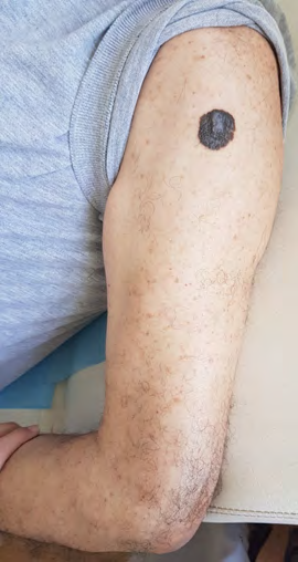
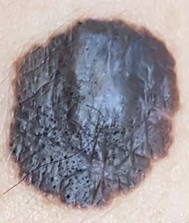
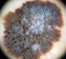
77-year-old patient, diabetic on insulin, who consulted for an increase in the size of a pigmented lesion in the left arm for 1 year asymptomatic. The dermatological examination found multiple seborrheic keratoses on the face, and a well limited blackish-brown pigmented plaque, slightly raised by place at the level of the left arm making about 3cm of long axis (Figure 1) whose clinical appearance resembled a pigmented seborrheic keratosis because it had prominent pseudofollicular openings on its surface (Figure 2). Dermoscopic examination revealed an asymmetrical and pigmented lesion with a bluish, dark brown to light brown and black coloration demonstrating a clearly visible bluewhite veil on the raised center of the lesion suggesting a malignant melanoma and a rainbow appearance, of more, a well-defined zone of seborrheic keratosis with multiple pseudo comedones (Figure 3). The examination of the lymph nodes was normal. A diagnosis of superficial spread malignant melanoma (MSS) was made histologically. An extension assesment was without particularities. A wide excision with margin of 0.5 cm was performed.
Figures
- Epithelioid Granuloma; 3cases with Different Clinical Features
- Advancing Representation in Dermatology Clinical Trials: Ethical, Scientific, and Regulatory Imperatives for Inclusion Across all Fitzpatrick Skin Types
- A Case of Atopic Dermatitis with Concurrent Psoriasis Vulgaris: Successful Treatment with Upadacitinib
- Innovation Lifting Eyeshadow: A Synthesis of Makeup and Optical Illusion
- Distinguishing Superficial Actinic Porokeratosis from Actinic Keratosis with UVF Dermoscopy: A Case Report
- High Mobility Group Box 1 (HMGB1) in Cutaneous Inflammation: An Immune Modulator Bridging Cellular Stress, Ferroptosis and Danger Signaling