Waardenburg Syndrome: Report of a Type III Family Case
Introduction: Waardenburg syndrome is an autosomal dominant condition, caused by changes in survival, proliferation, migration and/or differentiation of precursors of melanocytes. The most frequent detections are dystopia canthorum, sinofris, broad nasal base, pigmentary alterations of the iris and skin, congenital deafness and frontal white wick. It can be found in 4 types, the most common being types I and II. Case Report: We report a case of Waardenburg type III syndrome in a male patient, 13-years-old, with complaint of generalized pruritus for three months. The exam revealed clinical condition suggestive of Atopic Dermatitis, in addition to hypertelorism, bilateral dystopia canthorum, sinofris, iris heterochromia, frontal white wick, articular contractures in upper limbs and hypochromic lesions in the thorax, dorsum and abdomen. The mother had a similar condition, and both had congenital deafness. Conclusion: The patient was diagnosed with Waadenburg type III syndrome, in which there are musculoskeletal abnormalities of the upper limbs associated with the most frequent clinical. The diagnosis of the syndrome allows a multidisciplinary followup of the patients.
Introduction
Waardenburg syndrome is an autosomal dominant condition with variable penetrance and expressivity of its features. The most frequent findings are dystopia in the upper right corner, signori’s, broad nasal base, pigmentary changes in the iris and skin, congenital deafness and white frontal wick. It can be found in 4 types, the most common are types I and II.
About 90% of patients have an affected parent. It is a neurocristopathy in which occur changes in survival, proliferation, migration and / or differentiation of precursors of melanocytes to the inner ear, iris and skin.
The most frequent clinical signs are lateral displacement of the inner corners of the eyes, hyperplasia of the medial portion of the eyebrows, a prominent and enlarged nasal base, changes in the pigmentation of the iris and skin, congenital deafness, frontal white wick or early graying.
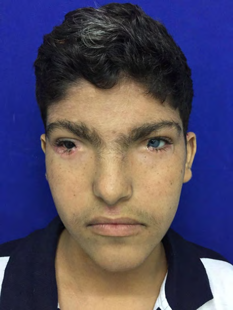
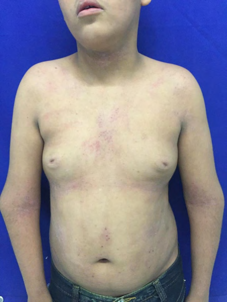
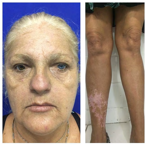
Male patient, 13 years old, brown, son of non- consanguineous parents, sought care complaining of generalized pruritus for 3 months. On examination, he had xerotic skin, scaly erythematous plaques on the trunk, back and limbs and lichenification in antecubital fossae, a clinical picture suggestive of Atopic Dermatitis. He also had hypertelorism, dystopia canthorum, sinofris, iris heterochromia, frontal white wick, joint contractures in the upper limbs and hypochromic lesions in the chest, back and abdomen (Figures 1 and 2). The patient’s mother had a similar clinical picture (Figure 3).
Mother and son were diagnosed with congenital deafness. Thus, the teenager was treated for atopic dermatitis and referred to the geneticist, being then diagnosed with type III Waardenburg Syndrome. He was also referred for otorhinolaryngological follow-up.
Conclusion
The case reported is a patient with type III Waardenburg Syndrome. There is no curative treatment, as it is a genetic disease, but physiotherapy sessions can be performed to improve muscle contractures.
The recognition of the disease by the dermatologist is very important, as it allows the early detection of sensorineural deafness, as well as the interdisciplinary monitoring of the various complications associated with the variants of this syndrome.
References
-
Zaman A, Capper R, Baddoo W (2018) Waardenburg syndrome: More common than you think. Clinical Otolaryngology 40(1): 44-48.
-
Imperato PJ, Imperato GH (2015) Clinical Manifestations of Waardenburg Syndrome in a Male Adolescent in Mali, West Africa. J Community Health 40: 103-109.
-
Llalliré JC, Park KY, Passarelli M, Petuaud G, Raffo G, et al. (2010) Síndrome de Waardenburg. Buenos Aires: Arch Oftal B Aires 81(2): 59-61.
-
Goenaga AM, Ferreira LC, Ferreira RC, Cestari TF (1996) Syndrome of Waardenburg: Report of two cases and literature review. Ann Bras Dermatol 71(5): 419-423.
- Epithelioid Granuloma; 3cases with Different Clinical Features
- Advancing Representation in Dermatology Clinical Trials: Ethical, Scientific, and Regulatory Imperatives for Inclusion Across all Fitzpatrick Skin Types
- A Case of Atopic Dermatitis with Concurrent Psoriasis Vulgaris: Successful Treatment with Upadacitinib
- Innovation Lifting Eyeshadow: A Synthesis of Makeup and Optical Illusion
- Distinguishing Superficial Actinic Porokeratosis from Actinic Keratosis with UVF Dermoscopy: A Case Report
- High Mobility Group Box 1 (HMGB1) in Cutaneous Inflammation: An Immune Modulator Bridging Cellular Stress, Ferroptosis and Danger Signaling