A Foot Mycetoma about One Case
Mycetomas are chronic subcutaneous infections that are mutilating and endemic in the dry tropics, they can be caused by fungal or bacterial agents. They sit preferentially on the ends. We report a case of mycetoma in a young woman living in urban areas and outside any endemic area of mycetoma.
Observation
A woman of 34 years had presented for a year a painless nodule sitting on the soles of the right foot. She had not been noted a particular antecedent, including a notion of trauma or residence in rural areas and she did not practice agricultural activities. Likewise, this patient had no previous history of moving to a rural area and was not used to walking barefoot.
The patient has been operated for this lesion on the level of the sole of the right foot, whose anatomopathological study was in favor of a granulomatous polymorphous inflammatory lesion evoking an infectious origin; the postoperative consequences were marked by the reappearance of the lesion in the following year, gradually increasing in size.
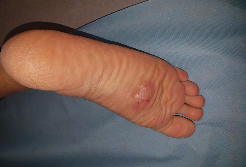
The dermatological examination revealed subcutaneous nodules, painless, confluent, producing a polylobed tumor appearance with an erythematous-purplish surface, strewn with openings in the sole of the right foot (Figure 1).
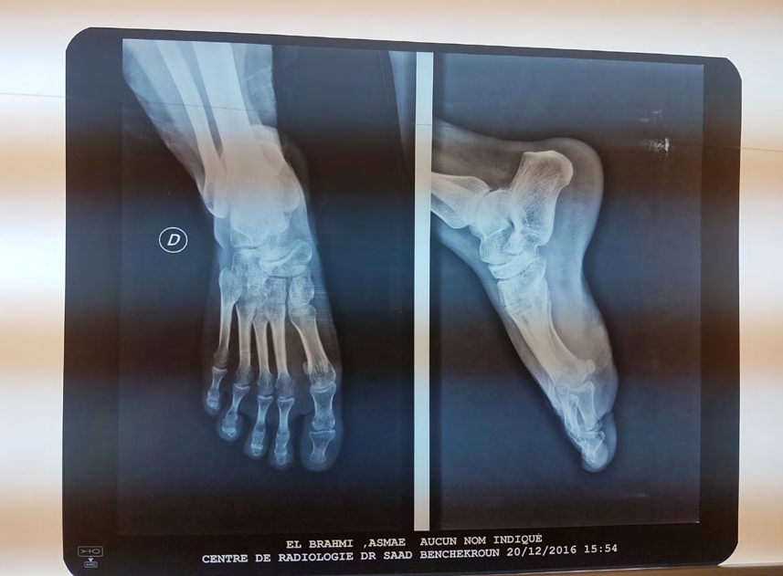
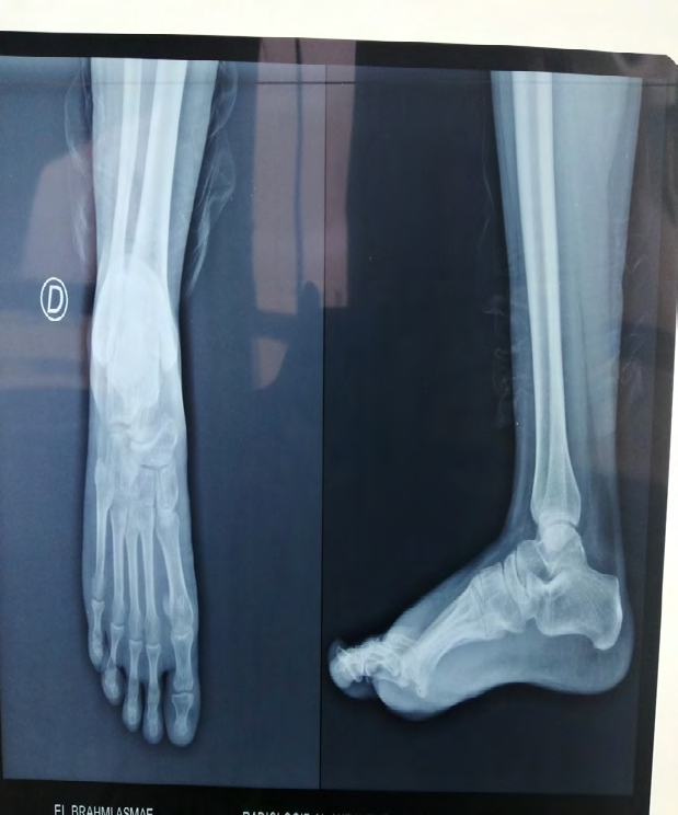
The patient had an X-ray of the limb showing a speckled aspect in the tarsal bones with poorly individualized joint lines and a thickening of the soft tissues facing it (Figure 2).
A biopsy with 3 fragments was performed. The analysis of the histological sections made and stained in turn with haematin-eosin and Gomori had found a moderately acanthotic and papillomatous epidermis surmounted by a compact hyperkeratotic hyperkeratosis. In the deep dermis and hypodermis, there were nodular infiltrates centered by foci of suppuration containing altered polynuclear debris surrounded by giant cells. The center of these micro-abscesses is occupied by grains composed of very fine entangled filaments surrounded by a fibrinoid fringe realizing the phenomenon of Splendore Hoeppli. Infectious agents are stained with positive PAS, gomorri crocott with a greyish, non-black appearance. They are also Gram positive and Ziehl neelson weakly positive.
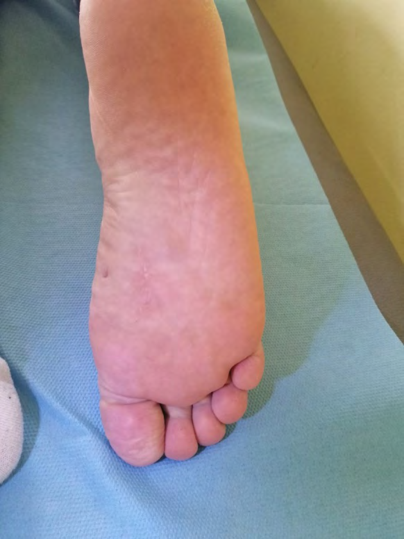
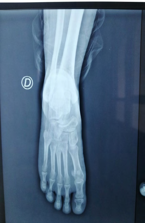
Bacteriological, mycological, parasitological examinations were negative. These examinations showed a deep fungal mycosis of bacterial origin, the histochemistry was in favor of norcardia. The patient was treated with the trimethropin-sulfamate-thoxazol combination for 6 months with good progress (Figure 3). The patient performed an x-ray of the control foot was normal (Figure 4).
Discussion
The mycetomas are a rare skin ailment that could be caused by actinomycetomas or eumycetomas which, in vivo, the developmental form is the grain [1]. They occur in the rural areas of tropical and subtropical countries [2, 3, 4], first described by Gill in 1842 in the Madura region of India. In Africa, the endemic areas are Sudan [5], Congo [6] and Somalia [7]. In July 2013, the mycetomas have been added to the neglected diseases list of WHO [8].
The mycetomas classically affect men of rural origin in the age group of 20-40 years old [9], farmers and people used to walking barefoot. In our case these facts contrasted with the data from literature. However it is shown that the contamination occurs during skin break-ins allowing the germ inoculation contained in the soil and plants [10]. The typical aspect of the mycetoma is a unilateral, painless swelling with productive fistulas whose emitted liquid, sero- hematic or contains grains visible or not to the naked eye. The fistulas can be unproductive [9]. Black grains point to a fungal etiology, red grains to an actinomycotic etiology. White or yellow grains are not discriminating [11].
The mycetomas preferentially sit on feet, like our case. Moreover, other extra-podal locations have been described particularly facial, nuchal and the upper limb [12]. The association with other pathologies is rare [8]. Bone complications are common types association image of destruction and reconstruction and must be systematically sought by radiology. The other complications are rarer: superinfection, lymph node metastases, local compressions, functional disability. Visceral invasions are exceptional [8].
The main differential diagnoses are bacterial osteomyelitis [13]. The confirmation of diagnosis is based on the microscopic examination coupled or not with the biological examination [7, 8] revealed the presence of several granulomas constituted of polymorphic inflammatory elements, bordering masses made of short and septated mycelial filaments, not massed. Whatever the etiology, the treatment should always be medical in the first place. The mycetomas’ treatment is difficult, long and expensive. Several therapeutic programs may be proposed, the choice will depend on the etiological agent [9].
The first-line treatment of actinomycetomas is the trimethropin-sulfamate-thoxazol combination. It must be given for a minimum of 1 year. Cotrimoxazol is the standard gold treatment, the association with amikacin is reserved for patients who do not respond to this treatment or who have severe forms. Other options are possible for the very rare cases where previous ones have failed [8]. There is also an interest in the amoxicillin-clavulanic acid and ketoconazole combination in the treatment of fungal mycetoma with superinfection by S.aureus [14]. Surgical indications have become exceptional and couldn’t be placed before a well led medical treatment. First-line surgery carries a risk of lymphatic lymph node metastasis [9]. For the moment, there is no biological criterion for healing. Prolonged monitoring after a clinical cure is therefore essential [8].
Conclusion
Madura’s foot, undiagnosed early, can be the cause of a functional and aesthetic damage. Although Morocco is not an endemic zone for these conditions this pathology must be known and evoked as a differential diagnosis in the face of any painful chronic suppuration of the foot.
References
-
Mendouga Menye CRB, Kouotou EA, Atangana PJA (2017) Mycétome : apport de l’histopathologie au diagnostic chez un commerçant camerounais, et possibilité d’une contamination urbaine. Journal de Mycologie Médicale 27(3): 417-420.
-
Welsh O, Vera-Cabrera L, Salinas-Carmona MC (2007) Mycetoma. Clin Dermatol 25(2): 195-202.
-
Van de Sande WWJ (2013) Global burden of human mycetoma: a syste- matic review and meta-analysis. PLoS Negl Trop Dis 7(11): e2550.
-
Magana M (1984) Mycetoma. Int J Dermatol 23(4): 221- 236.
-
Abbott P (1956) Mycetoma in the Sudan. Trans R Soc Trop Med Hyg 50(1): 11-30.
-
Vanbreuseghem R (1958) Épidémiologie et thérapeutique des pieds de Madura au Congo belge. Bull Soc Pathol Exot 51: 759-814.
-
Des tombes P, Mariat F, Rosati L, Segretain G (1977) Mycetoma in Somalia — results of a survey done from 1959 to 1964. Acta Trop 34(4): 355-373.
-
Develoux M (2016) Mycetoma and their treatment. J Mycol Med 26(2): 77-85.
-
Develoux M, Dieng MT, Kane A, Ndiaye B (2003) Prise en charge des mycétomes en Afrique de l’Ouest. Bull Soc Pathol Exot 96(5): 376-382.
-
Ravisse P (1994) Mycétomes. EMC-Maladies infectieuses [8- 606A-10, pp: 7].
-
Ferjani N, Litaiem N, Jones M, Harbaoui S, Midassi O, et al. (2018) Mycétomes en Tunisie: étude rétrospective de 41 ans (1976—2017). Annales de Dermatologie et de Vénéréologie 145(4): 36-37.
-
Denguezli M, Kourda M, Ghariani N, Belajouza C, Mokni B, et al. (2003) Les mycétomes en Tunisie (région du centre). Ann Dermatol Venerol 130: 515-518.
-
Ahmed AA, van de Sande WW, Fahal A, Bakker- Woudenberg I, Verbrugh H, et al. (2007) Management of mycetoma: major challenge in tropical mycoses with limited international recognition. Curr Opin Infect Dis 20(2): 146-151.
-
Mhmoud NA, Fahal AH, Mahgoub ES (2014) The combination of amoxicillin-clavulanic acid and ketoconazole in the treatment of Madurella mycetomatis eumycetoma and Staphylococcus aureus co-infection. PLoS Negl Trop Dis 8(6): e2959.
- Epithelioid Granuloma; 3cases with Different Clinical Features
- Advancing Representation in Dermatology Clinical Trials: Ethical, Scientific, and Regulatory Imperatives for Inclusion Across all Fitzpatrick Skin Types
- A Case of Atopic Dermatitis with Concurrent Psoriasis Vulgaris: Successful Treatment with Upadacitinib
- Innovation Lifting Eyeshadow: A Synthesis of Makeup and Optical Illusion
- Distinguishing Superficial Actinic Porokeratosis from Actinic Keratosis with UVF Dermoscopy: A Case Report
- High Mobility Group Box 1 (HMGB1) in Cutaneous Inflammation: An Immune Modulator Bridging Cellular Stress, Ferroptosis and Danger Signaling