The “Rainbow Pattern” and “Crystalline Structures” in a Post-Traumatic Scar
<p>Dermoscopy is a noninvasive diagnostic technique, which is performed in vivo and increases the possibility of diagnosis in both pigmented and non-pigmented lesions.</p> <p>We report the case of a patient with a scar of particular characteristics: the dermoscopic examination showed a “rainbow pattern†as well as “crystalline/chrysalis structuresâ€.</p> <p>The term "rainbow pattern" refers to the display of multiple colors and was described primarily in Kaposi's sarcoma (KS) although lately it has also been reported in other lesions, including scars.</p> <p>The “crystalline/chrysalis structures†are bright white lines, visible only with polarized light dermoscopy. They have been related to dermal fibroplasia, although this has not been properly determined yet.</p>
Ana María C1
Martín”, Argentina
“General San Martín”, Argentina
B1904CFU La Plata, Buenos Aires, Argentina, Tel: +54-0221 421-1190; E-mail: gbolomo@gmail.com in both pigmented and non-pigmented lesions.
pattern” as well as “crystalline/chrysalis structures”.
although lately it has also been reported in other lesions, including scars.
been related to dermal fibroplasia, although this has not been properly determined yet.
crystalline/chrysalis structures; Skin cancer
Introduction
Dermoscopy is a noninvasive diagnostic technique that is performed in vivo as a complement to clinical observation. In non-pigmented tumors it increases the possibility of diagnosis as it allows to identify vascular structures.
The dermatoscope or epiluminescence microscope combines a method that makes the corneal layer of the skin more translucent (either by the use of polarized light or immersion) with an optical aid that increases the size of the lesion four to ten times [1, 2].
After several years of use, many dermatoscopic patterns have been described for almost all the cutaneous lesions. As for scars, the most frequent one discribed in the bibliography is the "rope-ladder pattern", name based in the characterization of its vascularization. During the healing process of a wound, the vessels, which initially appear as short handles that arise from the edges of normal tissue, grow and lengthen until they contact with those arising from the contralateral side. These evolve into thin vessels from 20 to 30 micrometers across the white scar tissue, creating the previously mentioned “rope-ladder” appearance. In the case of keloids or hypertrophic scars, the dermatoscopic image is replaced by arboriform, linear and coma vessels. It was observed that keloids had vascular structures in more than 90% of cases while hypertrophic scars only in 27%. In a study conducted by Yoo et al in 2014, in which 41 lesions were analyzed, it was demonstrated that dermatoscopy was useful to differentiate these two entities and therefore direct the therapeutics, which according to these authors differed in each case [2, 3]. We present the case of a patient who consulted for a cicatrizal lesion of particular characteristics: it did not respond to any of the classic dermatoscopic patterns described.
Case Report
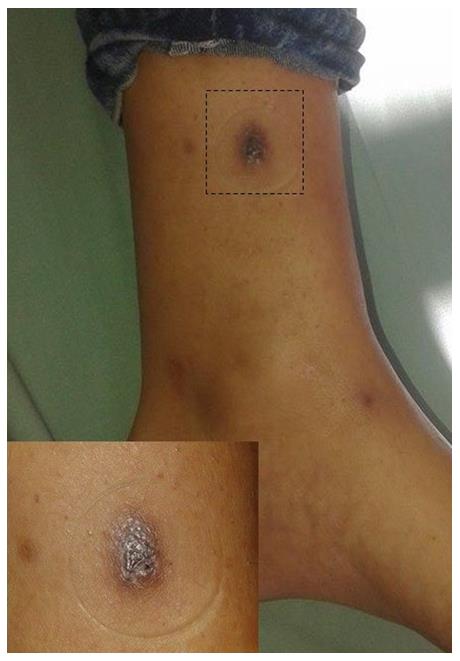
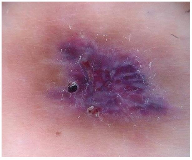
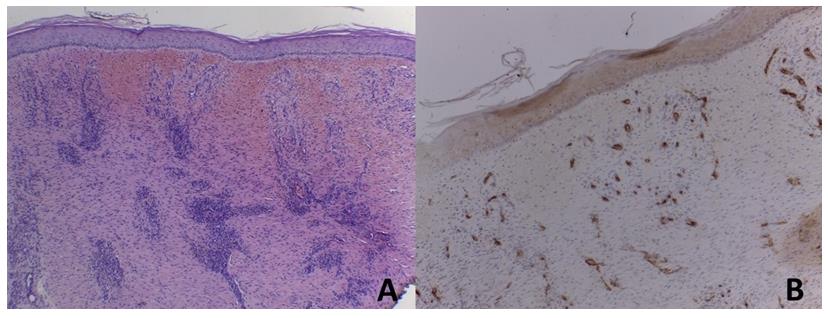
A 19-year-old female patient, with no relevant medical history consulted for a tumoral lesion on her left leg. She mentioned having a traumatic ulcer in this area about 2 months ago. Physical examination revealed a violaceous, firm lesion, size 12x10 mm and asymptomatic (Figure 1). In the dermatoscopic examen (Pro HR dermatoscope) we observed different colors, shades blue, violet and pink, which seemed to confirm the “rainbow pattern”, as well as elongated, shiny structures in the periphery, which were interpreted as crystalline structures (Figure 2). An incisional biopsy was performed, which reported dermal fibrosis with marked inflammatory component and hemorrhage (Figure 3). It was interpreted as a post- traumatic scar and we took an expectant conduct. It almost disappeard six months later.
Comments
The term "rainbow pattern" refers to the visualization of numerous colors in the shades of red, pink, violet, orange or light blue. It was first described as characteristic of Kaposi's sarcoma (KS) but it has now been also reported in other lesions, including scars. This multicolored pattern characterized by Hu et al. in 2009 as highly specific of SK, shows different colors, as it was a “rainbow”, and may either compromise the entire lesion or just a part of it. Its appearance is probably due to a complex physical phenomenon resulting from the interaction of polarized light with different elements of the tissue; it does not correspond to any specific histological structure. Cheng et al suggested that it was due to diffraction of the light through the dermis; however Vazquez-López et al. claimed that it was probably originated from a phenomenon of "dichroism" where light, in different states of polarization, experienced variable retardance and absorbance when interacting with tissue components. This absorption depended strongly on the nature of the object. In a material with multiple layers and structures as the dermis, this absorption varied, producing a wide range of colors. Cheng et al. observed that KS lesions showing the “rainbow pattern” had numerous vessels with very thin endothelium and little stroma, forming a structure similar to a honeycomb, with vascular lights arranged "back to back ". SK lesions that did not show the pattern had more stroma and more separated vascular lumens. The “rainbow” colors were only visible with polarized light, otherwise it was only seen red-blueish color [4, 5, 6].
As we previously mentioned, the optical effect responsible for this image is still unknown. It is commonly accepted that it would be a product of the interaction between polarized light and the "back to back" vessels and, although it was initially described as typical of KS, other authors have found it in lesions such as hemosiderotic dermatofibroma, basal cell carcinoma, pyogenic granulomas, melanomas, stasis dermatitis and lichen planus and others [1, 2, 7, 8]. In our patient's dermatoscopic exam we also described the “crystalline structures”. These elements are part of a group called "white lines", and are among those only visible with polarized light, the “bright structures” [9]. “Bright white structures” may be seen as bright white lines (formerly known as pupae), bright white areas and rosettes. It is postulated that these structures correlate with dermal fibroplasia. The birefringent properties of the disorganized collagen would result in a greater dispersion of the polarized light and that is why they would be seen in lesions with abundant dermal fibrosis. Nevertheless, a histopathological correlation has not yet been correctly determined [10]. Many benign and malignant tumors such as melanoma, Spitz nevus, dermatofibroma, actinic keratosis (AK), squamous cell carcinoma (SCC), basal cell carcinoma (BCC) and atypical fibrixanthoma (AF) had been reported for presenting theses “bright structures”. Balagula et al. recorded the presence or absence of crystalline structures in 265 biopsied lesions. They concluded that these were frequently observed in BCC and melanoma and were rarely seen in nevi. They also described their appearance on photo damaged skin on the bald scalp. In melanoma they were also a predictor of Breslow and progression of the neoplasia, a theory also proposed by Shitara et al who determined that in the preoperative evaluation of Breslow, the presence of “bright white structures” had 67.7% sensitivity and a 68% specificity for thick melanomas, which was comparable with other studies. They also demonstrated that these structures had a significant relationship with regression, since the melanomas that presented it were 3.2 times more likely to show the “bright white structures” in dermoscopy. In addition, in this study, the presence of these structures was associated with other criteria of dermal invasion, such as: blue-gray veil, milky red globules and polymorphic vessels in melanoma or whitish patch in dermatofibromas or homogeneous blue color in blue nevus. The theory that this phenomenon would be an optical artefact by structures found in the dermis took strength with these studies [10, 11, 12].
The aim of this case report was to make a brief review of these two dermatoscopic structures that, like any other pattern in dermoscopy, should be interpreted according to the context in which they are found. We believe that, in our case, since it was a recent scar, the abundant vascularization and fibrosis were responsible for such a controversial image.
References
-
Laureano A, Fernandes C, Cardoso J (2014) Morfologia e padrões vasculares em dermatoscopia - Parte II. SPDV 72(3): 307-324.
-
Martín JM, Bella-Navarro R, Jordá E (2012) Vascularization in dermoscopy. Actas Dermosifiliogr 103(5): 357-375.
-
Yoo MG, Kim IH (2014) Keloids and hypertrophic scars: characteristic vascular structures visualized by using dermoscopy. Ann Dermatol 26(5): 603-609.
-
Pérez-Pérez L, García-Gavín J, Allegue F, Zulaica A (2014) The rainbow pattern and rosettes in cutaneous scars. Actas Dermosifiliogr 105(1): 96-97.
-
Hu SC, Ke CL, Lee CH, Wu CS, Chen GS, et al. (2009) Dermoscopy of Kaposi’s sarcoma: Areas exhibiting the multicoloured ‘rainbow pattern’. J Eur Acad Dermatol Venereol 23(10): 1128-1132.
-
Cheng ST, Ke CL, Lee CH, Wu CS, Chen GS, et al. (2009) Rainbow pattern in Kaposi’s sarcoma under polarized dermoscopy: a dermoscopic pathological study. Br J Dermatol 160(4): 801-809.
-
Vázquez-López F, García-García B, Rajadhyaksha M, Marghoob AA (2009) Dermoscopic rainbow pattern in non-Kaposi sarcoma lesions. Br J Dermatol 161(2): 474-475.
-
Pitarch G (2014) Dermoscopic rainbow pattern in atypical fibroxanthoma. Actas Dermosifiliogr 105(1): 97-99.
-
Rosendahl C, Cameron A, Tschandl P, Bulinska A, Zalaudek I, et al. (2014) Prediction without Pigment: a decision algorithm for non-pigmented skin malignancy. Dermatol pract concept 4(1): 59-66.
-
Balagula Y, Braun RP, Rabinovitz HS, DuszaSW, Scope A, et al. (2012) The significance of crystalline/chrysalis structures in the diagnosis of melanocytic and non-melanocytic lesions. J Am Acad Dermatol 67(2): 194.
-
Shitara D, Ishioka P, Alonso-Pinedo Y, Palacios- Bejarano L, Carrera C, et al. (2014) Shiny white streaks: a sign of malignancy at dermoscopy of pigmented skin lesions. Acta Derm Venereol 94(2): 132-137.
-
Liebman TN, Rabinovitz HS, Balagula Y, Jaimes-Lopez N, Marghoob AA (2012) White shiny structures in melanoma and BCC. Archives of dermatology 148(1): 146-146.
- Epithelioid Granuloma; 3cases with Different Clinical Features
- Advancing Representation in Dermatology Clinical Trials: Ethical, Scientific, and Regulatory Imperatives for Inclusion Across all Fitzpatrick Skin Types
- A Case of Atopic Dermatitis with Concurrent Psoriasis Vulgaris: Successful Treatment with Upadacitinib
- Innovation Lifting Eyeshadow: A Synthesis of Makeup and Optical Illusion
- Distinguishing Superficial Actinic Porokeratosis from Actinic Keratosis with UVF Dermoscopy: A Case Report
- High Mobility Group Box 1 (HMGB1) in Cutaneous Inflammation: An Immune Modulator Bridging Cellular Stress, Ferroptosis and Danger Signaling