A Hypothesis of the Structure of Human Eyes
The combination of a tentacle and an eye forms a tentacle eye in a snail. A rod cell is a tentacle eye. A matrix of tentacle eyes (rod cells) create a stereoscopic compound on the retina. A lens in the eyeball plus the rod cells on the retina forms a telescope compound tentacle eye which is 3D stereoscopic.
Introduction
The Evolution of Animal Eyes
* Eyespot * Tentacle
* Tentacle eye * Compound eye * Simple camera eye * Telescope compound tentacle eye
![Figure 1: A Snail. Please see its tentacle and eye [1].](/fulltextimages/8096/fig_1.jpeg)
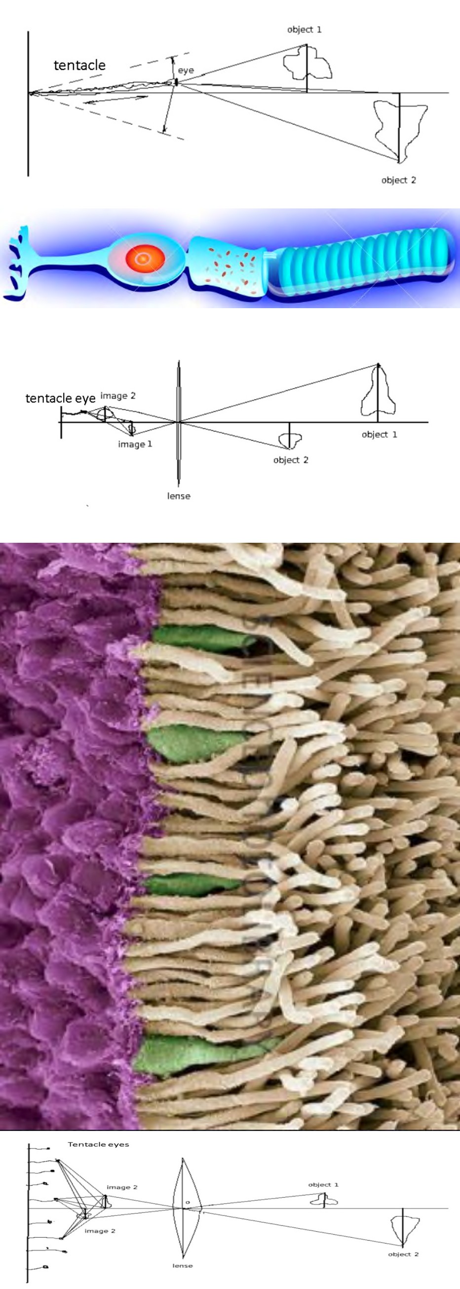
Figure 2A: My illustration of a tentacle eye.
Figure 2B: An image of a tentacle eye (a rod cell cited from reference [2]).
More images (Figure 3-5) are presented in references [3, 4]
Figure 4A: My illustration of a telescope compound tentacle eye. Figure 4B: Microscopic image of two types of photoreceptors: rods and cones [5].
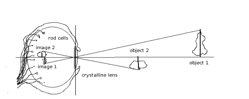
In a human eye, the lens and rod cells make a telescope system. The rod cells make a matrix, and the lens makes a 3D picture in front of the rod cells. The signals that the rod cells obtain are 3D holographic (stereo imaging). Please see [6] for the anatomy of a human eye.
The Evidence of My Guessing
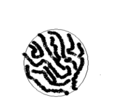
a. In a retina, the display of rod cells look like fingerprints (Figure 6). The density is sparser than the distinction rate of human eyes [7]. The pictures formed by the lens are in front of the rod cells instead of on the surface of the tips of rod cells.
b. The traditional opinion is that the lens of human eyes form 2D pictures on the retina [8, 9, 10, 11, 12], in the same way cameras form and sense images. Thus one eye cannot sense 3D images. However, when humans look at objects with only one eye, there is a strong feeling of protruding and sinking, and people can tell the surfaces of the objects are rough or smooth. When people see a 2D picture, they can have a 3D illusion. But lower animals have limited understanding of 2D pictures. For example, when a bird sees a picture of a bug, it does not show too much interest.
c. The images are holographic. The retina structure is not even. The local defect will not influence the completeness of the entire picture.
Materials and Methods
Tentacle movements were studied from a snail. It was then compared with rod cell microscopic images obtained from the internet (Figure 4). Fish eyes were collected from fish heads purchased in grocery stores. The fingerprint of the retina (Figure 6) was obtained from fish eyes after laying the retina on a piece of glass. The drawings of eye structures were made using the Paint software.
Discussion
There has been literature describing the movement of rod cells [13]. Even if there are microscopes designed to observe retinas in living eyes [14], and there are a lot of progresses in imaging retina activities in living eyes in recent years [15], it is still challenging to observe the detailed movements of rod cells. We should design an optical instrument to study the movement of rod cells during the process of looking at objects when the rod cells focus on a spot. In this way, we can have a better idea how they reach out for images.
Referring to the idea of tentacle eyes and this hypothesis of human eyes, we can design and construct artificial eyes for industrial usage. We can build matrices of tentacle light sensors, and then construct a telescope system including a lens in front of the tentacle light sensor matrices. The light sensors will pass electrical signals to the computers, which will apply statistics and machine learning approaches to process the holistic images. In this way, we can improve image quality and recognition.
Conclusion
A rod cell is a tentacle eye. Acknowledgement
I want to thank my grandparents for giving me my pet snails. The snails’ tentacle eyes inspired me to draft this paper. I also want to thank my school Buckingham Browne and Nichols for teaching me the necessary biology knowledge that makes this manuscript possible.
References
-
Schematic structure of the rod cell Peripheral vision Rod cells, or rods, are photoreceptor cells in the retina of the eye that can function in less intense light Vector scheme.123RF.
-
Plasma Membrane.
-
Eye and Vision Structure of the Retina Rods and Cones.123RF.
-
Electron microscope image of rod cells.
-
Anatomy of Eye. 123RF.
-
The Human Eye. Lumen.
-
NKCF, How Does The Human Eye Work?
-
(2020) Optics of the Human Eye. Anatomy & Physiology.
-
University Physics Volume 3.
-
Jin J, Jones GJ, Cornwall MC (1994) Movement of retinal along cone and rod photoreceptors. Vis Neurosci 11(2): 389-399.
-
Gerstner E (2011) Retinal rods resolved. Nature Physics 7: 521.
-
Hunter JJ, Merigan WH, Schallek JB (2019) Imaging Retinal Activity in the Living Eye. Annu Rev Vis Sci 5:15- 45.
- Genomic Landscape of Aggressive Penile Squamous Cell Carcinoma including TERT-p and NOTCH1 Mutations – An Institutional Experience
- Establishment of Baseline Haematological Values for Canine Population in North-Central Nigeria: A Cross-Sectional Study in the Federal Capital Territory
- Biochemical Assessment of Uroliths Extracted in Patients with Urolithiasis in a Tertiary Health Institution
- Update on Gastrointestinal Pecomas: Molecular Pathogenesis and Risk Stratification
- A Comparative Study of Serum C-reactive Protein Level Between Pre-eclampsia and Normal Pregnancy in Tertiary Level Hospital
- From Deformity to Alignment: Clinical Outcomes of the Schnepp Osteotomy in Hallux Valgus in 47 Feet