Maxillary Fourth Molar (Distomolar/Distodens) in Association with Rhyzomicroly and Pyramidal Molars in an Indian Patient – A Rare Case Report
Occurrence of dental anomalies in an Indian ethnic group is quite interesting from different aspect in order to enhance the existing research in the domain of dental anomalies. Presence of multiple anomalies in an individual is an uncommon finding. Therefore, the purpose of this manuscript is to present an interesting co-existence of three different dental anomalies such as supernumerary molar, rhyzomicroly and pyramidal molars which all together occurred in an Indian male patient.
Introduction
Odonto-stomatologic anomalies involving tooth number such as supernumerary tooth is the most commonly observable phenomenon. These represents the teeth which are more in number than the normal set of primary (20) or permanent teeth (32). Their prevalence in the general population varies from 0.1-3.8% [1, 2]. The most commonly seen supernumerary teeth are in the incisor region and they are termed as ‘mesiodens.’ Based on their position they are classified into four types as mesiodens [3], distomolars, paramolars and parapremolars as given by Primosch [4]. Supernumerary tooth occurring in the molar region are referred as paramolar or distomolar. ‘Paramolars’ are those teeth which seen either buccaly or palatally [5, 6] to the first, second or third molars, whereas ‘distomolars’ or ‘fourth molars’ or ‘distodens’ are those which are situated distal to the third molars. Between these two, occurrence of distomolars is quite uncommon. Their prevalence is estimated as just 0.32% based on a Japanese retrospective study [7] which found 87 distomolars among 26,721 cases evaluated. The gender prevalence was found as 1: 0.98, with most occurred in the maxillary arch (92%). Regarding shape assessed, 89.7% exhibited normal shapes being extremely smaller in size, constituting 82.8% of cases as impacted [7].
‘Short root anomaly’ (SRA) or ‘Rhyzomicroly’ is a rare condition affecting the root structure of the teeth characterized by abnormally short and blunt roots, with a crown to root ratio of 1:1 as described by Lind V [8]. Their prevalence in the general population ranges from 1.3% to 2.4%, and show a familial pattern of occurrence [9, 10]. The shortened roots seen in this condition is not due to resorption process or due to any external source or origin. Clinically crown of teeth having this condition usually appear normal in size, shape and colour with normal surrounding soft tissues. The most commonly affected teeth with this radicular phenomenon are maxillary incisors and premolars. Generalized occurrence involving all teeth is an extremely rare entity [8, 9, 10].
Pyramidal molars are anomalies involving radicular part in molars and are characterized by single rooted molars. This condition is also referred by various synonyms in the literature as ‘pyramidal,’ ‘tubular,’ ‘cylindrical,’ ‘cuneiform,’ ‘conical’ and ‘prismatic’ [11, 12]. Reports on occurrence of this root variation are quite infrequent.
Therefore, the aim of this paper is to present co- existence of fourth molar or distomolar, pyramidal molars and occurrence of rhyzomicroly in an Indian male patient.
Case Report
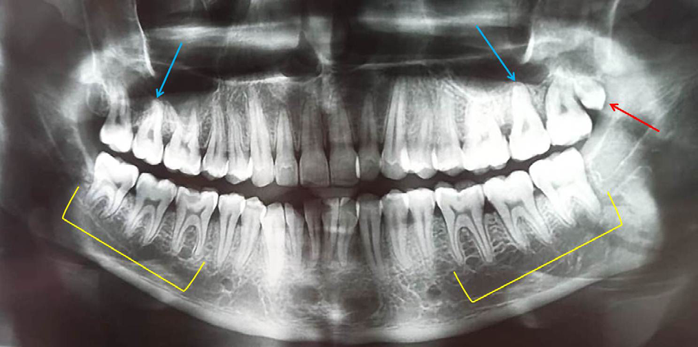
A 28-year-old male patient reported to a private dental clinic complaining of pain in the lower right back tooth from past one week. On physical examination patient appeared apparently normal with no history of systemic, metabolic or syndromic disorders. Patient built was moderate with moderately nourished and well-behaved behaviour. Examination of the oral cavity showed complete set of permanent teeth, all teeth erupted clinically including third molars. In the right quadrant of the mandibular arch, deep caries was observed in the mandibular first molar which was tender to percussion test. In the maxillary left quadrant, distal to the third molar, small tooth like structure was observed. There was no history of any discomfort or pain associated with this tooth. To rule out the status of complained tooth, a radiographic investigation was performed. Examination of the radiograph (orthopantomograph) showed presence of small, rudimentary tooth in the maxillary left quadrant distal to the third molar (Figure 1). Compared to third molar, this tooth was extremely small and was in a vertical position, with very short single/fused root. Further detailed examination of the radiograph showed extremely short roots involving the mandibular right and left first, second and third molars (Figure 1). The root length in all these teeth was almost equal to the crown length (1:1 ratio). Pyramidal shaped roots involving maxillary second molars in both right and left side was also observed (Table 1). Therefore, considering all these points, the present case was diagnosed as occurrence of fourth supernumerary molar (distomolar) in association with short root anomaly and pyramidal molars was diagnosed. Patient was explained about the presence of the extra tooth in the left upper region and its possible consequences and advised for extraction. As patient was not willing to go for extraction of this fourth molar, attention was given only to chief compliant of the patient.
| Age | Gender | Chief Complaint | Radiographic Features | Associated Anomalies |
|---|---|---|---|---|
| 28 years | Male | Pain in the right lower back tooth from past one week | Presence of fourth molar in the maxillary left quadrant distal to the third molar (small, conical shaped crown with short single root) Erupted in vertical position | Rhyzomicroly or Short root anomaly involving mandibular first, second and third molars (both right and left) with 1:1 crown root ratio. + pyramidal, single rooted maxillary right and left second molars |
Table 1: Patient Details with Fourth Molar/Distomolar Associated with Multiple Anomalies.
Discussion
Supernumerary teeth are classified based on their location in the oral cavity as mesiodens, paramolar, distomolar and parapremolars [13]. Nagaveni NB [13] published various research papers concerning paramolar, parapremolars and mesiodens occurring in Indian patients [1, 2, 3, 5, 6, 13]. The current paper is addition to this research list showing contribution from Indian country, as presentation from different ethnic group will greatly help to make guidelines, policies and protocols in the overall management of these anomalies concentrating on diagnostic and therapeutic aspects.
Fourth or distomolars exhibit normal morphology having completely formed crown, single root which is entirely distinct from the third molar. However, they have also reported to occur either in eumrophic or dysmorphic morphology. There are studies showing dysmorphic shaped distomolars characterized by tuberculated or molariform in shape and some showing peg shaped and conical shaped distomolars. According Arslan, et al. [14] distomolars exhibited three varieties in shape such as a. A Premolar Shaped with One Root b. A Premolar Shape with Only A Crown and No Root And c. A Rudimentary Conical Shape Most of the time they erupt in to the oral cavity fully and align themselves in the dental arch. Very rarely they become partial or complete impacted. In the present case, it appeared very small and conical in shape with short single root and was erupted normally into the oral cavity in vertical position.
Distomolars have encountered more in males than females with no side wise predilection, occurring equally on both right and left side. Compared to unilateral occurrence, bilateral presentation of distomolars is extremely rare. In this case, it was present in male patient and on left side (unilateral presentation) supporting the existing literature [14]. Asymptomatic impacted distomolars are usually detected on routine radiographic examination. Recently advanced imaging techniques like CBCT (Cone Beam Computed Tomography) have been used to evaluate their morphology, orientation, position in order to formulate therapeutic and diagnostic protocols. Those which have erupted usually show food lodgement or plaque accumulation due to insufficient space in the oral cavity. Surgical removal is the treatment option for symptomatic distomolars [15, 16, 17, 18].
Short root anomaly (SRA) or rhyzomicroly should be differentiated from idiopathic root resorption. In case of SRA, the root is short, closed and show blunt apex with equal crown and root ratio as described by Lind V [8]. As the teeth having this root anomaly appear clinically normal, this condition is always diagnosed on routine radiographic examination. To diagnose the tooth with SRA, the criteria given by Lind V [8] was followed. When the tooth is having equal proportion of crown and root length in the ratio of 1:1 with blunt closed apex then it is considered as short root anomaly. In the present case too, the crown and root length was 1:1 when measured using calibrated scale. Teeth with rhyzomicroly do not show any mobility whereas those with root resorption show pathologic mobility [19]. Regarding treatment, this condition has special significance pertaining to orthodontic and prosthodontic treatment. During orthodontic treatment, minimal pressure should be generated while moving these affected teeth compared to normal component. Therefore, radiographic evaluation is highly essential before initiating any dental treatment procedure during clinical practice.
Molars with multiple roots sometimes show variation in their root morphology exhibiting presence of single root due to fusion or deep taurodontism [20]. This condition is referred as ‘pyramidal,’ ‘tubular,’ ‘cylindrical,’ ‘cuneiform,’ ‘conical’ and ‘prismatic’ based on literature review. Occurrence of this condition is important as it depicts taurodontism, fused roots and pyramidal roots are the variations of a single heritable trait, with single pyramid-shaped root being the most severe expression of this trait. They may occur unilateral, bilateral, in primary or permanent, with no gender predilection and side affected. However, their occurrence in primary dentition is extremely low. They occur more commonly in the maxilla compared to mandibular teeth. There is no particular treatment required for this condition, however, during root canal treatment, various precautions should be considered, as management differs in these teeth.
Conclusion
An awareness and knowledge about the existence of fourth molars or distomolars, pyramidal molars and short root anomaly is highly essential among all clinicians as treatment varies compared to normal teeth. Reporting such unusual anomalies is also important for the research field to formulate new evidence pertaining to dental anomalies.
References
-
Nagaveni NB, Sreedevi B, Praveen BS, Praveen Reddy B, Vidyullatha BG, et al. (2010) Survey of mesiodens and its characteristics in 2500 children of Davangere city, India. Eur J Paediatr Dent 11(4): 185-188.
-
Nagaveni N, Shashikiran N, Reddy VS (2009) Surgical management of palatal placed, inverted, dilacerated and impacted mesiodens. Int J Clin Pediatr Dent 2(1): 30-32.
-
Nagaveni NB (2023) Inversion of impacted mesiodens: Report of Case series with literature review. Glob J Res Dent Sci 3(5): 7-12.
-
Primosch RE (1981) Anterior supernumerary teeth-- assessment and surgical intervention in children. Pediatr Dent 3(2): 204-215.
-
Nagaveni NB, Umashankara KV, Radhika NB, Praveen Reddy B, Manjunath S (2010) Maxillary paramolar: report of a case and literature review. Arch Orofac Sci 5(1): 24-28.
-
Nagaveni NB, Umashankar KV (2023) Report of a rare Odonto-Stomatologic Anomaly – Maxillary Paramolar. Series Clin Med Case Rep Rev 1(6): 1-3.
-
Bamgbose BO, Okada S, Hisatomi M, Yanagi Y, Takeshita Y, et al. (2019) Fourth molar: A retrospective study and literature review of a rare clinical entity. Imaging Sci Dent 49(1): 27-34.
-
Lind V (1972) Short root anomaly. Scan J Dent Res 80(2): 85-93.
-
Nagaveni NB, Umashankara KV, Vidhyllatha BG, Sreedevi, Radhika NB (2011) Permanent mandibular incisor with multiple anomalies – Report of a rare clinical case. Braz Dent J 22(4): 346-350.
-
Nagaveni NB (2023) Short Root Anomaly (SRA)/ Rhyzomicroly-Report of an Unusual Radicular Anomaly with Comprehensive Literature Review. Clin Pathol 7(1): 000171.
-
Nagaveni NB (2012) An Unusual Occurrence of Multiple Dental Anomalies in a Single Nonsyndromic Patient: A Case Report. Case Rep Dent pp: 1-4.
-
Klein U, Paimagham B, Blumhagen R, Kroehl M, Sain J (2017) Pyramidal and Taurodont Molars and Their Association With Other Tooth Anomalies. Pediatr Dent 39(1): 46-52.
-
Nagaveni NB (2023) Bilateral ‘Molarization’ of the mandibular second premolars in association with unusual dental variation – report of a rarest case. Glob J Res Dent Sci 3(5): 4-6.
-
Arslan A, Altundal H, Ozel E (2009) The frequency of distomolar teeth in a population of urban Turkish adults: A retrospective study. Oral Radiol 25: 118-122.
-
Moreira LM, Grillo R, de Almeida Júnior JC, Gonçalves Teixeira R (2023) Familial Fourth Molars In Non- Syndromic Patients: Case Report Of An Unusual Entity. Bulletin of Stomatology and Maxillofacial Surgery 19(4): 91-95.
-
Mitsea A, Vardas E, Papachatzopoulou A, Kalfountzos G, Leventis M, et al. (2015) The frequency of non-syndromic distomolar teeth in a Greek population sample?. J Clin Exp Dent 7(5): e589-e594.
-
Nagaveni NB, Umashankar KV (2023) Vertical, Intra- Osseous Impaction of permanent maxillary central incisor in association with multiple anomalies – Report of a rare case. EC Dent Sci 22(9): 1-4.
-
Nagaveni NB (2023) A rare combination of tooth agenesis in association with anomalous supernumerary tooth: Report of a rare case. Oral Health Dent 6.1: 18-21.
-
Lamani E, Feinberg KB, Kau CH (2017) Short Root Anomaly - A Potential “Landmine” for Orthodontic and Orthognathic Surgery Treatment of Patients. Ann Maxillofac Surg 7(2): 296-299.
-
Ramar K, Hariharavel VP, Nair MR (2016) Single-rooted pyramidal molars: A rare case report. Int J Pedod Rehabil 1: 72-74.
- Genomic Landscape of Aggressive Penile Squamous Cell Carcinoma including TERT-p and NOTCH1 Mutations – An Institutional Experience
- Establishment of Baseline Haematological Values for Canine Population in North-Central Nigeria: A Cross-Sectional Study in the Federal Capital Territory
- Biochemical Assessment of Uroliths Extracted in Patients with Urolithiasis in a Tertiary Health Institution
- Update on Gastrointestinal Pecomas: Molecular Pathogenesis and Risk Stratification
- A Comparative Study of Serum C-reactive Protein Level Between Pre-eclampsia and Normal Pregnancy in Tertiary Level Hospital
- From Deformity to Alignment: Clinical Outcomes of the Schnepp Osteotomy in Hallux Valgus in 47 Feet