Tamoxifen Induced Down Regulation of mTOR and UP Regulation of Exosomal miR-99b-5p and miR-100-5p in mcf-7 Breast Cancer Cells
Background: Breast cancer treatment has always been an important challenge in medicine. Recent studies have revealed the importance of microRNAs miR-99b-5p and miR-100-5p in breast cancer treatment. Moreover, however not fully understood, the impact of exosomal microRNAs in human cancer progress or inhibition has been highlighted yet. The present study aimed to investigate the effects of one of the most prescribed drugs in breast cancer chemotherapy, tamoxifen, on expression level of miR-99b- 5p, miR-100-5p and also mTOR gene in MCF-7 cell lines. Methods: MCF-7 cells were treated with 30 μM tamoxifen while being in a 70% surface density, then the expression level of mentioned microRNAs and mTOR gene were investigated in RNA extracts of the cells and their exosomes. Results: Results indicated the presence of a significant increase (P <0.01) in both miR-99b-5p and miR- 100-5p expression levels in cell lines following tamoxifen treatment. Also, the evaluation of mTOR gene expression in the same cells after treatment with tamoxifen showed significant (P <0.05) expression reduction. Conclusions: Interestingly, the decrease in mTOR expression level was completely consistent with increase in the expression of exosomal microRNAs level, which seems to be reasonable considering the targeting of the mTOR gene by the two mentioned microRNAs. The results of this study suggest that the therapeutic effects of tamoxifen on breast cancer cells, can be justified not only by its direct effect on cancer cells, but also by indirect impact on elements like microRNAs expression.
Introduction
Breast cancer, the most common cancer among women and the second leading cause of cancer associated death in world among women population, has always been one of the most serious challenges in medicine from disease diagnosis and treatment point of view [1]. The incidence and mortality caused by breast cancer have increased over the past three decades due to changes in the risk factors and also improvements in disease diagnosis. In 2020, more than two million new cases of breast cancer have been reported in the world [2].
In recent years, the increasing incidence pattern of this cancer has attracted attention in world and correspondingly in Iran [3]. Thus, progress and development of its treatment measures seems to have become a major necessity in recent years. In this regard, microRNAs (endogenous non-coding small RNA molecules) are one the most precious tools in molecular studies related to cancer treatment. It has been reported that the modification in quality and quantity of circulating miRNAs in patients is completely related to the initiation and progression of human cancer, and thus detection of which can help cancer therapy [4]. Despite their prediction and diagnostic value in human cancers [5, 6], the importance of some microRNAs in the treatment of human cancers like breast cancer has been highlighted yet [7, 8]. Also, it has been shown that microRNAs interact with chemotherapy drugs [9, 10, 11].
From this point of view, understanding the precise and detailed crosstalk between chemotherapy methods and microRNAs function may provide more efficient methods in breast cancer treatment. In recent years, in addition to the reports of modification in miR-99-5P and miR-100-5p expression levels in some human cancers like prostate and colon cancers, their vital role in proliferation, migration and invasion of tumors has been highlighted as well as their tumor suppressor role [12, 13, 14]. The issue that makes them a great choice for cancer therapy research.
Moreover, based on previous research, studying the effects of cancer treatment methods on mTOR (Mammalian Target of Rapamycin) gene expression have increased the current knowledge about chemical drugs’ mode of effect in cancer cells. It has already been demonstrated that the mTOR gene regulates cell proliferation, autophagy and apoptosis by participating in multiple signaling pathways in the body [15]. Thus, it is associated with cancer and tumor metabolism. Therefore, the mTOR signaling pathway has become a great target in anti -tumor treatment researches [16]. In recent years, a variety of newly discovered mTOR inhibitors have undergone clinical studies to help cancer progress prevention. As of today, mTOR gene is used as a favorable biomarker in breast cancer response to chemical drugs [17].
On the other hand, nowadays, tamoxifen, which interferes in receptors and inhibitors of estrogen in breast cancer cells, is considered as a gold standard treatment for millions of breast cancer patients across the world. As a hydrophobic anticancer agent and a selective estrogen modulator, tamoxifen has FDA approval to be used for hormone therapy of breast cancer [18]. Also, the World Health Organization has announced it as an essential drug needed for cancer treatment [19]. Thus, it goes without saying that tamoxifen as a frequently prescribed anticancer drug [20] is one the most efficient compounds against breast cancer even as complementary option alongside other treatment methods [21].
In context of beneficial elements for breast cancer treatment monitoring, exosomes are one the most helpful components that serve medicine. Exosomes as beneficial nano-vesicles in cellular communication, exist and are constantly moving in the blood, saliva, urine and some other body fluids and play an important role in intra-and intercellular communication and physiological events such as removing harmful or damaged cell components or stimulating immune cells [22, 23]. Exosomes are proven to play an important role in cancer cells [24, 25, 26]. Moreover, they are considered as reliable biomarkers of cancer progression and are helpful elements in cancer behavior prediction. As an important components of exosomes, the exosomal microRNAs are reported to help monitoring the cancer behavior [27, 28] and one of the most interesting observations that has attracted the attention of researchers is exosomal microRNAs benefits in monitoring the cancer patient’s response to chemical drugs [29].
In line with mentioned relationship between modification of miR-99-5P and miR-100-5p expression levels in cancers, also the presence of reports regarding the oncogenic function of mTOR gene in some human cancers [15], we aimed to investigate if tamoxifen treatment as one of the most common drugs in breast cancer chemotherapy induces any modification in expression level of miR-99-5P, miR-100-5p and their target mTOR gene, and also if this drug can modify exosome secreted microRNAs level in MCF-7 cell lines.
Materials and Methods
Cell Lines
MCF-7 cell lines obtained from Pasteur Institute of Iran were cultured in DMEM containing 10% fetal bovine serum (FBS), 2 mM L-Glutamine, 100 ugr/ml Streptomycin and 100 u/ml penicillin then were maintained in 37°C and 5% Co2. Cells passage from this source was performed several times using PBS and Trypsin to prepare needed cell lines for experiments.
MTT Assay
In order to cell survival evaluation of MCF-7 cells, tamoxifen treatment as doses of 10 to 40 µM were performed before MTT assay. In brief, cultured cells in their 80% density were treated with Trypsin and centrifuged 12000 rpm for 5 minutes, then the pellet (containing cells) was resolved in DMEM and 200 μl of them were transferred to each well of 96-
well plates. After a short incubation in 37°C and 5% Co2 for 5 minutes, an increasing concentration of tamoxifen was added to wells. Then 20 μl of MTT (5 mg/ml in PBS) were added and incubated in 37°C and 5% Co2 for 30 minutes. Finally, the liquid was removed and 200 μl DMSO were added into wells. Samples absorbance were measured in 490 nm wavelength in 96- well plates. The IC50 obtained from this experiment was used for the main experiment described below.
Cells Treatment with Tamoxifen
In order to perform the main experiment of tamoxifen effects on mentioned genes and microRNA expression level, MCF-7 cells in 70% density were treated with 30 μM tamoxifen Sigma-Aldrich (equal to IC50 from MTT assay result) while being in 12 well plates. Then, treated cells were incubated at 37°C for growth. Exosomes and RNA were isolated from these cell lines as explained in the following sections.
RNA Extraction, cDNA Synthesis and Real-Time PCR
In order to determine the expression level of mTOR gene in cell lines and miR-99b-5p and miR-100-5p in cell lines and exosome samples, total RNA extraction from samples was performed using TRIzol® reagent. In brief, MCF-7 cell lines were centrifuged and 50 μl TRIzol were added to the pellets and incubated for lysis process. The same TRIzol treatment process was done for exosomes suspension. Then, after 15 μl chloroform addition, vortex, incubation in room temperature for 2-3 minutes, and centrifuge at 12000 g for 10 minutes, the extracted RNA was collected from upper phase of supernatant. Finally, the RNA was precipitated with the same volume of isopropanol, then washed with ethanol and dried and resolved in 30 μl water. After extracting total RNA, its quality and quantity evaluation were assessed using Nanodrop. To do this, the samples absorbance was measured in 260 and 280 nm wavelength using NanoDrop® ND-1000 (NanoDrop Technologies, Wilmington, DE, USA).
Then, the cDNA synthesis was performed using miRNA Reverse Transcription Kit after polyadenylation reaction.
In brief, polyadenylation of 5 μg extracted RNA (miRNA) was performed using ATP (10mM) and polyA polemerase enzyme in 37°C for 10 minutes. Then, oligo dT was added and incubated in 65°C for 5 minutes. Finally, cDNA synthesis process was performed adding 5x Reaction buffer, DTT, Ribolock RNase, dNTP, RTase rxn buffer 10x and Hyperscript RTase enzyme. The final 20 μl was incubated in 42°C for 30 minutes, then in 25°C for 5 minutes. Then inactivation of RT enzyme followed in 70°C for 5 minutes.
At the same time, for cDNA synthesis from extracted mRNA, 30 minutes incubation at 37°C with DNAseI treatment was followed to remove possible DNA impurities from 1-2 µg RNA. Then, DNAseI was removed using 1 μl EDTA and incubation in 65°C for 10 minutes. Then, oligo dT addition was followed like the same for miRNA. Finally, cDNA synthesis process was done using 5x Reaction buffer, Ribo lock RNase, dNTP and RevertAid M-MuLV RT in 42°C for 60 minutes, then in 25°C for 5 minutes. A similar heat inactivation of RT enzyme followed in 70°C for 5 minutes as final step.
Finally, the expression level of mTOR and mentioned microRNAs were determined using Real-time PCR, and results were compared to the reference genes including GAPDH and U6snRNA. In brief, SYBR Green I Master mix kit was used to evaluate gene expression of 5ng cDNA samples loaded into 48 well Microamp, Applied Biosystema, Singapore plates, including 10 μl master mix, primers and water. The qPCR was performed in 40 temperature cycles of 95°C for 5 seconds denaturation step, 62-65°C for 20 seconds binding step, and 72°C for 30 seconds as extension step. Finally, ABI step one Real-Time PCR Software v2.0.2 (Applied Biosystems,UK) was used to analyze the data with Comparative Threshold Cycle method.
Results
MTT Assay Result
The results of MTT assay showed that half of the MCF- 7 cells were alive after tamoxifen treatment in 30 µM concentration; IC50=30 µM (Figure 1).

Bioinformatics and Statistical Analysis
The bioinformatics studies were also conducted to determine the chromosomal position of miR-99b-5p and miR-100-5p and mTOR (GeneCards database) and also to confirm the suppressor effect of microRNAs on mTOR (miRTargetLink database), augmented with investigation of microRNA Response Element (MRE) network (TargetScan database) and mTOR expression in various tissues (UCSC database). Meanwhile, two-sided paired student’s t-test was used to compare the average of expression level between control and treated cell lines. The SPSS software was used to analyze the data and P value was considered P<0.05 to determine statistically significant difference in results.
Bioinformatics Analysis
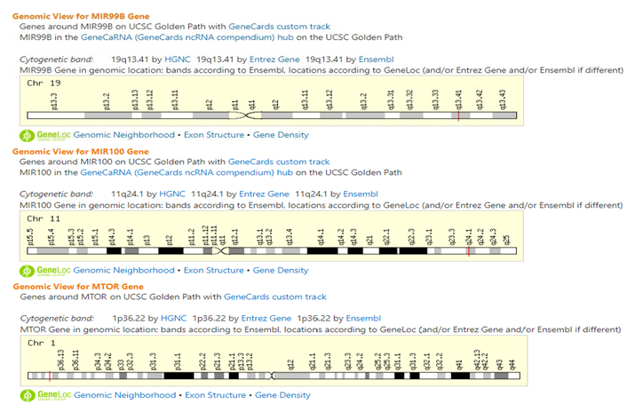
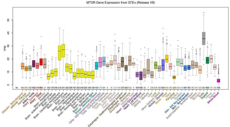
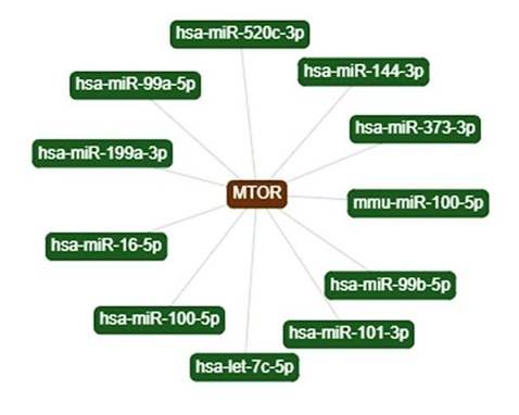
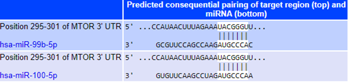
The genomic location studies (Chromosomal position and MRE network( were performed through https://www. genecards.org/ (The human gene database) showed, as presented in Figure 2, miR-99-5p and miR-100-5p are located in long arm of Chr 19 and 11 respectively (Red bands). mTOR belongs to the short arm on Chr 1. Meanwhile, bioinformatics investigation in https://genome.ucsc.edu/ (Genome bowser tool) for mTOR expression in tissues showed different expression level of mTOR gene in various tissues and organs including breast in human body. As shown in Figure 3, mTOR is highly expressed in testis and brain. Further bioinformatics analysis in https://ccb-compute. cs.uni- saarland.de/mirtargetlink2 (MicroRNA target tool) showed that several microRNAs can target mTOR gene (Figure 4). Eleven microRNAs are shown and among them the microRNAs, miR-99-5p, miR-100-5p, (used in this study) can be found. This result confirms the mTOR suppressor role of mentioned microRNAs. Meanwhile, the results of MRE investigation (by TargetScanH; prediction of microRNAs target tool) confirmed the presence of a common target sequence on 3’UTR region of mTOR gene for miR- 99-5p, miR-100-5p (Figure 5).
mTOR and MicroRNAs’ Gene Expression Modification
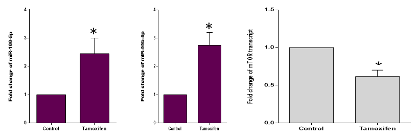
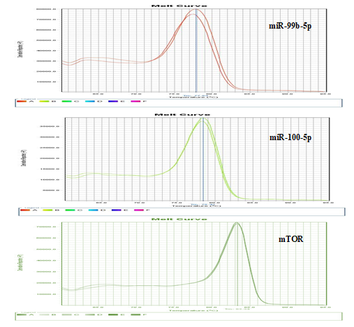
The results of the Real-Time PCR indicated a significant (P <0.01) increase in miR-99B-5P and miR-100- 5p expression levels (2.79 and 2.43 folds compared to the non-treat cells as control, respectively) following tamoxifen treatment (Figure 6). Evaluation of mTOR gene expression in the same cells after treatment with tamoxifen showed significant (P <0.05) expression reduction (0.63-fold compared to control) (Figure 6). Interestingly, the decrease in mTOR expression level was completely consistent with increase in the expression of exosomal microRNAs level. It should be noted that the Melt Curve Analysis showed the presence of a single specific distinct peak in Real-time PCR products (Figure 7).
Discussion
As the most common cancer among women, breast cancer is one of the most important challenges in medicine [1]. Successful treatment has always been a vital aim and thus various research has been performed to improve its treatment. As consequence, several therapy methods have been applied to treat breast cancer like surgery, chemotherapy, radiotherapy and immunotherapy [30, 31, 32, 33]. Among the mentioned methods chemotherapy seems to be the most effective and sometimes indispensable part of treatment [34]. Among the recently recommended drugs, tamoxifen is a well-known compound and as a non-steroidal anti-estrogenic compound with poor estrogenic effect is known as “gold standard” treatment for breast cancer. Tamoxifen inhibits estradiol and estrogen and thus breast cancer improvement, since estrogen feeds breast cancer cells. Although tamoxifen is not devoid of side effects, it is widely used as a complementary and efficient treatment method in breast cancer treatment [18, 21].
Along with chemical treatment, molecular research has always been ongoing to develop highly efficient treatment methods in breast cancer treatment. In this regard microRNAs and exosomal microRNAs have attracted attention. MicroRNAs regulate many physiological processes of the human body, including the regulation of gene expression. They play a prominent role in proliferation, differentiation and cellular death as well as cancer-related processes such as metastasis [4, 35, 36]. Reports indicate that breast cancer is affected by the expression of some microRNAs. Therefore, the investigation of their functions is of particular importance in diagnosing and treatment of breast cancer [7, 37]. Recent studies have shown the impact of two microRNAs, miR-99b-5p and miR-100-5p in several human cancers [12, 13, 14]. Since less is known about miR- 99b-5p and miR-100-5p influence from anti-breast cancer drugs like tamoxifen, we investigated their modification in breast cancer cell lines. In fact, we aimed to examine how tamoxifen treatment modifies the expression level of two mentioned microRNAs and also their target gene, mTOR in treated cell lines. It should be noted that the microRNAs were isolated from both cells and exosomes secreted by them. The reason why mTOR was investigated in this study was its expression role in different cancers (metastasis and tumor growth) and importance as a target in cancer therapy. In fact, the relationship between mTOR expression and predictions of different types of breast cancer is obvious. The different expression pattern of mTOR gene in healthy individuals and cancer patients, also its modification before and after treatment with anti-cancer drugs have attracted attentions [38, 39].
In overall view, the results of current study showed a meaningful modification in expression level of mTOR gene and miR-99b-5p and miR-100-5p in cell MCF-7 lines after tamoxifen treatment. To dive into more details, it should be noted that our bioinformatics studies confirmed the targeting of the mTOR gene by the two mentioned microRNAs. Also, a clear common target sequence on 3’UTR region of mTOR gene for miR-99-5p, miR-100-5p was recorded.
Moreover, results showed that the Tamoxifen treatment significantly (P <0.01) increased the expression level of miR- 99b-5p and miR-100-5p in MCF-7 cancer cells. Also, results indicated a significant (p <0.05) decrease in mTOR gene expression in cell lines as well. One step further, our studies indicated the same results of microRNAs expression increase in exosomes secreted from cell lines. Our results suggest that tamoxifen treatment not only has a direct effect on the cancer cells, but also is capable of inducing indirect effects on breast cancer by expression changes in exosomal microRNAs. Meanwhile, the decrease in mTOR gene expression and its reverse relationship with increased microRNA expression level seems quite reasonable. The results of this study clearly confirm the therapeutic effects of tamoxifen on cancer microenvironment. In other words, exosomes secreted by cancer cells are capable of trafficking their inside elements like microRNAs to the tumor microenvironment and can induce anticancer effects.
It should be noted that the results of the present study confirm previous reports regarding mTOR and miR-99- 5p, miR-100-5p significance in cancer treatment. Earlier, a relationship between decrease in mTOR gene expression and higher miR-100-5p activity in prostate cancer was reported [14]. Also, it has been reported that miR-99-5p inhibits mTOR gene expression and affects colon cancer [12]. Another similar study has recently indicated that the decrease in mTOR gene expression increases the success possibility of a breast cancer treatment [40].
Finally, in recent years, the increasing trend of breast cancer incidence has been recorded in the world. Despite the presence of several ways to prevent, screen, diagnose and treat, a great potential of treatment seems to be neglected in molecular area. MicroRNAs and exosomal microRNAs for their vital role in physiological events have shown promising horizon in fight with breast cancer. The result of present study indicates the importance of miR- 99-5p, miR-100-5p secreted from cells and exosomes in mTOR gene expression modification and their influence from chemotherapy with tamoxifen. Authors hope that this report will provide more detailed information not only about tamoxifen mode of action, but also regarding the exosomal microRNAs significance in breast cancer treatment.
Future Prospective
Since the importance of breast cancer treatment and mentioned history of previous research, it is recommended that some further experiments should be designed and performed to shed light on the unknowns of the area and increase the knowledge in the field. In this regard, authors suggest the investigation of other mTOR targeting microRNAs activity after treatment with chemical compounds. Also, it is highly recommended to study the gene expression in protein level for mTOR and other genes down- and upstream of its signaling pathway. Such experiments, especially in animal models can improve the current knowledge and may open new windows and promising road ahead of breast cancer treatment.
Funding Statement
This research did not receive any specific grant from funding agencies in the public, commercial, or not-for-profit sectors.
Declaration of Competing Interest
There is no conflict of interest.
References
-
Bray F, Ferlay J, Soerjomataram I, Siegel RL, Torre LA, et al. (2018) Global cancer statistics 2018: GLOBOCAN estimates of incidence and mortality worldwide for 36 cancers in 185 countries. CA: a cancer journal for clinicians 68(6): 394-424.
-
Łukasiewicz S, Czeczelewski M, Forma A, Baj J, Sitarz R, et al. (2021) Breast Cancer—Epidemiology, Risk Factors, Classification, Prognostic Markers, and Current Treatment Strategies—An Updated Review. Cancers 13(17): 4287.
-
Rezapour A, Nargesi S, Mezginejad F, Kemmak AR, Bagherzadeh R (2021) The economic burden of cancer in Iran during 1995–2019: a systematic review. Iranian Journal of Public Health 50(1): 35-45.
-
Cui M, Wang H, Yao X, Zhang D, Xie Y, et al. (2019) Circulating microRNAs in cancer: potential and challenge. Frontiers in genetics 10: 626.
-
Kim JE, Eom JS, Kim WY, Jo EJ, Mok J, et al. (2018) Diagnostic value of microRNAs derived from exosomes in bronchoalveolar lavage fluid of early‐stage lung adenocarcinoma: A pilot study. Thoracic cancer 9(8): 911-915.
-
Kashyap D, Kaur H (2020). Cell-free miRNAs as non- invasive biomarkers in breast cancer: Significance in early diagnosis and metastasis prediction. Life sciences 246: 117417.
-
Loh HY, Norman BP, Lai KS, Rahman NMANA, Alitheen NBM, et al. (2019) The regulatory role of microRNAs in breast cancer. International journal of molecular sciences 20(19): 4940.
-
Liu Q, Peng F, Chen J (2019) The role of exosomal microRNAs in the tumor microenvironment of breast cancer. International Journal of Molecular Sciences 20(16): 3884.
-
Si W, Shen J, Zheng H, Fan W (2019) The role and mechanisms of action of microRNAs in cancer drug resistance. Clinical epigenetics 11(1): 1-25.
-
Kulkarni B, Kirave P, Gondaliya P, Jash K, Jain A, et al. (2019) Exosomal miRNA in chemoresistance, immune evasion, metastasis and progression of cancer. Drug Discovery Today 24(10): 2058-2067.
-
Hussein GM, Mohammed SM, Faris M, Mohammed A, Kadhim MJ, et al. (2022) Find new channel for overcoming chemoresistance in cancers: Role of stem cells-derived exosomal microRNAs. International Journal of Biological Macromolecules 219: 530-537.
-
Li W, Chang J, Wang S, Liu X, Peng J, et al. (2015) miRNA- 99b-5p suppresses liver metastasis of colorectal cancer by down-regulating mTOR. Oncotarget 6(27): 24448- 24462.
-
Nabavi N, Saidy NRN, Venalainen E, Haegert A, Parolia A, et al. (2017) miR- 100-5p inhibition induces apoptosis in dormant prostate cancer cells and prevents the emergence of castration-resistant prostate cancer. Scientific reports 7(1): 4079.
-
Ye Y, Li SL, Wang JJ (2020) miR-100-5p downregulates mTOR to suppress the proliferation, migration, and invasion of prostate cancer cells. Frontiers in Oncology 10: 578948.
-
Popova NV, Jücker M (2021) The role of mTOR signaling as a therapeutic target in cancer. International Journal of Molecular Sciences 22(4): 1743.
-
Murugan AK (2019) mTOR: Role in cancer, metastasis and drug resistance. In Seminars in cancer biology. Semin Cancer Biol 59: 92-111.
-
Zou Z, Tao T, Li H, Zhu X (2020) mTOR signaling pathway and mTOR inhibitors in cancer: Progress and challenges. Cell & Bioscience 10(1): 1-11.
-
Day CM, Hickey SM, Song Y, Plush SE, Garg S (2020) Novel tamoxifen nanoformulations for improving breast cancer treatment: Old wine in new bottles. Molecules 25(5): 1182.
-
World Health Organization (2019) Model List of Essential Medicines.
-
Potkul RK, Unger JM, Livingston RB, Crew KD, Wilczynski SP, et al. (2016) Randomized trial of medroxyprogesterone acetate for the prevention of endometrial pathology from adjuvant tamoxifen for breast cancer: SWOG S9630. NPJ Breast Cancer 2(1): 1-5.
-
Bhide A, Datar S, Stebbins K (2021) Case Histories of Significant Advances: Tamoxifen, A Gold Standard Treatment for Breast Cancer. Harvard Business School General Management Unit Working Paper pp: 20-134.
-
Mathieu M, Martin-Jaular L, Lavieu G, Théry C (2019) Specificities of secretion and uptake of exosomes and other extracellular vesicles for cell-to-cell communication. Nature cell biology 21(1): 9-17.
-
Zebrowska A, Skowronek A, Wojakowska A, Widlak P, Pietrowska M (2019) Metabolome of exosomes: focus on vesicles released by cancer cells and present in human body fluids. International journal of molecular sciences 20(14): 3461.
-
Maia J, Caja S, Strano Moraes MC, Couto N, Costa-Silva B (2018) Exosome-based cell-cell communication in the tumor microenvironment. Frontiers in cell and developmental biology 6: 18.
-
Wan Z, Gao X, Dong Y, Zhao Y, Chen X, et al. (2018) Exosome-mediated cell-cell communication in tumor progression. American journal of cancer research 8(9): 1661-1673.
-
Liu SL, Sun P, Li Y, Liu SS, Lu Y (2019) Exosomes as critical mediators of cell-to-cell communication in cancer pathogenesis and their potential clinical application. Translational Cancer Research 8(1): 298-311.
-
He Y, Deng F, Yang S, Wang D, Chen X, et al. (2018) Exosomal microRNA: a novel biomarker for breast cancer. Biomarkers in medicine 12(2): 177-188.
-
Rahbarghazi R, Rezaie J (2019) Extracellular Vesicles And Their Therapeutic Applications: A Review. Studies in Medical Sciences 30(3): 187-206.
-
Sueta A, Yamamoto Y, Iwase H (2019) The role of exosomal microRNAs; focus on clinical applications in breast cancer. Cancer Drug Resistance 2(3): 847-861.
-
García-Aranda M, Redondo M (2019) Immunotherapy: a challenge of breast cancer treatment. Cancers 11(12): 1822.
-
Guanghui R, Xiaoyan H, Shuyi Y, Ju, C, Guobin Q (2019) An efficient or methodical review of immunotherapy against breast cancer. Journal of biochemical and molecular toxicology 33(8): e22339.
-
Haussmann J, Corradini S, Nestle-Kraemling C, Bölke E, Njanang FJD, et al. (2020) Recent advances in radiotherapy of breast cancer. Radiation oncology 15(1): 71.
-
Korde LA, Somerfield MR, Carey LA, Crews JR, Denduluri N, et al. (2021) Neoadjuvant chemotherapy, endocrine therapy, and targeted therapy for breast cancer: ASCO guideline. Journal of clinical oncology 39(13): 1485- 1505.
-
Claessens AK, Ibragimova KI, Geurts SM, Bos ME, Erdkamp FL, et al. (2020) The role of chemotherapy in treatment of advanced breast cancer: an overview for clinical practice. Crit Rev Oncol Hematol 153: 102988.
-
Shirjang S, Mansoori B, Asghari S, Duijf PH, Mohammadi A, et al. (2019) MicroRNAs in cancer cell death pathways: Apoptosis and necroptosis. Free Radical Biology and Medicine 139: 1-15.
-
Dilsiz N (2020) Role of exosomes and exosomal microRNAs in cancer. Future science OA 6(4): FSO465.
-
Abolghasemi M, Tehrani SS, Yousefi T, Karimian A, Mahmoodpoor A, et al. (2020) MicroRNAs in breast cancer: Roles, functions, and mechanism of actions. Journal of cellular physiology 235(6): 5008-5029.
-
Walsh S, Flanagan L, Quinn C, Evoy D, McDermott EW, et al. (2012) mTOR in breast cancer: differential expression in triple-negative and non-triple-negative tumors. Breast 21(2): 178-182.
-
Hare SH, Harvey AJ (2017) mTOR function and therapeutic targeting in breast cancer. American journal of cancer research 7(3): 383-404.
-
deGraffenried LA, Friedrichs WE, Russell DH, Donzis EJ, Middleton AK, et al. (2004) Inhibition of mTOR activity restores tamoxifen response in breast cancer cells with aberrant Akt Activity. Clinical cancer research 10(23): 8059-8067.
- Genomic Landscape of Aggressive Penile Squamous Cell Carcinoma including TERT-p and NOTCH1 Mutations – An Institutional Experience
- Establishment of Baseline Haematological Values for Canine Population in North-Central Nigeria: A Cross-Sectional Study in the Federal Capital Territory
- Biochemical Assessment of Uroliths Extracted in Patients with Urolithiasis in a Tertiary Health Institution
- Update on Gastrointestinal Pecomas: Molecular Pathogenesis and Risk Stratification
- A Comparative Study of Serum C-reactive Protein Level Between Pre-eclampsia and Normal Pregnancy in Tertiary Level Hospital
- From Deformity to Alignment: Clinical Outcomes of the Schnepp Osteotomy in Hallux Valgus in 47 Feet