Diffuse Large B-cell Lymphoma with BCL6::MYC Translocation due to a Rare t(3;8)(q27;q24) – Literature Review with Prognostic Data
Diffuse large B-cell lymphoma (DLBCL) is the most common histologic subtype of non-Hodgkin lymphoma. It comprises a heterogeneous group of diseases that varies in morphology, immunophenotype and molecular features. Evaluation of prognostic factors is important in determining the risk category of patients newly diagnosed with DLBCL. Analysis of MYC, BCL2 and BCL6 gene rearrangements is essential in identifying high-risk patients. Lymphomas with MYC and BCL2 and/or BCL6 gene rearrangements are known as high-grade B-cell lymphoma with MYC and BCL2 and/or BCL6 rearrangements and are associated with a poor clinical outcome. We report a rare t(3;8) BCL6::MYC gene rearrangement in a DLBCL patient and include survival information in these subsets of patients. This case also underscores the clinical utility of metaphase FISH in identifying such rare abnormalities which are indistinguishable by the interphase FISH assay using breakapart FISH probes.
Abbreviations
DLBCL: B-Cell Lymphoma; THL: Triple-Hit Lymphoma; DHL: Double-Hit Lymphoma; FISH: Fluorescence in Situ Hybridization.
Introduction
Diffuse large B-cell lymphoma (DLBCL) comprises a spectrum of diseases with varying morphology, immunophenotype, and molecular characteristics. Analysis of MYC, BCL2 and BCL6 gene rearrangements is the main method for identifying the aggressive and newly dubbed high-grade B-cell lymphoma with MYC and BCL2 and/or BCL6 rearrangement per the most recent updated terminology from both the World Health Organization and the International Consensus Classification groups [1, 2], previously known as double-hit lymphoma (DHL) or triple-hit lymphoma (THL). These high-grade B-cell lymphoma rearrangements confer a worse overall outcome with aggressive disease requiring intensive treatment regimens [3, 4]. The MYC, BCL2, and BCL6 genes often are seen rearranged with immunoglobulin genes with the typical rearrangements easily recognizable on conventional cytogenetic testing with G-banding.
Fluorescence in situ hybridization (FISH) with locus specific probes is a second and more rapid method to test for the rearrangements seen with these three oncogenes; however, not all cases exhibit classic rearrangements. t(3;8) is a rare rearrangement seen in DLBCL which functions as a “pseudo-double-hit”, equivalent to a single-hit MYC-activating rearrangement [5, 6, 7].
Herein, we report the case of a patient diagnosed with DLBCL with this rare t(3;8) BCL6::MYC gene rearrangement. The utility of metaphase FISH is further described in identifying the cryptic translocation not seen in interphase FISH. A literature review of similar t(3;8) rearrangements is provided with survival data and comparison to the case presented here.
Case Report
A 65-year-old female presented to her primary care provider with increasing abdominal pain, diarrhea, nausea, and weight loss. Upon evaluation, a CT was obtained and showed an irregular left abdominopelvic mass (24.5 cm), involving the peritoneum, omentum, small bowel, and splenic flexure of the colon. Her serum LDH level was elevated at 1,154 U/L (100-210 U/L). The remainder of her labs are significant for anemia with a hemoglobin of 10.9 g/dL (12.0-15.0 g/dL), platelet count of 580 K/µL (150-400 K/µL), mild leukocytosis with a white blood cell count of 13.4 K/µL (4.5-11.1 K/µL), and neutrophilic predominance.
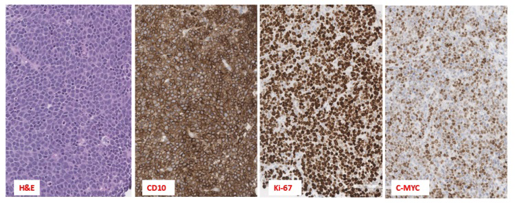
A core needle biopsy of the abdominal mass (Figure 1) was performed and showed DLBCL, germinal center type. Immunohistochemical stains performed demonstrated that the neoplastic lymphocytes were positive for CD79a and CD10, consistent with a germinal center phenotype of DLBCL (Figure 1). Stains for BCL2 and MYC proteins were both positive and the Ki-67 proliferation index was approximately 95% (Figure 1). Concurrent flow cytometric analysis identified a CD10(+) monoclonal lambda B-cell population. The PET CT scan showed widespread, FDG avid lymphadenopathy within the neck, chest, abdomen, and pelvis, hypermetabolic mural thickening about the splenic flexure of colon, and multiple FDG avid osseous lesions.
A bone marrow evaluation demonstrated a hypercellular bone marrow (50-60%) involved by DLBCL, comprising approximately 40-50% of the bone marrow cellularity. Immunohistochemical staining performed on the core biopsy was positive for CD10, CD20, BCL2, BCL6, and MYC, and negative for CD3, CD5, MUM1, cyclin D1, and EBV. The Ki-67 proliferation index was approximately 70%. A lumbar puncture with flow cytometry was negative for lymphoma involvement, and a dose of intrathecal methotrexate was given after the procedure. An MRI of the brain did not show any evidence of lymphoma in the CNS.
Conventional cytogenetics performed on the bone marrow specimen demonstrated a complex karyotype with multiple numeric and structural abnormalities, including t(3;8) and t(14;18) (Table 1). FISH using DNA probes specific for the MYC region at 8q24 and BCL6 region at 3q27 had an abnormal atypical signal pattern. Metaphase FISH confirmed a t(3;8) rearrangement along with additional rearranged signals of BCL6 and MYC on the homologous chromosome 8 (Figures 2 & 3). A FISH study performed on bone marrow clot specimen showed abnormal atypical signal patterns for MYC and BCL6 break apart probes, 2 signals for 5’MYC and 1 signal for 3’MYC, 1 fused BCL6 signal, 3 signals for 5’BCL6 and 1 signal for 3’BCL6, respectively. FISH showed typical signal pattern for t(14;18) IGH::BCL2 rearrangement. Together, both interphase and metaphase FISH results confirmed a very rare BCL6::MYC [t(3;8)] rearrangement. This result is consistent with high-grade B-cell lymphoma with BCL6::MYC, and IGH::BCL2 gene rearrangements. Molecular studies were not performed.
| CASE # | HANS | Age | Sex | IHC | CD10 | Ki-67 (%) | CD5 | CD20 | Conventional Karyotype | PCR | FISH | References |
|---|---|---|---|---|---|---|---|---|---|---|---|---|
| Study case | GC | 65 | F | MYC (+); BCL2 (+); BCL6(+) | + | 95 | - | - | 45-48,XX,add(1) (q42),del(2) (q33q35),t(3;8) (q27;q24.1),+7,+7, t(14;18) (q32;q21.3),-15, add(18)(q23),der(19) t(15;19)(q15;q13.3) [cp9]/46, XX[11] | NP | Interphase FISH: MYC nuc ish (5’MYCx2, 3’MYCx1) [77/200] BCL6 nuc ish(5’BCL6x4,3’BCL6x2) (5’BCL6 con 3’BCL6x1) [111/200] BCL2 nuc ish(IGH,BCL2)x3(IGH con BCL2x2)[78/100]; Metaphase FISH: ish der(8)t(3;8)(q27;q24.1) (5’MYC+,3’MYC-,5’BCL6- ,3’BCL6+)x2 ish der(3) ins(3)(p25)(3’BCL6+)t(3;8) (5’BCL6-,5’MYC-,3’MYC+) | Study case |
| 1 | GC | 53 | F | MYC (NR); BCL2 (+); BCL6(+) | + | 40 | - | + | 48,XX,t(3;8) (q27;q24.1), del(6) (q21q23),+12, +18[7]/46, XX[1] | IGH::BCL2 fusion transcript negative | t(3;8)(q27;q24.1) | Wang HY, et al. [8] |
| 2 | non-GC | 66 | F | MYC (>90% +); BCL2 (+); BCL6 (+) | - | NR | - | + | 47,XX,dup(1) (q25q32),?del(8) (q24),+13,del(13) (q13q21)x2,add(14) (q3?1)[cp11]/46, XX[15] | NR | Metaphase FISH: ish dup(1)(q25q32) (MEGF6+,ABL2++),t(3;8;14) (q27;q24;q32) (3’BCL6+,5’BCL6- ,5’IgH+;5’MYC+, 3’MYC- ,5’BCL6;3’IgH+,5’IgH- ,3’MYC+),del(13)(q13q21) (D13S319-, LAMP1+)x2 | De Paoli E, et al. [9] |
| 3 | non-GC | 37 | F | MYC (NR); BCL2 (-); BCL6(NR) | - | 100 | - | + | 46,XX,t(3;8)(q27;q24) [7]/46,XX[3] | NR | Interphase FISH: BCL6 nuc ish(BCL6x2)(5’BCL6 sep 3’BCL6x1)[12/100] MYC nuc ish(MYCx2) (5’MYC sep 3’MYCx1) [11/100] BCL6::MYC nuc ish(BCL6,MYC)x3(BCL6 con MYCx2)[14/100]; Metaphase FISH: ish t(3;8) (q27;q24)(5’MYC+;3’MYC+) | Sanders L, et al. [3] |
| 4 | GC | 72 | M | MYC (NR); BCL2 (-); BCL6(+) | + | 100 | - | + | 47,XY,inv(2) (p11q21),t(3;8) (q27;q24),del(6) (q23q25),+12,−13, t(14;18)(q32;q21) [9]/46,XY | NP | NR/NP | Motlló C, et al. [10] |
| 5 | GC | 44 | F | MYC (NR); BCL2 (-); BCL6(+) | + | 95 | NR | NR | 48,XX,t(3;8) (q27;q24),−4,add(7) (q21.2),+der(8)t(3;8) (q27;q24), add(11) (q22),add(12) (p13),t(14;18) (q32;q21),+mar1, +mar2 | IGH:: BCL2 fusion product. | NR/NP | Motlló C, et al. [10] |
| 6 | GC | 53 | M | MYC (NR); BCL2 (NR); BCL6(NR) | NR | NR | NR | NR | 48,XY,del(2)(q34),t(3;8) (q27;q24),+7,+der(8) t(3;8),t(14;18) (q32;q21) [18] | NR/NP | NR/NP | Bertrand P, et al. [11] |
| 7 | GC | 56 | F | MYC (NR); BCL2 (NR); BCL6(NR) | NR | NR | NR | NR | 49,XX,t(3;8) (q27;q24),+7,+der(8) t(3;8),del(10) (q24q25),+12,t(14;18) (q32;q21) [20] | NR/NP | NR/NP | Bertrand P, et al. [11] |
Table 1: ** Cases with BCL6::MYC rearrangement in DLBCL.
NP - Not performed; NR – Not reported. Table 1: Cases with BCL6::MYC rearrangement in DLBCL.
![Figure 2: Showing BCL6 (3q27) Break Apart Probe FISH Signal Translocation to Chromosome 8 Long Arm (Tel 5’BCL6 Red [R]; Cen 3’BCL6 Green [G]). A. G-band metaphase; B. Reverse G-band Metaphase; C. Karyogram with Representation of the BCL6 Rearranged Signals.](/fulltextimages/13083/fig_2.png)
![Figure 3: Showing MYC (8q24) Break Apart Probe FISH Signal Translocation to Chromosome 3 Long Arm (Cen 5’MYC Red [R]; Tel 3’MYC Green [G]). A. G-band metaphase; B. Reverse G-band; Metaphase C. Karyogram with Representation of the MYC Rearranged Signals.](/fulltextimages/13083/fig_3.png)
The patient began treatment with R-CHOP. Soon after beginning treatment the patient had a recurrence of diarrhea and abdominal pain and was re-admitted for C. difficile colitis. A CT performed demonstrated that there was significant lymphoma progression, and it was apparent that the lymphoma progressed through R-CHOP chemoimmunotherapy. The patient then started treatment with Tafasitamab and Lenalidomide as a subsequent therapy. She continued to decline and opted for palliative care, succumbing two weeks later.
Discussion
Analysis of MYC, BCL2 and BCL6 regions is essential in identifying high-risk patients. Lymphomas with MYC and BCL2, and/or BCL6 rearrangements are known as high- grade B-cell lymphoma with MYC and BCL2 and/or BCL6 rearrangements, previously called DHL or THL and are associated with a poor clinical outcome.
The t(3;8) is a rare rearrangement seen in DLBCL. The oncogenic effect of the t(3;8) rearrangement is heterologous activation of MYC by the BCL6 locus enhancer and therefore is equivalent to a single-hit MYC-activating rearrangement [5, 6, 7]. The FISH study performed on the bone marrow clot showed abnormal atypical signal patterns for MYC and BCL6 break-apart probes and typical signal pattern for t(14;18) IGH::BCL2 rearrangement. The identification of BCL6::MYC fusion using interphase FISH using break-apart probes is indistinguishable from more conventional DHL with independent MYC and BCL6 translocations.
The median overall survival of <2 years has been reported in DHL cases. A worse prognosis has been associated for DHL patients with additional clinicopathologic features including bone marrow involvement and IG::MYC rearrangement. Based on the literature review we identified seven cases of DLBCL with the reported BCL6::MYC gene rearrangement. Six of these cases showed t(3;8) by conventional cytogenetics. Only one had a cryptic three-way translocation involving chromosomes 3, 8, and 14. Our case is the first case which showed rare cryptic translocation involving chromosomes 3 and 8. Based on the data available for survival in some of the patients listed in Table 1, the median age was 59 years. Our patient had a significant mass effect with an abdominal mass over 20 cm, similar to Case #5 (Table 1) who developed a mass and had the worst outcome of the cases listed, with an average survival time of 3.5 months in these two cases. Overall survival data is limited due to few reported cases with this rearrangement.
Identifying more cases with similar rare translocations involved in DHL compared to typical DHL may help to better delineate such a rare entity. This case displays the crucial role of metaphase FISH in identifying specific chromosome partners involved in the translocation which are indistinguishable by the interphase FISH especially using FISH break apart probes. Although DLBCLs with DHL now known as high-grade B-cell lymphomas are associated with an overall survival at 3 years [12], presence of very rare BCL6::MYC with typical IGH::BCL2 gene rearrangements in DLBCL lymphomas along with additional comorbidities may have shorter survival rate.
References
-
Alaggio R, Amador C, Anagnostopoulos I, Attygalle AD, Araujo IBDO, et al. (2022) The 5th edition of the World Health Organization Classification of Haematolymphoid Tumours: Lymphoid Neoplasms. Leukemia 36(7): 1720- 1748.
-
Campo E, Jaffe ES, Cook JR, Quintanilla-Martinez L, Swerdlow SH, et al. (2022) The International Consensus Classification of Mature Lymphoid Neoplasms: a report from the Clinical Advisory Committee. Blood 140(11): 1229-1253.
-
Sanders L, Jayne S, Kennedy B, Miall F, Aukema SM, et al. (2014) A Double Hit CD10-Negative B-Cell Lymphoma with t(3;8)(q27;q24) Leading to Juxtaposition of the BCL6 and MYC Loci Associated with Good Clinical Outcome. Case Rep Hematol 2014: 120714.
-
Landsburg DJ, Falkiewicz MK, Maly J, Blum KA, Howlett C, et al. (2017) Outcomes of patients with double-hit lymphoma who achieve first complete remission. J Clin Oncol 35(20): 2260-2267.
-
Ryan RJH, Drier Y, Whitton H, Cotton MJ, Kauret J, et al. (2015) Detection of Enhancer-Associated Rearrangements Reveals Mechanisms of Oncogene Dysregulation in B-cell Lymphoma. Cancer Discov 5(10): 1058-1071.
-
Johnson SM, Umakanthan YM, Yuan J, Fedoriw Y, Bociek RG, et al. (2018) Lymphomas with pseudo-double-hit BCL6-MYC translocations due to t(3;8)(q27;q24) are associated with a germinal center immunophenotype, extranodal involvement, and frequent BCL2 translocations. Hum Pathol 80: 192-200.
-
Ohno H, Nakagawa M, Kishimori C, Fukutsuka K, Honjo G (2017) Cryptic t(3;8)(q27;q24) and/or MYC- BCL6 linkage associated with MYC expression by immunohistochemistry is frequent in multiple-hit B-cell lymphomas. Blood Cancer J 7(6): e578.
-
Huan-You W, Bossler AD, Schaffer A, Tomczak E, DiPatri D, et al. (2007) A novel t(3;8)(q27;q24.1) simultaneously involving both the BCL6 and MYC genes in a diffuse large B-cell lymphoma. Cancer Genet Cytogenet 172(1): 45- 53.
-
Paoli ED, Bandiera L, Ravano E, Cesana C, Grillo G, et al. (2018) A double-hit High-grade B-cell lymphoma with three-way translocation t(3;8;14)(q27;q24;q32) involving BCL6, MYC, and IGH. Clin Case Rep 6(12): 2411-2415.
-
Motlló C, Grau J, Juncà J, Ruiz N, José-Luis M, et al. (2010) Translocation (3;8)(q27;q24) in two cases of triple hit lymphoma. Cancer Genet Cytogenet 203(2): 328-332.
-
Bertrand P, Bastard C, Maingonnat C, Jardin F, Maisonneuve C, et al. (2007) Mapping of MYC breakpoints in 8q24 rearrangements involving non-immunoglobulin partners in B-cell lymphomas. Leukemia 21(3): 515-523.
-
Landsburg DJ, Falkiewicz MK, Maly J, Blum KA, Howlett C, et al. (2017) Outcomes of Patients With Double-Hit Lymphoma Who Achieve First Complete Remission. J Clin Oncol 35(20): 2260-2267.
- Genomic Landscape of Aggressive Penile Squamous Cell Carcinoma including TERT-p and NOTCH1 Mutations – An Institutional Experience
- Establishment of Baseline Haematological Values for Canine Population in North-Central Nigeria: A Cross-Sectional Study in the Federal Capital Territory
- Biochemical Assessment of Uroliths Extracted in Patients with Urolithiasis in a Tertiary Health Institution
- Update on Gastrointestinal Pecomas: Molecular Pathogenesis and Risk Stratification
- A Comparative Study of Serum C-reactive Protein Level Between Pre-eclampsia and Normal Pregnancy in Tertiary Level Hospital
- From Deformity to Alignment: Clinical Outcomes of the Schnepp Osteotomy in Hallux Valgus in 47 Feet