The Impact of Aircraft Noise Exposure on the Efficacy of Empagliflozin Therapy in an Animal Model of Obesity
Background: Obesity and type 2 diabetes mellitus (T2DM) are among the leading risk factors for mortality worldwide. Sodium-glucose co-transporter 2 Inhibitors (SGLT2i), such as empagliflozin, represent a last-generation drug class for T2DM treatment with cardiovascular benefits. This research project aims to investigate whether coexposure to aircraft noise affects the therapeutic efficacy of empagliflozin in an animal model of obesity. Methods and Results: To induce obesity, we fed C57BL/6JRj mice a Western (high fat) diet for 20 weeks. After 14 weeks, empagliflozin (10 mg/kg/d) treatment was applied via drinking water. To investigate the effects of aircraft noise, we additionally exposed the mice to aircraft noise for the last 4 days of the treatment period. Noise exposure alone interfered with glucose metabolism, increased blood pressure, impaired the endothelial function of the aorta, and increased vascular oxidative stress and inflammation. Six weeks of empagliflozin therapy improved blood glucose metabolism and vascular and oxidative parameters in high fat diet (HFD) animals. These therapeutic effects were attenuated or absent in animals exposed to additional aircraft noise. Conclusion: SGLT2i, such as empagliflozin, are widely used in clinical practice beyond the treatment of T2DM. Our study demonstrates that additional exposure to aircraft noise can attenuate the protective effects of SGLT2 inhibitors in a murine obesity model. Considering personalized medicine progress, it is crucial to understand better the interaction of several risk factors, especially environmental stressors, in order to improve the therapeutic outcome of established medications and thus enable better protection of vulnerable groups.
Keywords
Obesity; Aircraft Noise; Diabetes; Empagliflozin; Cardiovascular Complications, Metabolism; Oxidative Stress
Abbreviation
T2DM: Type 2 Diabetes Mellitus; SGLT2i: Sodium-Glucose Co- Transporter 2 Inhibitors; GLP-1: Glucagon-Like Peptide-1; eNOS: Endothelial Nitric Oxide Synthase; BP: Blood Pressure; DHE: Dihydroethidium; HPLC: High-Pressure Liquid Chromatography.
Introduction
Obesity and type 2 diabetes mellitus (T2DM) are among the leading causes of mortality worldwide [1]. Over the last decade, the pharmacotherapy of T2DM has been significantly improved by the clinical use of sodium-glucose co- transporter 2 inhibitors (SGLT2i) [2]. Due to their protective cardiovascular effects, SGLT2i are now part of standard therapy for heart failure and chronic renal insufficiency, also beyond T2DM [3, 4, 5]. In the treatment of obesity, glucagon- like peptide-1 receptor (GLP-1) agonists in particular have proven effective in recent years, although the beneficial effects of SGLT2i have also been described [6, 7].
In general, cardiovascular risk factors such as obesity and T2DM often occur in combination with other risk factors. Environmental factors including particulate matter or traffic noise, have become the focus of scientific attention in recent years. Transportation noise has been identified as an important cardiovascular risk factor, causing increased blood pressure, endothelial dysfunction, oxidative stress, and inflammation, mostly by activation of the phagocytic NADPH oxidase (NOX-2) and uncoupling of the endothelial nitric oxide synthase (eNOS) [8, 9]. Epidemiological studies have linked the incidence and mortality of T2DM and metabolic syndrome to noise exposure.
A meta-analysis of 15 studies found a 6% risk increase of diabetes for every 5 dB increase in noise exposure, primarily associated with air and road noise [10]. A cohort study performed in Taiwan showed a 13% and 24% risk increase in metabolic syndrome incidence after medium and high noise exposure respectively [11]. Moreover, our group has previously established an additive adverse effect of aircraft noise exposure on cardiovascular complications in animal models of diabetes and obesity [12]. We showed that noise interfered with glucose metabolism and insulin signaling, caused macro- and microvascular dysfunction as well as increased oxidative stress and inflammation in animals with pre-existing diabetes.
While "SGLT2i" have shown promising outcomes in the management of T2DM and obesity, especially in improving cardiovascular and renal health, the efficacy of the treatment in relation to coexposure to other risk factors, such as environmental noise has yet to be elucidated [13]. The rationale for this investigation is based on previous findings that SGLT2i therapy targets the main pathomechanisms of noise exposure, endothelial dysfunction, oxidative stress and inflammation, as shown for diabetic animals [14, 15]. Therefore, the present study aims to investigate whether coexposure to aircraft noise affects the therapeutic efficacy of the SGLT2i empagliflozin in an animal model of obesity and T2DM.
Materials and Methods
Animal Models
All animal experiments in this study were performed in accordance with the Guide for the Care and Use of Laboratory Animals, as adopted and promulgated by the US National Institutes of Health and were approved by the Ethics Committee of the University Hospital Mainz and the Landesuntersuchungsamt Rheinland-Pfalz (Koblenz, Germany, permit number 23 177-07/G15-1-094 and G20-1- 103).
For the study, we used male C57BL/6JRj mice ordered from Janvier (Le Genest-Saint-Isle, France). Animals (8 weeks old) were fed a high-fat diet (HFD, ssniff [#E15721- 34], 42% energy from fat (21.1% of total food composition), 0.21% cholesterol, ad libitum) for 20 weeks to induce obesity/type II diabetes mellitus [16]. From week 14, we started administering a sodium glucose co-transporter 2 inhibitor (SGLT2i), empagliflozin (Boehringer Ingelheim, 10 mg/kg/d via drinking water for a total of 6.5 weeks). For the last 4 days of treatment, we exposed the animals to aircraft noise [maximum sound pressure level of 85 dB(A), average sound pressure level of 72 dB(A)] as described previously [8]. For a detailed description of the treatment, see Figure 3A. At the end of the experiment, animals were sacrificed using ketamine anesthesia + xylazine analgesia, followed by cervical dislocation and sample collection (aorta, heart, brain and liver). In addition, blood samples were obtained by heart puncture using heparin and K2EDTA.
Non-Invasive Blood Pressure Measurements
Non-invasive blood pressure (BP) measurements were performed using plethysmography with the CODA system (Kent Scientific Corporation, Torrington, CT) as described previously [9]. To minimize the stress-induced effects on BP readings caused by restraint, we repeatedly trained the animals before the measurements. Animals were allowed to enter the restraining tube freely and after securing were placed on a preheated warming platform (32 °C), and allowed to rest for 15 min. For each animal, 15 BP values were obtained, with the first five discarded as acclimation cycles. The mean of the remaining 10 BP measurements was used for analysis. Measurements took place at a consistent time each day to account for diurnal variations in BP. The measurement schedule is illustrated in Figure 1A.
Blood Glucose, Oral Glucose Tolerance Test (OGTT), Glycated Hemoglobin (Hba1c), and Urine Glucose
Blood glucose levels were measured in the tail vein blood using a glucometer (Roche, ACCU-CHECK Aviva). OGTT was conducted on the final day of treatment in fasting animals (8 h). A glucose solution (1,5 g/kg D-(+)-Glucose in H2O) was administered via oral gavage (10 µl/g). Blood glucose levels were measured by glucometer (Roche, ACCU-CHECK Aviva) in tail vein blood at 0, 60 and 120 min post-administration [17]. Glycated hemoglobin (HbA1c) levels were determined using the A1CNow+Professional test kit (PTS Diagnostics, #3038) following the manufacturer’s protocol. Glycosuria was measured by CombiScreen® Glucose PLUS urine test strips (Analyticon Biotechnologies, #94501) per manufacturer’s instructions.
Plasma Insulin Levels
Plasma insulin levels were quantified using an Ultrasensitive Mouse Insulin Elisa-Kit (#90080 Crystal Chem) according to the manufacturer’s protocol.
Isometric Tension Studies
Endothelial function was assessed by isometric tension studies in aorta using an organ bath. Endothelium-dependent and independent relaxation was evaluated in intact mouse aortic rings (3 mm, perivascular fat, and connective tissues free) by applying acetylcholine (ACh 10−9 –10−5.5 M) and nitroglycerin (NTG 10−9 –10−4.5 M) upon pre-constriction with prostaglandin F2α, as described previously [18].
Oxidative Stress Measurement in Cerebral Arterioles
For dihydroethidium (DHE) staining, 10 μM cryosections were placed on Superfrost Plus slides (Thermo Fisher Scientific, Braunschweig, Germany), and 1 ml of 1 μM DHE solution was applied to each slide. The slides were then kept in a light-protected and humidified chamber, and incubated at 37 °C for 30 min. The fluorescence was recorded using an Eclipse TS 100 microscope (Nikon, Yurakucho, Tokyo, Japan) equipped with a DS–Fi1-U2 digital microscope camera (Nikon) and the imaging software NIS Elements (Nikon, Version 1.10 64 bit). Subsequently, fluorescence intensity in the vascular wall was evaluated using ImageJ (NIH, http:// rsb.info.nih.gov/ij/) as previously described [19, 20].
Oxidative Stress in Whole Blood
The leukocyte-dependent oxidative burst measures leukocyte-dependent hydrogen peroxide formation, primarily mediated by phagocyte-type NADPH oxidase (NOX- 2). Hydrogen peroxide is converted by myeloperoxidase to highly reactive oxygen-metal complexes, which lead to oxidation of L-012 to an intermediate that emits energy in the form of chemiluminescent light. Oxidative burst was assessed in fresh citrate blood diluted 1:50 and stimulated with zymosan A (50 µg/ml) or phorbol ester dibutyrate (PDBu, 10 µM) in PBS buffer containing Ca2+/Mg2+ (1 mM). The reaction was detected using L-012 (100 µM, 8-amino- 5-chloro-7-phenylpyrido[3,4-d]pyridazine-1,4-(2H,3H)- dione sodium salt, Wako Pure Chemical Industries, Osaka, Japan) enhanced chemiluminescence (ECL) using a Mithras2 chemiluminescence plate reader (Berthold Technologies, Bad Wildbad, Germany) as previously described [14].
Oxidative Stress Detection in Vascular and Cardiac Tissue
Oxidative stress in aortic and cardiac tissues was measured by a modified high-pressure liquid chromatography (HPLC)-based method, quantifying the total 2-hydroxy ethidium (2-HE) levels as described previously [21]. Additionally, the mitochondria-specific superoxide production was determined in mitochondria isolated from cardiac tissue using the mitoSOX/HPLC method as described previously [22]. Vascular superoxide formation in aortic rings (3 mm, perivascular fat, and connective tissues free) was determined by lucigenin(5 µM)-enhanced chemiluminescence (CL) using a Lumat LB9507 single photon counter as described [23]. The CL was registered at intervals of 60 s over 20 min and was normalized to the dry weight of aortic tissue.
Dihydroethidium (DHE)-dependent fluorescence was employed to assess reactive oxygen species (ROS) formation in aortic cryosections, as described previously [24]. Vascular rings (3 mm) were embedded in Tissue-Tek® O.C.T compound, and cryosectioned into 8 µm slices. These sections were incubated with 1 µM DHE for 30 min at 37 °C. Images were captured using a Zeiss Axiovert 40 CFF microscope equipped with a Axiocam MRm camera and quantified as integrated optical density (IOD) using ImageJ software.
Immunohistochemistry of Aortic Tissue
Immunohistochemistry has been performed following previously established protocols [9]. Briefly, aortic segments, including adventitial and perivascular tissue, were fixed in 4% formaldehyde, embedded in paraffin, and sectioned into 5 µm slices. After deparaffinization, samples were blocked with normal horse blocking solution (Vector) and stained with a primary antibody against NOX-2 (1:200, #LS-B12365, LS Bio) or endothelin-1 (ET-1, Abcam #117757: 1:1000), biotinylated with a secondary antibody (Thermo Fisher Scientific, Waltham, MA). Quantification of the stained images was performed using ImageJ software.
Detection of Plasma Triglycerides and Cholesterol
Plasma triglyceride, cholesterol, LDL and HDL levels were analysed in the Department of Clinical Chemistry and Laboratory Medicine, University Hospital Mainz, Germany, using the daily routine facilities for in-patient care.
Dot Blot Protein Analysis
The protein content in blood plasma was determined by Bradford assay, and samples were then diluted (in PBS containing 0.1% SDS) to achieve uniform protein concentration.
Dot blot analysis was performed as previously described using a Minifold-I vacuum Dot Blot system (Schleicher & Schuell, 10484138CP) [9]. Following two washing steps with 200 µl PBS, 20 µg of protein was loaded into each well of the system and transferred onto a nitrocellulose membrane via vacuum. The membrane was washed twice with PBS, dried at 60 °C for 60 min to fix the protein, and then stained with Ponceau S solution [9]. After removing the stain, the membrane was blocked at RT for 1 h with 3% BSA in PBS-T.
The membrane was incubated overnight at 4 °C with a primary anti-IL-6 antibody (dilution: 1:1000, ab6672) from Abcam (Cambridge, UK). Following a washing step (4 x 5 min in PBS-T), the membrane was incubated with an anti-rabbit peroxidase coupled secondary antibody (dilution 1:2000, PI- 1000, Vector Lab, Newark, CA, USA) at RT for 1 h. The signal was detected using an ECL development kit (#1859701 and #1859698, Thermo Fisher Scientific, Waltham, MA, USA) and Chemostar M6 imager from Intas Science Imaging. Densitometric quantification was performed using GelProAnalyser software (Version 3.0.00.00).
Statistical Analysis
Statistical analysis was conducted using Prism for Windows, version 9, GraphPad Software Inc. (GraphPad Software LLC, La Jolla, CA). Statistical comparisons were performed using unpaired t-test or two-way ANOVA with a repeated measures approach with Tukey correction as appropriate. The presented results are expressed either as mean ± SD or percentage of change ± SD. The n for every independent experiment is indicated. Statistical analysis was performed at a significance level α = 5%.
Results
Effects of Aircraft Noise Exposure on Glucose Metabolism and Vascular Function in HFD Mice
Aircraft noise exposure over 4 days induced a reduction in plasma insulin levels in HFD mice (Figure 1A-B). Analogously, an additive noise-associated increase in non- fasting blood glucose was found in obese animals (Figure 1C). Acute exposure to aircraft noise increased systolic blood pressure after 4 days in HFD mice (Figure 1D). By isometric tension studies, we could show that additive aircraft noise exposure also reduced the maximum relaxation capacity of the aorta in animals with experimental obesity (Figure 1E).
Impact of Aircraft Noise Exposure on Oxidative Stress in HFD Mice
In HFD mice, noise exposure increased reactive oxygen species (ROS) formation in cerebral arterioles, indicating a corresponding microvascular dysfunction (Figure 1F). Using a DHE-based HPLC assay, we could show that both aortic and cardiac superoxide formation were (non-significantly) increased by aircraft noise exposure (Figure 1G,1H). In isolated cardiac mitochondria, we detected increased superoxide production in HFD mice exposed to aircraft noise (Figure 1I).
![Figure 1: The effects of aircraft noise exposure on glucose metabolism, vascular function, and oxidative stress in diabetes. (A) Scheme of the treatment protocol and blood pressure measurements. (B) Plasmatic insulin levels were determined using an ELISA Assay Kit. (C) Non-fasting blood glucose levels were measured by a glucometer from the tail vein blood. (D) Systolic blood pressure measured using non-invasive plethysmography at D4 of noise exposure. (E) Endothelium-dependent (acetylcholine) relaxation of the aorta was measured using the isometric tension method. The results represent the maximal precentral relaxation of pre-constriction. (F) The ROS formation measured by in situ dihydroethidium (DHE) fluorescence in cerebral arteriole cross-sections, including representative pictures (scale bar 20µm). (G) Aortic superoxide formation measured by 2-HE HPLC analysis, including representative chromatogram. (H) Superoxide levels in cardiac tissue measured by 2-HE HPLC analysis, including representative chromatogram. (I) Mitochondria-specific superoxide formation in cardiac tissue measured by mitoSOX HPLC analysis, including representative chromatogram. (B) Each data point represents a measurement from blood plasma pooled from three to six animals (n = 8–9). (C-F, H) Each data point represents an individual animal [C (n = 8–15), D (n = 12–17), E (n = 19–22), F (n = 8), H (n = 10)]. (G) A pair of data points represents a sample pooled from three to six animals [G (n = 4–5)]. (I) A triplet of data points represents a sample obtained by pooling tissue from three to six animals [I (n = 5–6 pooled mitochondrial preparations)]. For statistical analysis, we used an unpaired t-test, and p < 0.05 were considered significant. (*p < 0.05, ****p < 0.0001). The treatment scheme was generated with biorender.com. All figures were adapted from [12].](/fulltextimages/14049/fig_1.png)
Figure 1: The effects of aircraft noise exposure on glucose metabolism, vascular function, and oxidative stress in diabetes. (A) Scheme of the treatment protocol and blood pressure measurements. (B) Plasmatic insulin levels were determined using an ELISA Assay Kit. (C) Non-fasting blood glucose levels were measured by a glucometer from the tail vein blood. (D) Systolic blood pressure measured using non-invasive plethysmography at D4 of noise exposure. (E) Endothelium-dependent (acetylcholine) relaxation of the aorta was measured using the isometric tension method. The results represent the maximal precentral relaxation of pre-constriction. (F) The ROS formation measured by in situ dihydroethidium (DHE) fluorescence in cerebral arteriole cross-sections, including representative pictures (scale bar 20µm). (G) Aortic superoxide formation measured by 2-HE HPLC analysis, including representative chromatogram. (H) Superoxide levels in cardiac tissue measured by 2-HE HPLC analysis, including representative chromatogram. (I) Mitochondria-specific superoxide formation in cardiac tissue measured by mitoSOX HPLC analysis, including representative chromatogram. (B) Each data point represents a measurement from blood plasma pooled from three to six animals (n = 8–9). (C-F, H) Each data point represents an individual animal [C (n = 8–15), D (n = 12–17), E (n = 19–22), F (n = 8), H (n = 10)]. (G) A pair of data points represents a sample pooled from three to six animals [G (n = 4–5)]. (I) A triplet of data points represents a sample obtained by pooling tissue from three to six animals [I (n = 5–6 pooled mitochondrial preparations)]. For statistical analysis, we used an unpaired t-test, and p < 0.05 were considered significant. (*p < 0.05, ****p < 0.0001). The treatment scheme was generated with biorender.com. All figures were adapted from [12].
Based on the results from our previous publication, we were able to clarify the correlation between aircraft noise exposure and oxidative stress in HFD mice, using further experiments. Accordingly, we found elevated levels of vascular ROS in whole blood after exposure to aircraft noise, which could be demonstrated with both the faster- acting PDBu-based (Figure 2A-B) and the delayed-acting Zymosan-associated (Figure 2C,D) stimulation of leukocytes. Maximum kinetics values were each displayed as separate bar graphs with scatter plots (Figure 2B,D).
![Figure 2: Aircraft noise exposure increases vascular oxidative stress and inflammation. (A-D) Leukocyte-derived ROS in whole blood by L-012 enhanced chemiluminiscence (ECL). Oxidative burst kinetics upon PDBu (A) and zymosan A (C) stimulation and quantification at 20 min upon PDBu (B) and at 55 min upon zymosan A (D) stimulation. (E) Protein expression of interleukin 6 (IL-6) was assessed by dot blot analysis. (F) Quantification of aortic superoxide formation by lucigenin ECL. (A, C) Data represent the mean of n = 4 independent measurements of blood samples pooled from four animals. (B, D, E) Each data point represents a measurement from blood plasma pooled from three to six animals [B, D (n = 4), F (n = 6)]. (F) Each data point represents an individual animal (n = 8). For statistical analysis, we used an unpaired t-test (B, D, E, F) or two-way ANOVA (A, C). p < 0.05 were considered significant. (*p < 0.05, **p < 0.01, ***p < 0.001, ****p < 0.0001) All figures were adapted from [12].](/fulltextimages/14049/fig_2.png)
Figure 2: Aircraft noise exposure increases vascular oxidative stress and inflammation. (A-D) Leukocyte-derived ROS in whole blood by L-012 enhanced chemiluminiscence (ECL). Oxidative burst kinetics upon PDBu (A) and zymosan A (C) stimulation and quantification at 20 min upon PDBu (B) and at 55 min upon zymosan A (D) stimulation. (E) Protein expression of interleukin 6 (IL-6) was assessed by dot blot analysis. (F) Quantification of aortic superoxide formation by lucigenin ECL. (A, C) Data represent the mean of n = 4 independent measurements of blood samples pooled from four animals. (B, D, E) Each data point represents a measurement from blood plasma pooled from three to six animals [B, D (n = 4), F (n = 6)]. (F) Each data point represents an individual animal (n = 8). For statistical analysis, we used an unpaired t-test (B, D, E, F) or two-way ANOVA (A, C). p < 0.05 were considered significant. (*p < 0.05, p < 0.01, *p < 0.001, ****p < 0.0001) All figures were adapted from [12].
This correlated with increased IL-6 expression in plasma associated with aircraft noise (Figure 2E). Using a lucigenin- based electroluminescence assay, we detected increased aortic superoxide in HFD mice exposed to aircraft noise (Figure 2F).
Aircraft Noise Exposure Changes the Effects of Empagliflozin on Glucose Metabolism in HFD Mice
The additive treatment with empagliflozin in the last 6 weeks of the (20 weeks) high-fat diet did not cause relevant changes in animal body weight independently of noise exposure (Figure 3A-B). On the other hand, a reduction in fasting blood glucose was observed in both the HFD and the HFD+noise group under empagliflozin treatment (Figure 3C). Like fasting blood glucose, the oral glucose tolerance test displayed a blood glucose-lowering effect in both groups in response to the additive empagliflozin therapy (Figure 3D, E). However, these changes were only significant in the HFD mice not exposed to aircraft noise. Empagliflozin caused a substantial reduction in the HbA1c in HFD mice, not in those exposed to aircraft noise (Figure 3F). Empagliflozin is known to induce glucosuria in mice; therefore, we assessed urine glucose levels as a proof-of-concept for the therapy [25]. The test showed markedly increased glycosuria in the empagliflozin-treated animals compared to the controls (Figure 3G), suggesting successful administration of the therapy.
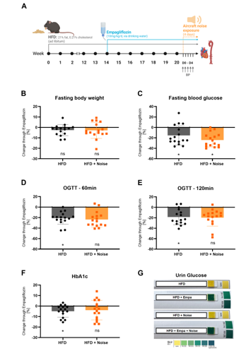
Figure 3: The effects of empagliflozin treatment on body weight and glucose metabolism in diabetic animals with and without aircraft noise coexposure. (A) Scheme of the treatment protocol and blood pressure measurements. (B) Fasting body weight was measured at the last day of the treatment period. (C) Fasting blood glucose levels were measured by a glucometer from tail vein blood. (D, E) The oral glucose tolerance test (OGTT) was measured at the last day of treatment at 60 (D) and 120 min (E) after glucose administration. (F) Glycated hemoglobin (HbA1c) was measured from tail vein blood using a commercially available kit. (G) Representative pictures of urine glucose levels collected on the last day of treatment. (B-F) Each data point represents the change (in %) caused by empagliflozin in individual animals (n = 16). For statistical analysis, we used two-way ANOVA, and p < 0.05 were considered significant. (*p < 0.05). The treatment scheme was generated with biorender.com.
Effects of Empagliflozin on Lipid Metabolism and Liver Weight in Response to Aircraft Noise Exposure in HFD Mice
The additive treatment with empagliflozin did not significantly alter lipid metabolism regarding triglycerides, cholesterol, HDL and LDL or their ratio in obese animals (Figure 4A-E). This was also the case with animals that had been additionally exposed to aircraft noise (Figure 4A-E). Liver weight was reduced in both groups by empagliflozin treatment, although the difference was only significant in the HFD mice without noise exposure (Figure 4F).
![Figure 4: The effects of empagliflozin treatment on plasma lipids, organ weight, and mitochondrial oxidative stress. (A-D) Lipid content was measured in blood plasma: triglycerides (A), cholesterin (B), high-density lipoprotein (HDL) (C), low- density lipoprotein (LDL) (D). (E) LDL to HDL ratio. (F, G) Liver and heart weights were measured on the last day of the treatment period. (H) Mitochondria-specific superoxide formation in cardiac tissue measured by mitoSOX HPLC analysis, including representative chromatogram. (A-G) Each data point represents the change (in %) caused by empagliflozin in individual animals (n = 8). (H) A triplet of data points represents the change (in %) caused by empagliflozin in a sample obtained by pooling tissue from four animals [(n = 4 pooled mitochondrial preparations)]. For statistical analysis, we used two-way ANOVA, and p < 0.05 were considered significant (*p < 0.05).](/fulltextimages/14049/fig_4.png)
Figure 4: The effects of empagliflozin treatment on plasma lipids, organ weight, and mitochondrial oxidative stress. (A-D) Lipid content was measured in blood plasma: triglycerides (A), cholesterin (B), high-density lipoprotein (HDL) (C), low- density lipoprotein (LDL) (D). (E) LDL to HDL ratio. (F, G) Liver and heart weights were measured on the last day of the treatment period. (H) Mitochondria-specific superoxide formation in cardiac tissue measured by mitoSOX HPLC analysis, including representative chromatogram. (A-G) Each data point represents the change (in %) caused by empagliflozin in individual animals (n = 8). (H) A triplet of data points represents the change (in %) caused by empagliflozin in a sample obtained by pooling tissue from four animals [(n = 4 pooled mitochondrial preparations)]. For statistical analysis, we used two-way ANOVA, and p < 0.05 were considered significant (*p < 0.05).
Effects of Empagliflozin on Heart Weight and Cardiac Oxidative Stress in HFD Mice with and without Aircraft Noise Exposure
Neither in the HFD mice nor in the HFD mice exposed to aircraft noise did the additional therapy with empagliflozin result in a relevant change in heart weight (Figure 4G).
Using the mitoSOX/HPLC assay, we demonstrated that empagliflozin caused reduced superoxide formation in cardiac mitochondria only in HFD mice (Figure 4H). Additional exposure to aircraft noise inhibited this empagliflozin-induced decrease in mitochondrial superoxide formation (Figure 4H).
The Effect of Aircraft Noise Exposure on Vascular Function and Oxidative Stress in Empagliflozin- Treated Obese Mice
Endothelium-dependent (acetylcholine-based) aorta dilation was improved by empagliflozin treatment in the HFD animals without aircraft noise exposure (Figure 5A). In contrast, empagliflozin therapy had no relevant effect on endothelium-independent (nitrate- or muscle cell- mediated) dilatation in both groups (Figure 5B). Using HPLC- and fluorescence-staining-based DHE assays, aircraft noise exposure did not alter the aortic superoxide levels in empagliflozin-treated, HFD mice (Figure 5C,D,G). There was a tendency towards a lower amount of oxidative stress-associated enzymes, such as NOX-2 and ET-1, under empagliflozin treatment in HFD mice (Figure 5E,F and H,I). These effects were diminished (without significance) or completely absent in the animals exposed to aircraft noise (5E,F and H,I).
![Figure 5: The effects of empagliflozin treatment on vascular function and oxidative stress. (A, B) Endothelium-dependent (acetylcholine) (A) and endothelium-independent (nitroglycerine) (B) relaxation of the aorta was measured using the isometric tension method. (C) Aortic superoxide formation measured by 2-HE HPLC analysis, including representative chromatogram. (D) Aortic ROS levels were analyzed with DHE staining. The respective representative pictures (scale bar 50µm) are shown in (G). (E, F) The levels of NOX2 (E) and ET-1 (F) expression were determined by immunohistochemical analysis of aortic paraffin sections. The respective representative pictures (magnification 40x) are shown in (H, I). (A, B, D-F) Each data point represents the change (in %) caused by empagliflozin in an individual animal [A, B (n = 16), D-F (n = 5-8)]. (C) A pair of data points represents the change (in %) caused by empagliflozin in a sample pooled from four animals [G (n = 4)]. For statistical analysis, we used two-way ANOVA, and p < 0.05 were considered significant (*p < 0.05).](/fulltextimages/14049/fig_5.png)
Figure 5: The effects of empagliflozin treatment on vascular function and oxidative stress. (A, B) Endothelium-dependent (acetylcholine) (A) and endothelium-independent (nitroglycerine) (B) relaxation of the aorta was measured using the isometric tension method. (C) Aortic superoxide formation measured by 2-HE HPLC analysis, including representative chromatogram. (D) Aortic ROS levels were analyzed with DHE staining. The respective representative pictures (scale bar 50µm) are shown in (G). (E, F) The levels of NOX2 (E) and ET-1 (F) expression were determined by immunohistochemical analysis of aortic paraffin sections. The respective representative pictures (magnification 40x) are shown in (H, I). (A, B, D-F) Each data point represents the change (in %) caused by empagliflozin in an individual animal [A, B (n = 16), D-F (n = 5-8)]. (C) A pair of data points represents the change (in %) caused by empagliflozin in a sample pooled from four animals [G (n = 4)]. For statistical analysis, we used two-way ANOVA, and p < 0.05 were considered significant (*p < 0.05).
Discussion
Health risk factors can be divided into environmental, occupational, behavioral, and metabolic risks [1]. For decades, medicine and pharmacotherapy have focused in particular on the group of metabolic risk factors, which is why a wide range of therapeutic strategies have been developed. The introduction of SGLT2i as treatment for T2DM has been a groundbreaking development over the last decade, with this group of drugs also being used beyond diabetes due to their beneficial effects. In contrast, there are hardly any sufficient therapeutic strategies available for the other two risk factor groups mentioned, i.e., environmental and occupational, as well as behavioral risk. Human
individuals are typically exposed to multiple, varied risk factors. It is therefore essential to understand the effects of the simultaneous presence of different risk factors and to review established pharmacotherapies in this context. In recent years, our research group has demonstrated additive cardiovascular-damaging effects of the environmental factor aircraft noise in combination with other risk factors such as arterial hypertension or particulate matter [20, 26]. We recently showed that exposure to aircraft noise in mice with pre-existing diabetes mellitus aggravated inflammation, oxidative stress, and endothelial dysfunction [12].
In this study, we aimed to build upon these results and expand on them in the HFD model. This model has the advantage over more specific diabetes models in that it represents a combination of the risk factors T2DM and obesity. This makes it particularly representative of the population of industrialized countries with the associated cardiovascular risk profile [27]. We demonstrated that exposure to aircraft noise in HFD mice led to increased oxidative stress and inflammation in the blood and vasculature. In addition, we aimed to investigate the extent to which empagliflozin treatment can have protective effects in this context. This showed that the noise-exposed animals benefited less overall from empagliflozin treatment than the mice on HFD alone. In both groups of HFD mice, with and without exposure to aircraft noise, glucose metabolism was improved by empagliflozin. However, this benefit was less pronounced under the influence of aircraft noise, whereas empagliflozin treatment showed no relevant effects on lipid metabolism in either group. Only in HFD mice without aircraft noise exposure did the treatment reduce oxidative stress in cardiac mitochondria and the aorta, leading to improved endothelial function. If the HFD mice were additionally exposed to aircraft noise, the effect of the therapy was diminished for these parameters.
Other groups have also demonstrated the protective cardiovascular effects of empagliflozin in HFD mice. However, different dosages or application forms of empagliflozin were used, which is why the results only appear to be comparable to a limited extent [28, 29, 30]. In this context, similar experiments using different antidiabetic drugs, such as metformin, would be highly interesting to evaluate general or substance-specific effects on the vasculature, ROS production, and inflammatory cells. Here, Liu et al. described different impact on pathways in a model of HFpEF, where metformin and empagliflozin both downregulated TGF-ß, whereas empagliflozin alone downregulated HSP90 mRNA and protein expression [31].
To date, there is no established pharmacotherapy for the damaging effects of traffic noise. Appropriate mitigation strategies must be differentiated for the various types of transportation noise [32]. For road traffic noise, recommendations such as speed limits, noise barriers, or investments in infrastructure to promote cycling paths, car sharing, or public transportation can be made [33]. Optimized flight routes, night flight bans, and other measures would reduce aircraft noise exposure [34]. Measures such as rail grinding reduction, regular maintenance, brake upgrading, and nighttime operation bans appear to have a protective effect on railway noise [32]. In an animal model, we have recently demonstrated that activation of the enzyme 5’ adenosine monophosphate-activated protein kinase (AMPK) through fasting, endurance training, or pharmacological stimulation attenuates the cardiovascular damage caused by aircraft noise [35]. In this context, the work of our group also highlighted the protective effects of propranolol and phenoxybenzamine [36].
The novelty of this study lies in demonstrating that the clinically established pharmacotherapy with empagliflozin for obesity, respectively, T2DM became less effective under the simultaneous influence of aircraft noise. On the one hand, this highlights the relevance of traffic noise and other environmental factors such as extreme weather, particulate matter, and light, among others. Furthermore, in large-scale randomized controlled trials, environmental factors should also be taken into account in addition to the usual baseline characteristics such as age, gender, ethnicity, medication, and concomitant diseases, as these can have a significant influence on the respective outcome.
Conclusion
Empagliflozin has become an essential drug for patients suffering from type 2 diabetes mellitus due to a significant reduction in cardiovascular complications and mortality. Underlying mechanisms remain partially elusive. Here, we could demonstrate that empagliflozin treatment improves HFD-induced vascular dysfunction and vascular ROS production. Obviously, the underlying pathophysiologic mechanisms of HFD and aircraft noise-induced vascular dysfunction and inflammation are diverse, as evidenced by the loss of beneficial effects of empagliflozin on endothelial- dependent vascular dysfunction and NOX-2 expression. Therefore, specific “protective” strategies should be implemented that reduce noise exposure in individuals suffering from obesity and diabetes mellitus.
Acknowledgements
Empagliflozin was kindly gifted from Boehringer Ingelheim GmbH & Co. KG, Germany. We thank Angelica Karpi, Nicole Glas, Alexandra Rosenberger, and Jörg Schreiner for excellent technical assistance. This publication and the included results are part of the doctoral thesis of D. Gillenkirch.
Authors Contributions
P.S., T.J., A.D., T.M., and D.M. contributed to the conception or design of the work. P.S., A.D., T.M., T.J., A.G., D.M., and L.S. contributed to the analysis or interpretation of data for the work. D.M., L.S., A.C., A.G., H.U., and D.G. contributed to the acquisition of data for the work. D.M., P.S., A.D., and T.J. drafted the manuscript. D.M., P.S., T.J., A.D., T.M., P.L., A.G., M.K., M.O., M.M., and Z.G. critically revised the manuscript. All gave final approval and agreed to be accountable for all aspects of the work, ensuring integrity and accuracy.
Funding
T.M. is P.L. and A.D., M.K. and P.S. are (Young) Scientists of the DZHK (German Center for Cardiovascular Research), Partner Site Rhine-Main, Mainz, Germany. The work was supported by the environmental network EXPOHEALTH funded by the state Rhineland-Palatinate, Germany. T.M. was supported by the Mainzer Wissenschaftsstiftung. T.M., L.S., M.K. and A.D. were supported by the environmental research consortium MARKOPOLO under the HORIZON call HLTH-2024-ENVHLTH-02-06 (Grant Agreement Number 101156161) funded by the European Union and the Swiss State Secretariat for Education, Research and Innovation (SERI). Views and opinions expressed are, however, those of the author(s) only and do not necessarily reflect those of the European Union, the European Health and Digital Executive Agency (HADEA) or the SERI. Neither the European Union nor the granting authorities can be held responsible for them. References
1. Collaborators GBDRF (2024) Global burden and strength of evidence for 88 risk factors in 204 countries and 811 subnational locations, 1990-2021: a systematic analysis for the Global Burden of Disease Study 2021. Lancet 403(10440): 2162-2203.
2. ElSayed NA, Aleppo G, Aroda VR, Bannuru RR, Brown FM, et al. (2023) Comprehensive Medical Evaluation and Assessment of Comorbidities: Standards of Care in Diabetes-2023. Diabetes Care 46: S49-S67.
3. Marx N, Federici M, Schutt K, Muller-Wieland D, Ajjan RA, et al. (2023) 2023 ESC Guidelines for the management of cardiovascular disease in patients with diabetes. Eur Heart J 44(39): 4043-4140.
4. McDonagh TA, Metra M, Adamo M, Gardner RS, Baumbach A, et al. (2024) 2023 Focused Update of the 2021 ESC Guidelines for the diagnosis and treatment of acute and chronic heart failure: Developed by the task force for the diagnosis and treatment of acute and chronic heart failure of the European Society of Cardiology (ESC) With the special contribution of the Heart Failure Association (HFA) of the ESC. Eur J Heart Fail 26: 5-17.
5. Kidney Disease: Improving Global Outcomes CKDWG (2024) KDIGO 2024 Clinical Practice Guideline for the Evaluation and Management of Chronic Kidney Disease. Kidney Int 105(4S): S117-S314.
6. Neeland IJ, McGuire DK, Chilton R, Crowe S, Lund SS, et al. (2016) Empagliflozin reduces body weight and indices of adipose distribution in patients with type 2 diabetes mellitus. Diab Vasc Dis Res 13: 119-26.
7. Wilding JPH, Batterham RL, Calanna S, Davies M, Van Gaal LF, et al. (2021) Once-Weekly Semaglutide in Adults with Overweight or Obesity. N Engl J Med 384: 989-1002.
8. Munzel T, Daiber A, Steven S, Tran LP, Ullmann E, et al. (2017) Effects of noise on vascular function, oxidative stress, and inflammation: mechanistic insight from studies in mice. Eur Heart J 38: 2838-2849.
9. Kroller Schon S, Daiber A, Steven S, Oelze M, Frenis K, et al. (2018) Crucial role for Nox2 and sleep deprivation in aircraft noise-induced vascular and cerebral oxidative stress, inflammation, and gene regulation. Eur Heart J 39: 3528-3539.
10. Zare Sakhvidi MJ, Zare Sakhvidi F, Mehrparvar AH, Foraster M, Dadvand P (2018) Association between noise exposure and diabetes: A systematic review and meta-analysis. Environ Res 166: 647-657.
11. Huang T, Chan TC, Huang YJ, Pan WC (2020) The Association between Noise Exposure and Metabolic Syndrome: A Longitudinal Cohort Study in Taiwan. Int J Environ Res Public Health 17.
12. Mihalikova D, Stamm P, Kvandova M, Pednekar C, Strohm L, et al. (2024) Exposure to aircraft noise exacerbates cardiovascular and oxidative damage in three mouse models of diabetes. Eur J Prev Cardiol 32(4): 301-314.
13. Braunwald E (2024) Gliflozins in the Management of Cardiovascular Disease. N Engl J Med 386: 2024-2034.
14. Oelze M, Kroller Schon S, Welschof P, Jansen T, Hausding M, et al. (2014) The sodium-glucose co-transporter 2 inhibitor empagliflozin improves diabetes-induced vascular dysfunction in the streptozotocin diabetes rat model by interfering with oxidative stress and glucotoxicity. PLoS One 9: e112394.
15. Steven S, Oelze M, Hanf A, Kroller Schon S, Kashani F, et al. (2017) The SGLT2 inhibitor empagliflozin improves the primary diabetic complications in ZDF rats. Redox Biol 13: 370-385.
16. Xia N, Horke S, Habermeier A, Closs EI, Reifenberg G, et al. (2016) Uncoupling of Endothelial Nitric Oxide Synthase in Perivascular Adipose Tissue of Diet-Induced Obese Mice. Arterioscler Thromb Vasc Biol 36: 78-85.
17. Pedro PF, Tsakmaki A, Bewick GA (2020) The Glucose Tolerance Test in Mice. Methods Mol Biol 2128: 207-216.
18. Munzel T, Sayegh H, Freeman BA, Tarpey MM, Harrison DG (1995) Evidence for enhanced vascular superoxide anion production in nitrate tolerance. A novel mechanism underlying tolerance and cross-tolerance. J Clin Invest 95: 187-194.
19. Bayo Jimenez MT, Gericke A, Frenis K, Rajlic S, Kvandova M, et al. (2023) Effects of aircraft noise cessation on blood pressure, cardio- and cerebrovascular endothelial function, oxidative stress, and inflammation in an experimental animal model. The Science of the total environment 903: 166106.
20. Kuntic M, Kuntic I, Krishnankutty R, Gericke A, Oelze M, et al. (2023) Coexposure to urban particulate matter and aircraft noise adversely impacts the cerebro-pulmonary- cardiovascular axis in mice. Redox Biol 59: 102580.
21. Frenis K, Kalinovic S, Ernst BP, Kvandova M, Al Zuabi A, et al. (2021) Long-Term Effects of Aircraft Noise Exposure on Vascular Oxidative Stress, Endothelial Function and Blood Pressure: No Evidence for Adaptation or Tolerance Development. Front Mol Biosci 8: 814921.
22. Kalinovic S, Oelze M, Kroller Schon S, Steven S, Vujacic Mirski K, et al. (2019) Comparison of Mitochondrial Superoxide Detection Ex Vivo/In Vivo by mitoSOX HPLC Method with Classical Assays in Three Different Animal Models of Oxidative Stress. Antioxidants (Basel) 8(11): 514.
23. Wenzel P, Schulz E, Oelze M, Muller J, Schuhmacher S, et al. (2008) AT1-receptor blockade by telmisartan upregulates GTP-cyclohydrolase I and protects eNOS in diabetic rats. Free Radic Biol Med 45: 619-626.
24. Stamm P, Oelze M, Steven S, Kroller Schon S, Kvandova M, et al. (2021) Direct comparison of inorganic nitrite and nitrate on vascular dysfunction and oxidative damage in experimental arterial hypertension. Nitric Oxide 113- 114: 57-69.
25. Nguyen AT, Amigo Z, McDuffie K, MacQueen VC, Bell LD, et al. (2024) Effects of Empagliflozin-Induced Glycosuria on Weight Gain, Food Intake and Metabolic Indicators in Mice Fed a High-Fat Diet. Endocrinol Diabetes Metab 7: e00475.
26. Steven S, Frenis K, Kalinovic S, Kvandova M, Oelze M, et al. (2020) Exacerbation of adverse cardiovascular effects of aircraft noise in an animal model of arterial hypertension. Redox Biol 34: 101515.
27. Piche ME, Tchernof A, Despres JP (2020) Obesity Phenotypes, Diabetes, and Cardiovascular Diseases. Circ Res 126: 1477-1500.
28. Sun X, Han F, Lu Q, Li X, Ren D, et al. (2020) Empagliflozin Ameliorates Obesity-Related Cardiac Dysfunction by Regulating Sestrin2-Mediated AMPK-mTOR Signaling and Redox Homeostasis in High-Fat Diet-Induced Obese Mice. Diabetes 69: 1292-1305.
29. Jhuo SJ, Lin YH, Liu IH, Lin TH, Wu BN, et al. (2023) Sodium Glucose Cotransporter 2 (SGLT2) Inhibitor Ameliorate Metabolic Disorder and Obesity Induced Cardiomyocyte Injury and Mitochondrial Remodeling. Int J Mol Sci 24(7): 6842.
30. Shi Y, Zhao L, Wang J, Liu X, Bai Y, et al. (2024) Empagliflozin protects against heart failure with preserved ejection fraction partly by inhibiting the senescence-associated STAT1-STING axis. Cardiovasc Diabetol 23: 269.
31. Liu X, Zhao H, Liu S, Wen S, Fan W, et al. (2025) Comparison of the effects of metformin and empagliflozin on cardiac function in heart failure with preserved ejection fraction mice. Front Cardiovasc Med 12: 1533820.
32. Munzel T, Sorensen M, Daiber A (2021) Transportation noise pollution and cardiovascular disease. Nat Rev Cardiol 18: 619-636.
33. Munzel T, Molitor M, Kuntic M, Hahad O, Roosli M, et al. (2024) Transportation Noise Pollution and Cardiovascular Health. Circ Res 134: 1113-1135.
34. Munzel T, Steven S, Hahad O, Daiber A (2021) Noise and cardiovascular risk: nighttime aircraft noise acutely triggers cardiovascular death. Eur Heart J 42: 844-846.
35. Kvandova M, Rajlic S, Stamm P, Schmal I, Mihalikova D, et al. (2023) Mitigation of aircraft noise-induced vascular dysfunction and oxidative stress by exercise, fasting, and pharmacological alpha1AMPK activation: molecular proof of a protective key role of endothelial alpha1AMPK against environmental noise exposure. Eur J Prev Cardiol 30: 1554-1568.
36. Kuntic M, Kuntic I, Zheng J, Nardi L, Oelze M, et al. (2025) Interventions by Cardiovascular Drugs Against Aircraft Noise-Induced Cardiovascular Oxidative Stress and Damage. Antioxidants (Basel) 14(1): 59.
- Investigation of Polymorphisms in PPAR-Ɣ and TRHR Genes and their Impact on Turkish Diabetic and Obese Individuals
- Rooibos Mitigates Metabolic and Inflammatory Dysfunctions in Mice Fed a High-Carbohydrate Diet
- Synergistic Effect of Combined Leaf Extract of Vernonia amygdalina, Ocimum gratissimum, and Zingiber officinale Tuber on Phytochemical Profile, Antioxidant Activity, Serum Insulin, and Biochemical Parameters in Streptozotocin-Induced Diabetic Rats
- Investigation of Cardiovascular Responses to Aerobic Exercise in Obese University Students
- A Look at the Phase Angle Obtained by Electrical Bioimpedance
- Metabolic Syndrome and Pre-Metabolic Syndrome among Health Care Workers in Yemen: Prevalence and Associated Risk Factors