Quality Assurance in Forensic Biology Inspection: A Validation Study
Forensic biology inspection is a specialized activity which aims to investigate biological evidence present on items found on a crime scene. This type of inspection has its application-time before genetic analysis and deals with processing input-elements such as the items in output-elements such as biological samples obtained from the detected evidence. The inspection takes place using the most modern technologies and innovative validated procedures that allow the identification of latent evidence. Quality assurance, an essential element of Forensic Science, is the maintenance of a specific level of quality within a working procedure. It includes all those planned and systematic actions designed to ensure that the procedure is free of discrepancies. The generation, documentation, and transmission of data allows the procedure to be performed correctly. Quality assurance and quality control checks together constitute the key quality systems. Therefore, it is necessary for biological inspection to satisfy quality standards to ensure accuracy, reproducibility, and repeatability of results; these features are only possible through the presence of reference procedures. This means that the development of protocols and validated procedures are essential to ensure and preserve scientific value. The aim of this research project is to show that the standardization of the technical procedure is essential to guarantee the maintenance of high-quality standards. The data obtained shown an enhancement of the detection efficiency of latent biological evidence and a minimization, as far as possible, of the variability between forensic operators during the forensic biology inspection analysis.
Introduction
The purpose of this research work is to create the reference for developing an operational protocol that ensures the maintenance of quality criteria in the reporting of biological evidence on what may be seized items. In such a way as to standardize the methods used in the pre-analytical phase going to determine the quality of the laboratory itself, a necessary condition to make the forensic analysis reliable. In this study we dealt exclusively with biological inspection through a technical procedure designed to ensure quality and accuracy, regulating the phase of search, identification, and documentation of latent biological evidence on various types of findings of biological- forensic interest.
Quality performance is an essential component for
obtaining reliable results and for reducing the chance of error. A well-developed quality assurance program provides for quality products or services and reliable results. A good quality assurance system allows for identifying limitations, focusing on minimizing risk of error, and instituting methods of detecting error [1]. The main entities that enable the definition and improvement of quality are the standardization bodies, certification bodies, and accreditation bodies. Standards bodies oversee the definition of technical standards, i.e., consensus or voluntary standards that are issued by private national, community or international bodies. There is no one specified set of rules for the development of a forensic quality management system; several are acceptable. Competent accreditation programs exist based on the International Standard of ISO⁄IEC 17025, expressing “General Requirements for the Competence of Testing and Calibration Laboratories” supplemented with appropriate forensic requirements [2] already provide forensic laboratories with the requirements to develop a robust and comprehensive quality system. This ISO standard is so essential in the forensic field, especially since 2009, when Italy joined the Prüm treaty [3], a convention that allows signatories to exchange data on DNA, fingerprints, vehicle registration of persons involved and to cooperate against terrorism [4, 5].
There is a prevailing lack of validation of protocols according to ISO 17025 in the pre-genetic phase, i.e. for the performance of the “Beginning of Technical Operations” phase on forensic biology findings. This gap must be filled to ensure accuracy, reproducibility, and repeatability of results.
Why do we need to standardize? A criticism that is raised consistently is that subjectivity can affect reliability [5]. Subjectivity could impact the reliability of a result, so there is a need to standardize. Indeed, many forensic science laboratories are taking the necessary steps to regulate themselves by employment of Quality Assurance programs that are appropriate to their operations and stringent [6].
Materials and Methods
Materials
The Exhibit: The biological inspection carried out in the forensic biology laboratory Bio Forensics Research Center (BFRC) was performed on a specimen corresponding to light-coloured jeans pants, with dimensions of 64 cm high, 76 cm of major base (lower side) and 41 cm of minor base (upper side). The specimen had 26 evidence known only to a neutral part.
Forensic Lights: Inspection of the item and photo- documentation were carried out utilizing two different alternative light sources and the Wood’s Lamp:
- Alternative Light Source (ALS) model Rook 380 + 395 nm Forensic Light System p/n 940-311 brand “FoxFury”.
- Alternative Light Source (ALS) model Rook 450 + 470 nm Blue Forensic Light System p/n 940-312 brand “FoxFury”.
- Wooden Lamp (WL) Rosh, voltage: 100V–240V/50Hz– 60Hz.
Methods
Methods of Observation and Documentation of Biological Evidence: The item was observed and photographed primarily in the naked eye condition with artificial white light. Subsequently the forensic lights mentioned above, were used, in acondition of total darkness, for the detection of the so-called latent evidence that can escapes from the view of the naked eye. These instruments emit radiation at different wavelengths, which will be absorbed or re-emitted from the biological evidence with a wavelength that falls within the visible spectrum, thus making it visible to the operator. Electromagnetic radiation makes up what we understand as “light”. Each radiation has its own wavelength (λ) which is the distance between two points of two adjacent waves and itsown frequency (ν) which is the number of waves that pass a given point in time of one second [7]. It is also known that the human eye has its peak sensitivity around 550 nm (green/yellow region), on the contrary in the range below 450nmand above 650nm sensitivity is greatly reduced [8].
Before proceeding to the observation of the find with ALS 380 nm, each operator had to wear specific glasses with a yellow filter, to filter out radiation and protect against possible visual damage. In addition, the camera had to be equipped with a special yellow filter to allow proper documentation of the signal emitted by the evidence. In the same way we proceeded for the observation using ALS 450 nm, for which protective glasses and an orange filter for the camera had to be used.
Wood’s Lamp is a light source, also called black light, which emits electromagnetic radiation mainly in the spectrum of ultraviolet rays with wavelengths between 320 and 420 nm, and in a much smaller way in the field of visible light [9]. This black light in dark environments illuminates showing a purple coloration [10]. The working principle of Wood’s lamp is based on an arc of mercury which is filtered at high pressure through a compound of barium silicate with 9% nickel oxide; this allows the projection of ultraviolet radiation from the WL [11].
Experimental Phases: During this research work, three experimental phases were carried out, each, respectively, with its own technical procedure (T.P.) as is shown in Table 1.
| Experimental phases | Technical procedures | Notes |
|---|---|---|
| First stage | NSP* | No internal protocol |
| Second stage | T.P. 1 | T.P. 01 Rev**.0 of 26/11/2021 |
| Third stage | T.P. 2 | T.P. 02 Rev.0 of 02/12/2021 |
| *The acronym “NSP” means “No standardized procedure” **The abbreviation “Rev” means “Revision” |
Table 1: Overview table.
A technical procedure is a fundamental tool for such laboratory activity, this document provides the detailed instructions to perform all the operations necessary for the operator to carry out a process. This in fact, provides to describe the functions, responsibilities, and any interactions of the operators but also the activities, materials and instrumentation needed to perform a certain activity.
First Stage: A double analysis of the item was carried out. The analysis was performed by two external operators, who adopted their own protocol for the “Beginning of Technical Operations”, hence a no standardized procedure. At the end of the two operations only the neutral part was able to judge both the final reports, to identify the detection efficiency according to the number of the observed evidence and the quality of the two reports.
Second Stage: Four operators were performed the analysis on the same finding of the first stage according to the internal procedure of the BFRC, Rev.0 of 26/11/2021, so the T.P.
Second Stage: Four operators were performed the analysis on the same finding of the first stage according to the internal procedure of the BFRC, Rev.0 of 26/11/2021, so the T.P. 1. T.P. 1 had the aim to fill the gap present in T.P.0, those key- points were: • Internal referral procedure (PPE, clean-up), • Format for verbalization, • Nomenclature format, • Operating methodologies (use of technical tools), • Photo-documentation procedure, • Integrity of the photo-files, • Objectivity conducting inspection, • Track marker positioning format (arrows, metric, letter), • Description (general / detailed) find, • Methodology of extracting the find from the safety bag.
At the end of the four operations the neutral part was able to judge all the final reports, to verify the similarity between all of them and to demonstrate the reproducibility of the internal procedure of the BFRC laboratory.
Third Stage: Six operators belonging to BFRC were performed the analysis on the same finding of the other stages according to the internal procedure of the BFRC, Rev.0 of 02/12/2021, the T.P. 2. Compared to T.P.1, T.P.2 presents the declaration of the total number of evidence found, in addition, improvements have been made in the following key points:
- Format for reporting
- Format for nomenclature
- Photo-documentation procedure
- Objectivity in conducting inspection At the end of the six operations the neutral part was able to judge all the final reports.
In addition, a further technical procedure (T.P. 3) has been considered, but no experimental phase has been carried out, since it extends T.P. 2 regarding the declaration total number of photos of the photographic file.
Data Analysis: Data analysis was conducted using Excel calculation software. It was possible to create graphs with double ordinates, because there is a linear correspondence between the number of traces and the percentage of efficiency since the latter derives from the following calculation: % Efficiency = (number of tracks / total tracks) x 100.
Result and Discussion
Once all the reports were produced, it was possible to evaluate the efficiency of detection of the different methods of observation used. The phenotypic evaluation was qualitative; therefore, tables were produced which indicated only the visibility (+) or non-visibility (-) of the evidence. Consequently, the tables were plotted in graphs with two variables: performance efficiency and total number of evidence detected by each operator. Performance efficiency was calculated through the ratio of positive samples to total evidence. In addition, a quantitative analysis based on the number of total evidence each operator identified was also performed; to have an average efficiency for each of the three phases. This chapter describes the results obtained from the three experimental phases: first stage, second stage and third stage.
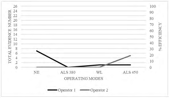
First Stage
The first stage, performed by two operators, not aware the internal procedure of the BFRC (Figure 1). The first operator showed a rather low detection efficiency with whole observation modes (evidence visibility NE> forensic lights). Second operator’s results present 0% for NE, ALS 380, WL. In this stage was not possible to analyze the dissimilarity between the two reports, as it was not possible to implement a comparison of the latent evidence identified by the operators given the different nomenclature they used. Both the first and the second operators inspecting the find without any standardized procedure. This stage had an average detection efficiency of 7% and 4.75% respectively for the first and second operators, hence very low. In addition, the average percentage of detection efficiency (based on quantitative analysis) was 27%. The comparison between first stage reports showed a clear lack of standardized guidelines and this meant that the two operators were not able to reach a suitable standard of quality and reproducibility. It is also clear that it is difficult for operators who do not follow a clear technical procedure to have a precise and repeatable workflow in several performances.
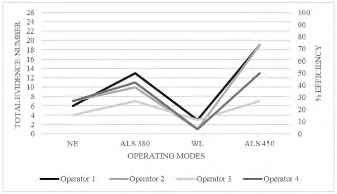
Second Stage
The second stage carried out by four operators, aware with the internal procedure at BFRC: particularly, the indications of the first technical procedure of BFRC (T.P. 01 Rev.0 of 26/11/21). As can be seen from Figure 2, the performances of all four operators were uniform with respect to the operating modes; moreover, it can be observed a better detection efficiency using ASL 450. As the first stage in this one it was not possible to analyze the dissimilarity between reports and it was not possible to compare latent evidence identified by the operators because of the different nomenclature they used. This is a strong indicator of no repeatability of operations and highlights the need to make changes to the same internal procedure to get closer to high standards of quality and reproducibility. As can be seen from Figure 2, there was an increase in detection efficiency compared to the performances carried out in first phase. In particular, the increase can be seen in the reports of the first, second and fourth operators, 17%, 17% and 15% respectively. In this stage the average percentage of detection efficiency (based on quantitative analysis) was equal to 55%, it was therefore appreciable the increase between the first two stages of the experimental phase. The comparison between the four reports has clearly shown the presence of a standard procedure usable by all operators and this has made them increase the standards of quality and reproducibility for the performance of the phase “beginning of technical operations” on forensic biology specimens. The operators were able to follow a specific workflow, and therefore everyone had a clear interpretation of the steps of the process to be applied to increase the detection rate. However, this is not enough to reach the high- quality standards, especially because of the absence of dissimilarity value.
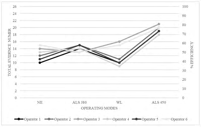
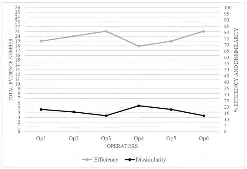
Third Stage
The third stage carried out by six operators, aware with the internal procedure at BFRC; particularly, following the indications of the second technical procedure of BFRC (T.P. 02 Rev.0 of 02/12/21). As the Figure 3 shows, the performances of six operators were uniform regarding the operating modes; only the use of Wood’s Lamp has been created dissimilarity between operators’ reports. It should be observed that in this stage, unlike the other two previous stages, it was possible to analyze the dissimilarity between the six reports; the dissimilarity is the ratio between the number of traces that showed different visibility in each of the six reports and the total number of evidence with different visibility in the whole of the six performances; particularly, the average percentage efficiency has risen to 75% and the dissimilarity is only 17% (Figure 4). In addition, it was possible to implement a comparison of the latent evidence identified by the operators because of the same nomenclature they used during the inspection. This is a strong indicator of repeatability of operations that are increasingly close to high standards of quality and reproducibility. In this stage, it was evident that the operators were able to follow a specific workflow, and therefore all of them had a clear idea of the steps of the procedure to be realized to increase the detection percentage. Furthermore, it has been possible to observe how the quality of the work was ensured using the T.P. 2, since the average percentage of dissimilarity between the six operators, equal to 17%, is rather low, indicating good reliability in the visibility of the evidence, consequently making the recorded efficiency, which has an average value of 75%, very reliable. Recording low values of dissimilarity supports the reliability of scientific data achieved during the observation of the evidence.
Conclusion
The data obtained through this validation study have demonstrated that the standardization of the technical procedure inherent to the phase of “beginning of technical operations” of forensic biology, leads to an increase in the efficiency of detection of latent traces that by their nature are difficult to detect and, to the minimization of variability between operators in the phase preceding the laboratory. Alternatively, this study confirmed that operating in the absence of a standardized protocol does not guarantee the efficiency and quality of technical operations. Therefore, if a standardized protocol were applied to the phase of “beginning of technical operations”, it would be straightforward to demonstrate the quality of operational procedures, homogeneity, and reproducibility of technical and scientific data.
References
-
Budowle B, Bottrell MC, Bunch SG, Fram R, Harrison D, et al. (2009) A perspective on errors, bias, and interpretation in the forensic sciences and direction for continuing advancement. J Forensic Sci 54(4): 798-809.
-
(2006) Supplemental requirements for the accreditation of forensic science testing and calibration laboratories. ASCLD⁄LAB -International.
-
Walsch C (2008) Europeanization, and democracy: Negotiating the Prüm treaty and the Schengen III agreement. Croat Polit Sci Rev 45(5): 81-90.
-
(2008) Council Decision 2008/616/JHA of 23 June 2008 on the Implementation of Decision 2008/615/JHA on the Stepping up of Cross-border Cooperation, Particularly in Combating Terrorism and Cross-border Crime. Official Journal of the European Union 29: 214-274.
-
Pádár Z, Nogel M, Kovács G (2015) Accreditation of forensic laboratories as a part of the “European Forensic Science 2020” concept in countries of the Visegrad Group. Forensic Science International: Genetics Supplement Series 5: e412-e413.
-
Byrd JS (2006) Confirmation bias, ethics, and mistakes in forensics. J Forensic Identif 56: 511-25.
-
Houck MM, Siegel JA (2009) Fundamentals of forensic science. Academic Press.
-
Lennard C, Stoilovic M (2004) Application of forensic light sources at the crime scene. In: Horswell J (Ed.), The practice of crime scene investigation. CRC Press, pp: 123-156.
-
Santucci K, Nelson D, McQuillen K, Duffy S, Linakis J (1999) Wood’s lamp utility in the identification of semen. Pediatrics 104(6): 1342-1344.
-
Al Aboud DM, Gossman W (2021) Wood’s Light. StatPearls.
-
Sharma S, Sharma A (2016) Robert Williams Wood: pioneer of invisible light. Photodermatology, Photoimmunology and Photomedicine 32(2): 60-65.
- Forensic Implications of Adverse Drug Reactions in Schizophrenia A Case Series
- Narcotics and Digital Forensics: Bridging Crimes in the Digital Age
- Ethics in Forensic Psychiatry: Principles, Dilemmas, and Human Rights
- Impact of Acute Stress on Attentional Orienting to Social Cues
- Head Injury and Intracranial Hemorrhage in Western Region of Libya
- A Forensic Study on Handedness: Examination of Handwriting Features in Right and Left Handed Writers