Challenges and Innovations in Forensic Identification: Case Study of Decomposed Remains in a Crocodile's Body for Missing Child Identification
Identifying a missing child from decomposed remains in a crocodile's body poses significant challenges and complexity. Forensic investigations involving human remains within animal’s body can present unique difficulties apart from conventional investigative scenarios. Decomposed remains added an additional layer of difficulty as the digestive process of a crocodile can significantly degrade the body and its features. Forensic anthropology examinations were impeded due to the limited availability of tiny bone samples. In this scenario decomposed bone pieces were the only remaining material for DNA analysis, the choice of an efficient DNA recovery procedure becomes crucial for successful outcome. In the study, prior to the DNA extraction process, a decontamination technique was employed, followed by decalcification method on the bone sample. The resulting, clean and decalcified bone sample was then subjected to extraction using a silica-based method, and the extracted DNA was subsequently typed through STR amplification. The derived DNA profile information from the bone sample pertaining to the child is compared with the profile details of parents for identification purposes.
Introduction
In paternity cases, complications may arise when analyzing DNA from bones or limited remaining materials. While DNA in skeletal muscles is relatively more stable compared to other tissues, degradation is still a notable factor [1]. The case involves a missing child found within a crocodile’s body and DNA analysis was conducted to facilitate identification with the parents. The only remaining material available for analysis was a small piece of bones discovered inside the crocodile. Recovering human DNA from bones within an animal’s body, living in the water is a complex process due to degradation of bone DNA by environmental or biological factors. Such conditions can impact the stability of DNA in skeletal remains. Furthermore, the combination of the animal’s digestive process and a considerable duration since the death of the individual introduces additional complexities to the recovery of viable DNA. Adhering to well- established protocols is essential to maintain the integrity and reliability of the results in such cases [2, 3].
Purifying DNA from bones often demands modified DNA extraction techniques compared to other biological samples. However, there is considerable variation among laboratories in the methods employed for isolating DNA from hard tissues. Many of these processes begin with a step focused on eliminating contamination [4]. To minimize surface contamination, we employed a decontamination technique by scraping off flesh and dirt followed by immersion in sodium hypochlorite (bleach).
After surface decontamination, traditional DNA extraction methods typically involve grinding the bone into powder, before proceeding with DNA purification [5]. Although bone powdering is widely employed as an effective technique for extracting DNA from skeletal samples, it does have drawbacks. Powdering bones makes it more susceptible to contamination and the process may not completely eliminate mineral impurities, impacting DNA extraction and subsequent analyses [6].
Bone tissue consists mainly of protein and minerals and when certain areas of the bone become highly mineralized, it creates barriers for extracting DNA. In the case study, we employed an optimized technique of decalcification method by incubating entire bone fragments in a 0.5M EDTA solution. The EDTA solution both demineralizes the bone by removing minerals and simultaneously inactivates DNAs by chelating to bivalent cations like Magnesium or Calcium [7].
Following demineralization, the bone sample and parent’s reference blood samples underwent extraction using a silica column following standard protocol and monitored Via Real-time PCR and the amplification of Short Tandem Repeat (STR) loci using the AmpFlSTR Identifiler plus PCR amplification kit.
Case History
In the evening, a young girl accompanied by her parents and several others went for a bath at a lake located in rural area of North Central Province in Sri Lanka. Tragically, during the bath, a crocodile suddenly dragged her and disappeared into the middle of the lake. Surrounding people and her parents witness to the distressing event as the girl was pulled away by the crocodile. Despite thorough searches by the police and divers, they couldn’t find the girl’s body or any clues. Villagers are aware of the presence of a crocodile in the lake and there have been occasional sightings in the vicinity. However, there have been no reported instances of missing persons or eyewitness accounts of humans being dragged by the crocodile. Two days later, the police was informed that angry villagers had captured and killed the crocodile believed to be involved. The police subsequently reported the incident to relevant authorities for the postmortem of crocodile. Following this, careful retrieval of the remains from the crocodile’s digestive system was conducted, and the two pieces of bones obtained were promptly dispatched to the forensic laboratory under the authority of a court order.
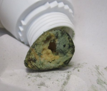
Figure 1 represents images of two bone fragments discovered within the crocodile’s body.
Materials and Methods
Sample Information
To eliminate potential contaminants and dirt, the surfaces of two bone samples (each approximately 3 to 5 cm in length) were scraped using a variable speed dental milling drill (Dermal, Racine, WI, USA). The samples were then washed twice with 10% sodium hypochlorite for 5 minutes, followed by twice rinsing with water for 5 minutes each. The samples were left to dry overnight. Subsequently, two bone pieces were placed into a plastic container, submerged in 0.5M EDTA pH 8.0, and incubated for two weeks on a shaking rotator at 56°C. After the first week, the EDTA solution was replaced with an equal volume of 0.5M EDTA solution. Following a two-week incubation period, the softened bone pieces were washed with distilled water and allowed to dry using blotting papers. Subsequently, the dried bone pieces were finely cut into small fragments for DNA extraction.
In accordance with the court-issued paternity order, the parents were brought to a DNA laboratory to undergo blood collection. The obtained blood samples were preserved for subsequent analysis, facilitating the execution of DNA fingerprinting.
DNA Analysis
Finely cut bone pieces (approximately 2 grams) were subjected to extraction using a QIAmp DNA Investigator kit, along with an appropriate quantity of reference samples obtained from the child’s parents. DNA quantification and PCR amplification were conducted on bone and reference samples. DNA quantification was performed with Quantifiler™ Human DNA Quantification Kit (Applied Biosystems, USA) according to manufacturer’s protocol. A total of fifteen STR markers, along with a marker for gender identification, were investigated for human identification using the AmpFlSTR Identifiler Plus PCR Amplification Kit from Applied Biosystems, USA. Separation of amplified PCR products was carried out on Genetic Analyser 3500 (Applied Biosystems, Life Technologies, Foster City, CA, USA) and analyzed with GeneMapper ID, version 3.2, from Applied Biosystems using standard procedures.
Results and Discussion
In the current investigation, the objective of STR typing was to address uncertainties regarding the parentage of the bone analyzed, specifically whether it belongs to the child of the father and mother produced by court order to the department.
The DNA obtained from the bone was of satisfactory quality and quantity, suitable for generating a comprehensive DNA profile. The results of autosomal genetic markers derived from DNA profiles of Mother, Child (Bone sample) and Father are summarized in the following table.
| Loci | Mother | Child (Bone) | Father |
|---|---|---|---|
| D8S1179 | 10,14 | 10,14 | 13,14 |
| D21S11 | 32,33.2 | 30,32 | 30 |
| D7S820 | 8,12 | 8,12 | 8,10 |
| CSF1PO | 10,12 | 10,12 | 10 |
| D3S1358 | 16,18 | 15,16 | 15 |
| TH01 | 6,9 | 6,9 | 9 |
| D13S317 | 8,14 | 12,14 | 10,12 |
| D16S539 | 9,12 | 9,11 | 11,12 |
| D2S1338 | 22,24 | 19,22 | 19,20 |
| D19S433 | 13,14 | 14 | 14 |
| vWA | 15 | 15,17 | 15,17 |
| TPOX | 9,11 | 9,10 | 10 |
| D18S51 | 16,17 | 11,16 | 11,14 |
| D5S818 | 12 | 11,12 | 11,12 |
| FGA | 22 | 22,23 | 19,23 |
| Amel | X | X | XY |
Table 1: The Identifiler Plus STR DNA Profiles for Mother, Child (Bone Sample) and Father.
Successful DNA typing was achieved for the child, mother, and father at all autosomal STR loci. The genotype profile of bone sample from the child revealed a shared allele with the mother’s genotype profile. Additionally, all non-maternal alleles at the amplified identifier STR loci in the bone sample from child were consistent with one of the alleles present in the genotype profile of the father. It can be concluded the female child’s alleles were found to be exclusively contributed by both father and mother.
Combined Paternity Index was calculated using eDNA 2.3 softwear package. In paternity testing, a probability of 99% or higher is typically regarded as conclusive evidence of a biological relationship, supporting the claim of paternity.
The table below presents a Summary Matrix for the distribution of corresponding alleles of Mother, Child (Bone sample), and Father, along with the calculated Paternity indices as per the software analysis.
| Summary Matrix | ||||||||||
|---|---|---|---|---|---|---|---|---|---|---|
| System | Mother | Child | Alleged Father | PI | Mutation Code | Pattern | Rule | |||
| D8S1179 | 10 | 14 | 10 | 14 | 13 | 14 | 1.2712 | PQ/PQ/QR | 1[2(P+Q)] where P equals. 1983 and Q equals. 1950 | |
| D21S11 | 32 | 32 | 30 | 32 | 30 | 5.8824 | PQ/PR/R | 1/R where R equals .1700 | ||
| D7S820 | 8 | 12 | 8 | 12 | 8 | 10 | 1.2396 | PQ/PQ/PR | 1[2(P+Q)] where P equals .2267 and Q equals .1767 | |
| CSF1PO | 10 | 12 | 10 | 12 | 10 | 1.7143 | PQ/PQ/P | 1/(P+Q) where P equals .1867 and Q equals .3967 | ||
| D3S1358 | 16 | 18 | 15 | 16 | 15 | 3.3898 | PQ/PR/R | 1/R where R equals .2950 | ||
| TH01 | 6 | 9 | 6 | 9 | 9 | 1.7442 | PQ/PQ/Q | 1/(P+Q) where P equals .2367 and Q equals .3367 | ||
| D13S317 | 8 | 14 | 12 | 14 | 10 | 12 | 2.5000 | PQ/QR/RS | 1/2R where R equals .2000 | |
| D16S539 | 9 | 12 | 9 | 11 | 11 | 12 | 1.4925 | PQ/PR/QR | 1/2R where R equals .3350 | |
| D2S1338 | 22 | 24 | 19 | 22 | 19 | 20 | 3.4090 | PQ/PR/RS | 1/2R where R equals .1467 | |
| D19S433 | 13 | 14 | 14 | 14 | 1.0000 | PQ/Q/Q | 1/Q where Q equals 1.0000 | |||
| vWA | 15 | 15 | 17 | 15 | 17 | 1.8293 | P/PQ/PQ | 1/2Q where Q equals .2733 | ||
| TPOX | 9 | 11 | 9 | 10 | 10 | 11.5380 | PQ/PR/R | 1/R where R equals .0.867 | ||
| D18S51 | 16 | 17 | 11 | 16 | 11 | 14 | 29.9940 | PQ/PR/RS | 1/2R where R equals .0167 | |
| D5S818 | 12 | 11 | 12 | 11 | 12 | 1.4151 | P/PQ/PQ | 1/2Q where Q equals .3533 | ||
| FGA | 22 | 22 | 23 | 19 | 23 | 3.0303 | P/PQ/QR | 1/2Q where Q equals .1650 | ||
| CRI Pop = 3,244,325.9936 | ||||||||||
| Probability = 99.9999% |
Table 2: The Summary Matrix for Paternity Indices for Mother, Child (Bone sample) and Father.
According the calculated Probability value in the table, the probability of Paternity was 99.9999%. The given result conclusively establishes that the questioned father and mother are the biological parents of the child associated with the bone sample taken from the crocodile’s body.
Conclusion
Analyzing DNA from bones or limited biological materials in paternity cases can introduce complexities. Challenges may arise from factors such as DNA degradation, resulting in fragmented and damaged genetic material. Despite technological advancements, careful consideration and specialized techniques are necessary for accurate analysis in these challenging scenarios. This is important because the presence of DNAs can interfere with the extraction process, and by inactivating it, ensure that the extracted DNA is clean and free from contaminants.
The case study reveals that utilizing the complete demineralization technique, coupled with STR analysis, is a successful approach for DNA analysis in bones. This method provided a precise match with the DNA of the parents, even when the bone samples were collected two days later, from inside the crocodile’s body. This effectiveness extended to the analysis of remaining small bone fragments.
The robustness of the findings can also be enhanced by running these samples through various PCR Amplification kits containing different types of autosomal STR markers. Furthermore, X chromosome markers, inherited from both the father and mother, provide valuable insights into biogeographic ancestry, making them useful for additional confirmation studies. Additionally, maternal linkage can be assessed through markers present in mitochondrial DNA.
In summary, establishing the paternity of the child in such cases can be conclusively accomplished through the thorough demineralization of skeletal materials. This process, when combined with the utilization of autosomal STR markers from various amplification kits, X-STR markers, and mitochondrial DNA analysis, proves to be a comprehensive and effective approach for human identification in such complicated events.
Ethical Issues
The study adhered to the guidelines set forth by the Government Analyst’s Department and was conducted in accordance with principled standards.
References
-
Ozcan SS, Petridis G, Yukseloglu EH, Kocias Y, Kalfoglu EA, et al. (2009) A new approach in the identification of degraded paternity samples. Forensic Science International Genetics Supplement Series 2(1): 174-175.
-
Bertolini E, Grignani P, Bertoglio B, Marrubini G, Mazzarelli D, et al. (2022) Dead migrants in the Mediterranean: genetic analysis of bone samples exposed to seawater. Forensic Science International 340: 111421.
-
Latham KE, Miller JJ (2019) DNA recovery and analysis from skeletal material in modern forensic contexts. Forensic Sci Res 4(1): 51-59.
-
Kemp BM, Smith DG (2005) Use of bleach to eliminate contaminating DNA from the surface of bones and teeth. Forensic Sci Int 154(1): 53-61.
-
Duijs FE, Sijen T (2020) A rapid and efficient method for DNA extraction from bone powder. Forensic Science International Reports 2: 100099.
-
Rancourt AC, Sainte-Marie S, Blackmore V, Currie KA (2023) Evaluation of low-cost bone and teeth processing methods for automated DNA extraction. Forensic Science International Reports 8: 100328.
-
Loreille OM, Diegoli TM, Irwin JA, Coble MD, Parsons TJ (2007) High efficiency DNA extraction from bone by total demineralization. Forensic Sci Int Genet 1(2): 191-195.
- Forensic Implications of Adverse Drug Reactions in Schizophrenia A Case Series
- Narcotics and Digital Forensics: Bridging Crimes in the Digital Age
- Ethics in Forensic Psychiatry: Principles, Dilemmas, and Human Rights
- Impact of Acute Stress on Attentional Orienting to Social Cues
- Head Injury and Intracranial Hemorrhage in Western Region of Libya
- A Forensic Study on Handedness: Examination of Handwriting Features in Right and Left Handed Writers