Major Drawbacks of Blood Species Analysis Using Human Polyclonal MNS Antisera: The Turin Shroud as a Case Example
The Turin Shroud is a linen cloth that has been suggested to represent either the burial wrapping of the historical Jesus of Nazareth, or a clever medieval forgery. Previously, the observation was made that blood fibers taken from the Shroud reacted with human polyclonal antisera raised against the S antigen, located on glycophorin B. As expression of the S antigen is exclusive to humans, this finding could support the idea that human blood is present on the Shroud, a notion often promoted in various books and websites. A modern assessment of the experimental design, however, shows that such antisera were particularly prone to cross-reactivity with blood from a bountiful number of other species. Indeed, it is now established that anti-alpha galactose 1,3 antibodies are highly abundant in human sera, which recognize red blood cells of all non-primate mammals. Thus, such human polyclonal antisera could not be used to distinguish blood species of origin as the cross-reactivity potential is quite vast and would confound any potential binding observed with anti-S specific antibodies. These findings underscore the necessity of using more current serological tools in any future investigation of blood-stained artifacts such as the Shroud, particularly in relation to species determination.
Commentary
Approximately fifty antigens are part of the MNS blood group system, with the most well-known being M,N,S, and s. S and s are located on the glycophorin B molecule (CD 235), a major sialoglycoprotein expressed on human erythrocytes. S and s antigens differ by a single amino acid substitution at position 29 (Figure 1), [1, 2, 3].
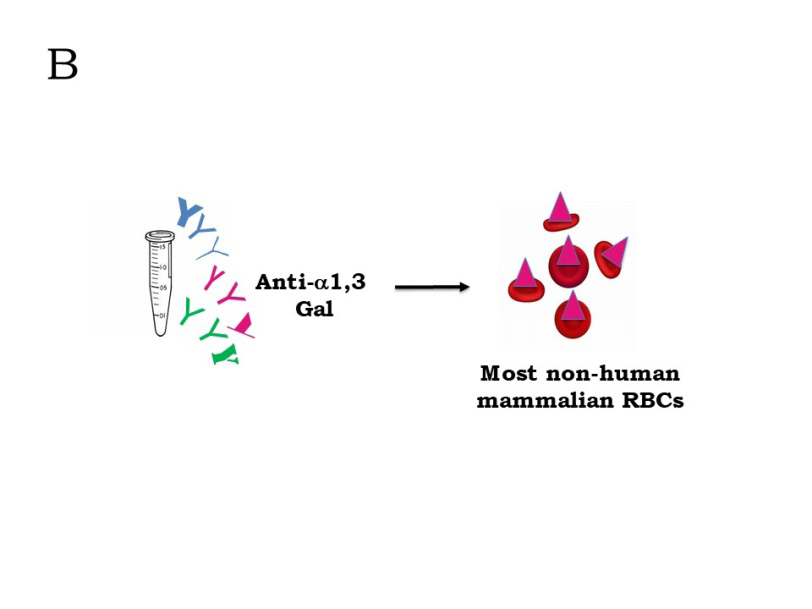
Prior to the development of monoclonal antibody technology, companies typically obtained S antisera from individuals who had been immunized via transfusion or pregnancy. Although such antisera may have been generally specific for the S antigen, due to its polyclonal nature, it would also contain antibodies that recognized other antigens Commentary (Figure 2A). Indeed, scientific results within the past several decades have established that all human antisera contain antibodies reactive with alpha 1,3 galactose residues (α1,3 Gal), which are present on erythrocytes of all non-primate mammals (Figure 2B), [4, 5, 6]. As recent studies have shown, such antibodies are a major component of human antisera and represent the primary barrier to xenotransplantation with organs from other animal species to humans. Erythrocytes from all species except humans, higher apes, and Old-World monkeys express α1,3 Gal epitopes on their surface [4, 5, 6]. Thus, such human polyclonal antisera could not be used to distinguish blood species of origin as the cross-reactivity potential is quite vast and would confound any potential binding observed with anti-S specific antibodies.
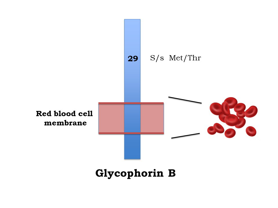
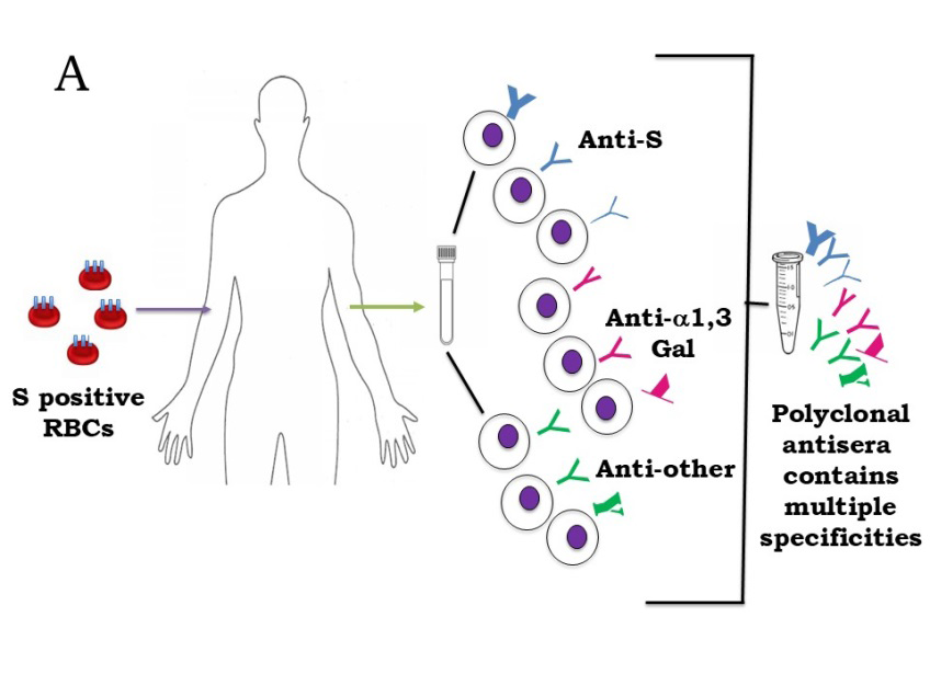
Figure 2: Preparation of Human Polyclonal Antisera Directed against the S Antigen. A: Individuals Lacking S Antigen that are Immunized with S Positive Red Blood Cells Will Contain Three Types of Antibody-Producing B Cells in their Blood: Those Directed Against the Immunogen (Anti-S), Endogenous Clones Producing Antibodies Specific for α1,3 Galactose(Gal) Structures (Which Humans Lack), and Endogenous Clones Specific for a Variety of Other Environmental Antigens. When Polyclonal Sera are Prepared, all Three Types of Antibodies will be Present; Anti-α1,3 Gal Antibodies have been Shown to be Particularly Abundant in Human Sera. B: Antibodies Specific for α1,3 Galactose (Gal) Antigens that are Present in Polyclonal Preparation of Human Anti-S Antisera Will Recognize Red Blood Cells of All Mammalian Species Except those from Humans, Great Apes, and Old-World Monkeys.
The original observation of anti-S reactivity was published in the Shroud-specialty periodical Sindon and was principally related to general blood characterization in the context of ethnicity [7]; years later, as the restricted expression of the S antigen was realized [8], the potential importance of this observation increased relative to blood species characterization on the Shroud. However, as the further understanding of the robust reactivity of α1,3 Gal antibodies present in human sera has been more recently
and more fully elucidated, it is evident that previous observations do not justify any scientific conclusions regarding blood species origin. It should also be noted that no photographs were presented for these preliminary findings of anti-S reactivity with Shroud fibers, only the written description that “fairly good” binding was observed; as previously noted, it is difficult to interpret any such results without a detailed presentation of the data [8].
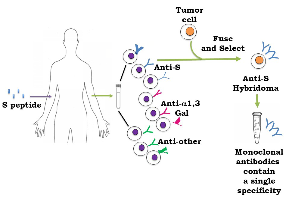
Figure 3: Preparation of Human Monoclonal Antibodies Specific for the S Antigen. Individuals Lacking S Antigen that are Immunized with Peptides Corresponding to the S Antigenic Region Will Contain Three Types of Antibody-Producing B Cells in their Blood: Those Directed against the Immunogen (Anti-S), Endogenous Clones Producing Antibodies Specific for α1,3 Galactose Structures (Which Humans Lack), and Endogenous Clones Specific for a Variety of Other Environmental Antigens. Individual B Cell Clones are Fused with a Myeloma (Tumor) Cell to Produce a Hybridoma Cell Line, which Produces a Single Type of Monoclonal Antibody.
Moreover, this example underscores the necessity and importance of utilizing more modern serological techniques in blood analysis of aged artifacts such as the Shroud. While such antisera could prove useful in the analysis of blood from a known source (human), modern evidence indicates that it is not suitable for species characterization of blood from an unknown origin. Certainly, the potential for reactivity with blood from a huge array of animal types expressing α1,3 Gal antigens exists.
Contemporary methods involving the production of monoclonal antibodies circumvent potential problems with endogenous, broadly reacting α1,3 Gal antibodies present in polyclonal human antisera created in the past (Figure 3).
Indeed, anti-S reagents can now be selectively created using human antisera (Figure 3) or other species [9, 10, 11, 12]. Moreover, the use of selected peptide regions on glycophorin B (or A) as an immunogen negates relatively cruder methods of immunization via whole erythrocytes [9, 10, 11, 12].
Scientific characterization of the blood species origin on the Shroud of Turin is an important, fundamental step in the further understanding of this historical artifact. The current report, together with other recent findings [13, 14] show that claims of human origin for the blood on the Shroud of Turin must be revaluated in light of contemporary knowledge, which has greatly expanded since the time such original observations were made. Given the relative uniqueness and enigmatic origins of such an object, it is important to carefully consider the potential merits and disadvantages of any future immunological or molecular approaches to determine the origin of blood species present.
Finally, it should be noted that despite being “the most studied artifact in the world” there have never been any publications of original data regarding the blood species origin on the Shroud in a peer-reviewed scientific journal. Only partial glimpses of (brief) works in progress exist, the majority of which were done some forty or so years ago before the advent of more modern serological and molecular techniques. Indeed, current technology allows a more thorough evaluation of this issue and can verify what type of blood may be present (or absent) on the cloth.
Acknowledgements
A special thank you to the various immunohematologists who provided helpful discussion, especially Drs. Liz Storry and Christine Francis.
References
-
Lopez GH, Hyland CA, Flower RL (2021) Glycophorins and the MNS blood group system: a narrative review. Ann Blood 6: 39.
-
Dean L (2005) Blood Groups and Red Cell Antigens. The MNS blood group, National Center for Biotechnology Information, USA.
-
GYPB orthologs, NCBI (nih.gov).
-
Eisenson DL, Hisadome Y, Yamaa K (2022) Progress in Xenotransplantation: Immunologic Barriers, Advances in Gene Editing, and Successful Tolerance Induction Strategies in Pig-To-Primate Transplantation. Front Immunol 13: 899657.
-
Boulet J, Cunningham JW, Mehra MR (2022) Cardiac Xenotransplantation: Challenges, Evolution, and Advances. JACC: Basic to Translational Science 7(7): 716- 729.
-
Kreft L, Schepers A, Hils M, Swiontek K, Flatley A, et al. (2022) A novel monoclonal IgG1 antibody specific for Galactose-alpha-1,3-galactose questions alpha-Gal epitope expression by bacteria. Front Immunol 13: 958952.
-
Baima Bollone PL (1985) Search for M, N and S antigens in blood traces on the Shroud. Sindon 34: 9-13.
-
Kearse KP (2012) Blood on the Shroud of Turin: An immunological review. Shroud com, pp: 1-22.
-
Kothari M, Wanjari A, Acharya S, Karwa V, Chavhan R, et al. (2024) A Comprehensive Review of Monoclonal Antibodies in Modern Medicine: Tracing the Evolution of a Revolutionary Therapeutic Approach. Cureus 16(6): e61983.
-
Halverson GR, Reid ME, Sutherland J, Rhodes M (1999) Evaluation and comparison of three human monoclonal anti-S, two human polyclonal anti-S, and one murine anti-GPB. Immunohematology 15(4): 163-166.
-
Halverson GR, Tossas E, Velliquette RW, Lobo CL, Reid ME, et al. (2009) Murine monoclonal anti-s and other anti- glycophorin B antibodies resulting from immunizations with a GPB.s peptide. Transfusion 49(3): 485-494.
-
Alejandra WP, Miriam Irene JP, Fabio Antonio GS, Rocio Patricia RG, Elizabeth TA, et al. (2023) Production of monoclonal antibodies for therapeutic purposes. Int Immunopharmacology 120: 110376.
-
Kearse KP (2020) Unanticipated issues in the serological analysis of blood species-The Shroud of Turin as a case example. Forensic Science International: Reports 2: 100073.
-
Kearse KP (2023) Validation and limitations in the DNA analysis of aged bloodstains: The Shroud of Turin as a sample case. Forensic Science International: Report 8: 100335.
- Forensic Implications of Adverse Drug Reactions in Schizophrenia A Case Series
- Narcotics and Digital Forensics: Bridging Crimes in the Digital Age
- Ethics in Forensic Psychiatry: Principles, Dilemmas, and Human Rights
- Impact of Acute Stress on Attentional Orienting to Social Cues
- Head Injury and Intracranial Hemorrhage in Western Region of Libya
- A Forensic Study on Handedness: Examination of Handwriting Features in Right and Left Handed Writers