Fire and Blood: The Dynamic Reconstruction of a Double Murder with Different Attempts to Conceal the Corpses
This case report describes the forensic investigations performed after the discovery of two bodies under suspicious circumstances. The first body was almost completely burned and therefore unrecognizable, while the second one had been concealed inside a dwelling and showed craniofacial injuries by a blunt force trauma. Numerous bloodstains and bloody stones were found at the crime scene which were carefully photographed and inventoried. Their analysis allowed an initial reconstruction of the dynamics of the events, suggesting a double murder with two different attempts to conceal the bodies. Forensic investigations were conducted on the remains using traditional forensic methods (necropsy, fingerprinting, histology) along with modern radiological (virtopsy) and toxicological exams. The results made it possible to determine the causes, means, and time of death, as well as a detailed reconstruction of the dynamics of murder and concealment of the bodies. Thus, the uniqueness of this case lies in demonstrating how the combined use of various forensic sciences can significantly contribute to judicial investigations. Moreover, it highlights the importance of the most modern techniques in solving complex cases such as the one described in this article, especially in view of the extremely different methods used to conceal the two bodies right after death.
Abbreviations
BPA: Bloodstain Pattern Analysis;
Introduction
This case report describes the forensic investigations performed on two corpses found during the research for two missing brothers. The crime scene was preserved until the arrival of the Investigative Unit and the forensic pathologist, who conducted the site examination. The first body found (F) was almost entirely burned and so unrecognizable, hence specific measures for handling a corpse in such conditions were taken [1]. During the inspection of the surrounding area, a pool of blood from which a blood trail led to a nearby building was noticed.
Inside of this construction the second body (C) was found, identified as one of the missing brothers, with clear signs of head and face blunt force injuries. On the crime scene, there were several stones with multiple traces of blood, from both projection and rubbing, which were photographed and collected for blood analysis.
At the end of the forensic examination, the bodies were taken to the morgue for forensic radiology investigations, necropsy, and sample collection for further laboratory and histological tests. Personal identification procedures, including fingerprinting, and comparing dental and radiological records along with anamnestic information provided by the relatives, were also conducted on the charred body. Furthermore, specific toxicological tests were run on the victims’ blood samples.
All these investigations allowed the determination of the causes, means, and time of death of the two deceased and an accurate reconstruction of the murder dynamics.
Materials and Methods
The following elements, collected during various stages of forensic and criminal investigations, were examined:
- Photographic evidence from the crime scene inspection and autopsy examination.
- Results of the Bloodstain Pattern Analysis (BPA) [2] on various biological traces found at the scene.
- Images obtained through CT scans with 3D and MPR reconstructions using the modern forensic technique “Virtopsy” [3]: On body C: of the craniofacial region exclusively; On body F: of the remaining burned body parts and specific scans of the craniofacial region.
Death Scene and Bloodstains
• Histological samples processed in paraffin with standard hematoxylin-eosin staining: For C’s body: Biventricular section of the heart, brain, lungs, liver, spleen, kidneys, and skin fragments of the chest. For F’s body: Brain, cerebellum, heart walls, lungs, liver, kidneys, skin; some of those with burn marks.
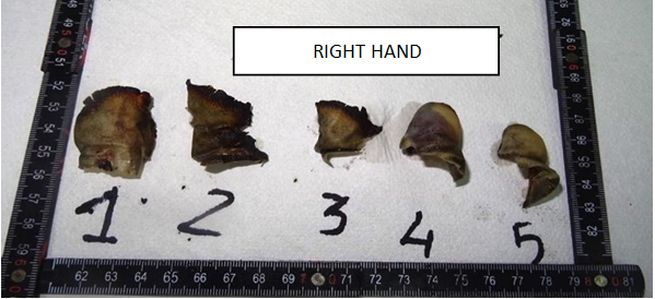
- Fingerprint analysis on F’s body for personal identification using properly processed carbonized skin residues from the right hand [4].
- Results of first-level toxicology screening on vitreous humor and urine of C’s body.
- Toxicology reports for body F:
- Targeted carbon monoxide search on kidney and brain samples [5].
- Generic search for non-volatile organic substances (drugs, psychoactive substances like benzodiazepines, antidepressants, antipsychotics, and most pharmacologically active substances) in liver, kidney, brain, and lung samples [6].
- Quantitative determination of Amisulpride and Sertraline on biological samples of liver, kidneys, brain, and lungs [7].
Evidence Analysis and Discussion
Examination of the elements acquired during the various steps of the forensics and criminal investigation produced the results discussed below.
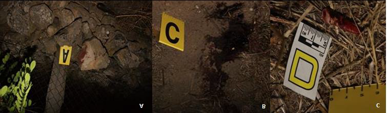
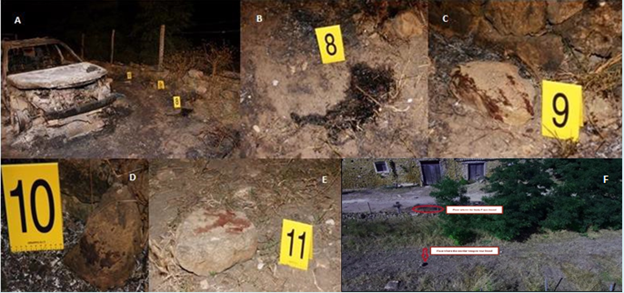
The inspection of the crime scene was carried out starting from the elements related to body C. The first hints found (Figure 1) were the blood traces that documented drag marks leading to the inside of the building where C was found.
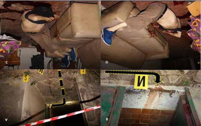
Corpse C was a male subject, about 70-75 years old, with identifying features that matched one of the two missing brothers. The body was lying supine with the shoulders on the ground and the legs on the sofa. In the space between the left shoulder and the left shoulder blade, soil with dried plant residues and stones was found. Bloodstains were visible on the sofa, with blood trickling to the floor and other blood spots. On the left side of the body, multiple blood pools lead to the base of the table and under the chair (Figure 2).
On the other hand, there was no biological material projected on the room walls.
In the aforementioned room, the indoor temperature, with the window and door open and the light on, was 20.3°C (double measurement); whereas the outdoor temperature was 32.6 °C at 02:30 AM. Regarding the consecutive abiotic phenomena: the rectal temperature was 32.25 °C at 3:05 AM; scarce hypostases, at stage I, easily compressible and paling, consistent with the position the corpse was found;
rigor mortis was present, even at the finger joints, but easily reduced mechanically. All of these signs made it possible to formulate an initial estimate of the time of death between 3:00 PM and 5:00 PM on the day of discovery. External signs of craniofacial injury, conveyed in Figure 3, were noted during the cadaveric inspection.
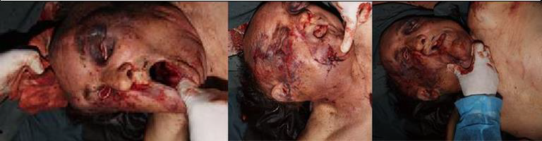
Figure 3: Photographic Evidence of the Cadaveric Inspection on Body C. The Face was Completely Destractured, With Disarrangement of the Bony Structures of the Facial Massif. Contused-Lacerated Wounds on the Frontal-Paretial Region and Chin Region were Visible, with Sunken Eyeballs. The Entire Right Side of the Face Showed Ecchymotic Mass from the Right Mandibular Angle to the Right Supraorbital Region. The Entire Left Side of the Face Presented an Extensive Excoriated Ecchymotic Complex.
Thus, the cause of death was attributed to severe blunt trauma with cranio-brain dislocation, and the medium was a blunt tool of considerable mass and weight. Moreover, there was a post-mortem excoriation complex on the back referable to dragging, so it was possible to assume that the attempted corpse concealment had occurred subsequent to death [8, 9, 10, 11].
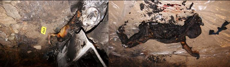
Figure 4: Photographic Surveys of Judicial Inspections. A: The Corpse was Found Lying Supine, in a Wrestler-Type Position Next to an Off-Road Vehicle, Completely Burned. B: The Body was Placed on a Plastic Sheet in Order to Complete the Cadaveric Inspection and to avoid the Dispersion of Organic Material. The Physiognomic Features were not Identifiable Due to 4th Degree Face Burns. The Entire Left Hemithorax, the Left Side of the Abdomen, the Pelvis and the Left Lower Limb from the Middle- Distal Third of the Thigh were Missing. Regarding the Right Lower Limb, It was Disarticulated at the Knee Level and a Calcified Tibial Fragment was Found next to the Body. The Right Thoracic Wall, the Central Wall B the Trunk and the Volar Surface of the Right Limb were Intact.
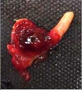
Not far from the first crime scene the charred F’s corpse was lying. The male gender was easily distinguished by the presence of a charred penis stump.
Due to the condition of F’s body, it was not possible to provide any indication of the time, cause and means of his death. The only clues that led to the suspicion that his death was also caused by a skull and brain blunt force trauma were a preternatural motility of the frontal region and the blood pool and two stones with traces of blood found near the body (Figure 5), which tested positive in the Combur3test [12].
However, the findings on the crime scene suggested a homicidal dynamic.
Forensic Haemogenetics on Bloodstains
The forensic blood analysis conducted on the numerous
Virtopsy on C Body
biological traces found at the crime scene confirmed that they belonged to corpses C and F.
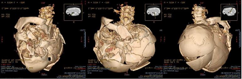
Figure 6: Cranium and Brain CT Scan with 3D Reconstruction. This Technique Revealed the Multiple Fractures of the Skull and Facial Massif; Those Complex Fractures had Decomposed and Overlapping Edges, Resulting from the Application of a Crushing Force on the Left Anterolateral Side of the Face. There are Subarachnoid Blood Collections, Soft Tissues Subcutaneous Emphysema of the Facial Massif and Left Temporo-Occipital Epidural Pneumocephalus.
Virtopsy on F Body
Forensic Radiology Findings
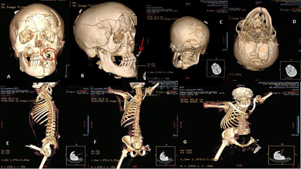
The injuries of the skull and brain shown in the CT scan performed on both bodies were attributed to a blunt tool consistent with the boulders found at the crime scene (it may be possible that some of these were used to bludgeon both the subjects).
The injuries on F’s body were possibly compatible with a combined action from different blunt elements (e.g., stick and stone), which could not, however, be confirmed due to the corpse conditions.
Autopsy on C’s Body
The full autopsy examination of body C revealed the following elements useful in defining the murder dynamics. The corpse was of a male subject, 76 years old, identified as one of the two missing brothers.
The algor and rigor were of little thanatological significance due to the presence of the corpse in a refrigerated environment. The hypostases were poorly represented, fixed, at the 2nd stage, in a light red wine hue and distributed to the back and lower limbs, in a position compatible with that found at the time of the discovery, and, at the same time, the presence of pale striations from compression at the gluteal region and between the two shoulder blades.
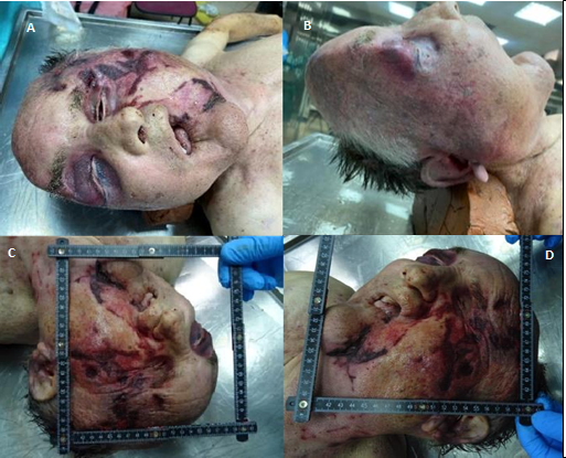
Figure 8: Judicial Autopsy Photographic Findings on C Body. Both the Eyeballs, Especially the Left One, were Sunken in their Respective Orbits with Bilateral Periorbital and Orbito-Zygomatic Ecchymosis and Hematoma (A). The Skull’s Anatomic Shape was Deformed due to Multi-Fragmentary Fractures of the Bones of the Facial Massif, Most Appreciable on the Left Side. There was a Group of Ecchymotic Abrasions with Some Lacerated Wounds on the Left Chin, In the Left Frontal Region and the Left Orbito- Zygomatic Region which were Heavily Bruised and Contused (B, C and D).
On the right parotid area, an ecchymotic complex expanded to the whole right half of the face up to the frontal region and the lateral orbital and right orbital-zygomatic region. There was a fracture of the inferior side of the maxilla with a large mucosal laceration along the lower mandibular border with diastasis of the chin symphysis with associated full-thickness laceration of the third distal portion of the tongue. At the neck level, multiple ecchymotic abrasions were present as well as in the dorsal region and on the right upper limb.
During the Autopsy
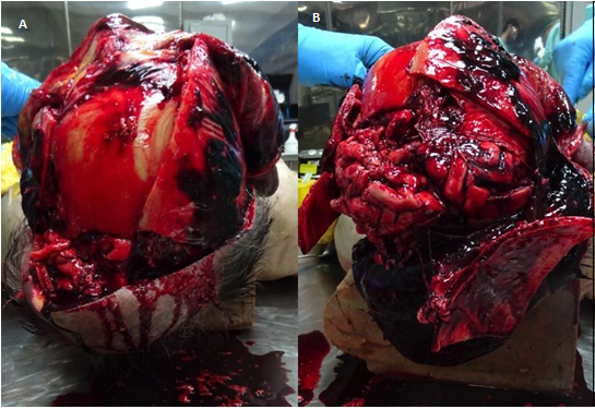
Figure 9: Photographic Findings of the Judicial Autopsy on C Body. A: On the Head Section, Diffuse Blood Infiltration from the Internal Layer of the Scalp and Periosteum with Multiple Cranial Vault Fractures from which the Encephalic Parenchyma Herniated. B: Focusing on the Encephalon, there was Diffuse Subarachnoid Hemorrhage and Scattered Intraparenchymal Bleeding Areas.
The skull base was completely destroyed, the damage involved the foramen magnum and the anterior, posterior and middle cranial fossae.
On the neck section, blood infiltration around the thyroid, the trachea, of the left carotid intima, and on the left greater horn of the hyoid bone was observed, corresponding perfectly to a right hand grasping action of the neck, as the thumb was placed on the right lateral region and the other four fingers on the left lateral region where an excised ecchymotic complex was present.
The upper airway showed a laryngeal opening coated in blood draining down the entire internal surface of the trachea up to the right and left bronchial branches.
The post-mortem changes observed during the judicial inspection and the necropsy, and especially the stomach content, allow to place the time of death (PMI) between 4:00 PM and 6:00 PM on the day the body was found.
Using the Henssge Method Henssge C, et al. [13] (calculations provided by the websites www.pathguy.com and www.swisswuff.ch), it was possible to narrow the time of death presumably between 5:30 PM and 6:30 PM on the day it was found.
Concerning the cause and means of death, the observations suggest that the subject was alive when violently strangled as the left hyoid bone was broken and bloody. He fell to the ground still alive and his head was then compressed between the ground and a blunt object (possibly one of the boulders found on the scene), as the presence of inhaled blood in the upper airways due to the destruction of the facial structure conveys. Due to this severe head trauma, the subject went through a rapidly worsening cardio-respiratory failure. The body was then dragged by the feet (evidenced by a series of parallel abrasions between the shoulder blades) into the building, where the assailant tried to place it on a sofa. However, the body fell off, as shown by the downward bloodstains found on the piece of furniture.
Autopsy on F’s body
The full autopsy examination on F’s burned remains also revealed elements that were useful in defining the murder dynamics. The head was still in place when found, it was immediately with a ‘Self Fix’ elastic bandage to prevent the loss of any organic material. The post-mortem changes were not visible due to the carbonized skin. The external examination of the head and face area is shown in Figure 11
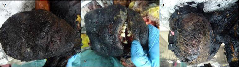
The corpse was found supine with the whole right upper limb and partially the right lower limb, with a stump just above the knee with burned and calcined bone at the distal extremity. The left side of the body was partially missing, represented only by a residue of the left hemithorax, abdomen and pelvic girdle and a charred left lower limb consisting of a thigh stump.
The right hemithorax was fairly well preserved, with thickened and dehydrated skin tissue with a leopard-like- skin texture (a characteristic that allows us to state that F was set on fire post-mortem), which was best appreciated at the root of the right lower limb and on the pelvic girdle.
The head soft tissues were carbonized, adhering to the external skull plate that was also affected by a complex frontal fracture, with the sinking of the frontal sinuses and the vault of the bilateral orbital wall, with extradural hematoma. On the contrary, the dura mater was intact but with visible burning signs, especially on the frontal- parietal-temporal area bilaterally. The encephalon was fairly preserved in shape and small in volume, with frontal-temporal-parietal subarachnoid hemorrhage. Moreover, hemorrhage of the brainstem and scattered intraparenchymal bleeding sites were seen as well as the flooding of the anterior and posterior horns of both ventricles.
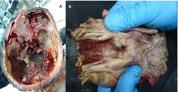
Figure 12: Photographic Findings of the Judicial Autopsy on F’s Body. A: Complex Fractures of the Skull Base and of the Anterior Cranial Fossa Bilaterally, of the Left Middle Cranial Fossa, with Structural Loss of the Posterior Cranial Fossa where there was a Conspicuous Thickened Hematoma. B: After Excision and Analysis of the Tongue-Hypopharynx-Larynx Block, there was no Endoluminal Carbonaceous Material, But only Cooked Blood Stratification Indicative of Cadaveric Charring.
Regarding F’s time of death, it is easily deduced that it was about the same as his brother C, as this was a double homicide occurring as part of a single criminal act.
Concerning F’s cause and means of death, it can be reasonably assumed that there was repeated blunt force trauma (two or three strikes) to the victim’s head with a boulder (specifically the one shown in Figure 5E, as the other stones in Figure 5B,C,D were not moved from the ground as there were no of removal marks on the soil). However, the impossibility of identifying any specific injury patterns or defensive wounds on the surface of the body, due to the carbonized tissues, does not allow to exclude the likelihood that, besides the stones, a second blunt object (such as a stick) might have been used, or that the back of the head may have been compressed against one of the stones found next to the car (Figure 5.B, C, D). Nevertheless, it is undeniable that the severe blunt force trauma to the head and face led to acute cardiorespiratory failure.
The body was then placed supine with the left side along the left side of the vehicle, which was ultimately set on fire; the contact with the burning vehicle caused the abovementioned body signs. Identification of the Bodies The recognition of corpse F was carried out by comparing the orthodontic structures (described in Figure 7A&B), the anamnestic information acquired from the next of kin and using the dactyloscopy (Figure 12).
Histological Investigations on C’s Samples
The histopathological investigations revealed the following findings • Brain: Congestion of the meninges, brain, and cerebellum with micro-hemorrhagic blood leakages through the fissures and multiple intravascular blood clots. • Lungs: Widespread not ventilated areas due to structural damage with extensive blood leakage and alveolar hemorrhagic edema; extensive bleeding obstructing medium-sized bronchial branches. • Spongy Bone, Periosteum, and Skeletal Muscle Samples: Discontinuity in the muscle structures, bone trabeculae fractures, bone marrow and spongy bone extensive bleeding. Histological investigations on F’s samples The histopathological investigations revealed the following findings:
• Brain: Sections of brain tissue with normal-sized neurons, almost entirely surrounded by Wirchow-Robin spaces filled with abundant blood cells. In the granular layer of the cerebellum, many Purkinje cells showed liquefactive neuronal necrosis. • Skin: Burned material adhered to the eroded epidermal surface; coagulative necrosis of the underlying dermis, both covered in an extensive fibrin-hemorrhagic exudate. Widespread blood infiltration in the hypodermis. • Lungs: Predominantly showed poorly ventilated areas, characterized by pulmonary edema and hemorrhagic edema; these patterns alternated with subacute emphysematous dilation of respiratory spaces. In fact, this confirms that F was set on fire post-mortem, as there was no microscopic evidence of particulate matter aspiration in the tracheobronchial tree. Toxicological Screening on C’s Samples The transpubic collection of urine performed during the autopsy allowed a rapid toxicology screen for eleven different substances (Amphetamine, Barbiturates, Buprenorphine, Benzodiazepines, Cocaine, MDMA/XTC Ecstasy, Methamphetamine, Opiates, Methadone, Tricyclic Antidepressants, Cannabinoids), all resulted negative. The same toxicological screening was conducted on vitreous humour and resulted negative, therefore it could ruled out the possibility that C had taken substances able to alter his defense before his death.
Toxicological Investigations on F’s Samples
• Carbon Monoxide: negative result, which further confirms that F was set on fire postmortem. • General Research for Non-volatile Organic Substances: positive for Sertraline (an antidepressant, part of the selective serotonin reuptake inhibitors – SSRI – category) and Amisulpride (an antipsychotic drug, commonly used for treating schizophrenia). Both medications were regularly prescribed to F by his doctor. • Quantitative Determination of Amisulpride and Sertraline: both concentrations were within therapeutic ranges and did not contribute to F’s death.
• Therefore, the cause of death can be solely and directly related to the severe head trauma from a blunt object.
Reconstruction
All the forensic and criminal investigations described above allowed for a detailed reconstruction of the events (time, cause, and means of C’s and F’s death) and provided beneficial information for the judicial investigations. Regarding the dynamics and the nature of the double homicide, it is important to highlight that the action was accomplished by one or more attackers, given the complexity of the crime scene and the two different locations. Considering the sites of the two bodies, it is highly probable and logically plausible that F was the first injured by a blunt object.
Subsequently, C reached the road where he was firstly choked and then as well hit by a blunt object. C’s body was then dragged and lifted by the ankles into the stable, where he was found. During an attempt to place the body on the sofa, possibly to better conceal it, it slipped from the hands of the attacker(s).
After leaving C and closing the door, the attacker(s) returned to the initial crime scene, moving F’s body next to the left side of the vehicle, where it was then set on fire.
Conclusions
The double homicide case examined is of particular forensic interest because it perfectly illustrates how modern forensic pathology can benefit from integrating traditional methods (e.g., time-of-death diagnosis based on post- mortem changes, assessment of injuries during the crime scene inspection and autopsy) along with advanced laboratory tests (forensic toxicology) and radiological exams (e.g., Virtopsy). This combination of techniques allows not only the determination of time, cause, and means of the murder but also an extremely accurate reconstruction of the criminal dynamics Moreover, the case involves two bodies with nearly overlapping times and places of death, both killed by similar means but subjected to two different concealment attempts: one body was simply moved away from the murder site, while the other was set on fire, resulting in partial destruction. This peculiarity conveys how the same homicidal actions can lead to vastly different outcomes depending on what is done to the bodies immediately after death, especially if one is burned (a condition that requires more delicate handling and specific investigations).
In conclusion, this case demonstrates how modern practices can be used alongside traditional forensic pathology to solve otherwise inextricable medico-legal and criminal issues.
Declaration of Conflicting Interests
The authors declare no potential conflicts of interest concerning the research, authorship, and/or publication of this article.
References
-
D’Antonio G, Serinelli S, Albore M, Bolino G (2023) Medico-legal scene investigation in the case of burned bodies - a systematic review. Med Leg J 91(4): 226-230.
-
Rotter G, Raffino C, Burrascano G, Ventura Spagnolo E, Baldino G, et al. (2024) The Role of Bloodstain Pattern Analysis (BPA) in Reconstructing the Dynamics of Forensic Cases. Clin Ter 175(Suppl 2(4): 125-129.
-
Gascho D, Thali MJ, Niemann T (2018) Post-mortem computed tomography: Technical principles and recommended parameter settings for high-resolution imaging. Med Sci Law 58(1): 70-82.
-
Cattaneo C, De Angelis D, Porta D, Grandi M (2006) Personal identification of cadavers and human remains. Forensic Anthropology and Medicine, In Humana Press eBooks Chapter 15, pp: 359-379.
-
Popovic VM, Atanasijevic TC, Nikolic SD, Micic JR (2009) Concentration of carbon-monoxide in carbonized bodies-- forensic aspects. Leg Med (Tokyo) 11(S1): S318-S320.
-
Telepchak MJ, August TF, Chaney G (2004) Forensic and clinical applications of solid phase extraction. In Humana Press eBooks.
-
Wood M, Laloup M, Samyn N, Mar Ramirez Fernandez MD, De Bruijn EA, et al. (2006) Recent applications of liquid chromatography–mass spectrometry in forensic science. Journal of Chromatography A 1130(1): 3-15.
-
Dolinak D, Matshes EW, Lew EO (2005) Forensic Pathology-Principles and Practice, In: 1st (Edn.), Elsevier Academic Press, San Diego (CA).
-
Madea B (2014) Handbook of Forensic Medicine. Wiley BlackwelI, UK.
-
Saukko P, Knight B (2016) Knight’s forensic pathology, In: 4th (Edn.), CRC Press, Boca Raton.
-
Maloney MS, Housman DG, Gardner RM (2014) Crime Scene Investigation-Procedural Guide, In: 1st (Edn.), CRC Press, Boca Raton, pp: 1-348.
-
De Vittori E, Barni F, Lewis SW, Antonini G, Rapone C, et al. (2016) Forensic application of a rapid one-step tetramethylbenzidine-based test for the presumptive trace detection of bloodstains at the crime scene and in the laboratory. Forensic Chemistry 2: 63-74.
-
Henssge C, Madea B (2007) Estimation of the time since death. Forensic Sci Int 165(2-3): 182-184.
- Forensic Implications of Adverse Drug Reactions in Schizophrenia A Case Series
- Narcotics and Digital Forensics: Bridging Crimes in the Digital Age
- Ethics in Forensic Psychiatry: Principles, Dilemmas, and Human Rights
- Impact of Acute Stress on Attentional Orienting to Social Cues
- Head Injury and Intracranial Hemorrhage in Western Region of Libya
- A Forensic Study on Handedness: Examination of Handwriting Features in Right and Left Handed Writers