Clinical Assessment of Lung Cancer Using 18F-FDG PET-CT
<p>Cancer is a main threaten human health and lung cancer is responsible death individual worldwide. Early detection of cancers can diminish problem resulted from them and 18F-FDG PET-CT can be helpful to solve this problems. 8F-FDG PET-CT depicts metabolic and anatomical information about tumor. It facilitates treatment planning and enhances overall survival in patients with lung cancer. 18F-FDG-PET-CT has potential ability to detect early metabolic response to chemotherapy. 18F-FDG, PET/CT is a prominent and key tool for assessment and staging lung cancer and today it widely serve for demonstration of lung cancer. In addition, it has pivotal role in personalized patient management. Here, we reviewed important of 18F-FDG PET-CT in detection of lung cancer. This study was designed as chronological order based on studies conducted by 2014 to 2017. The papers were searched from valid databases such as Wiley; Scopus; Science Direct; Springer and PubMed.</p>
Seyyed Hossein Hassanpour1* and Seyyedeh Zeinab Karami2
Direct; Springer and PubMed.
Keywords: Lung cancer; 18F-FDG PET-CT; Early detection; Treatment planning; Overall survival
Introduction
Cancer burdens enormous problem to health society so that is main responsible human death worldwide. Therefore, today effort for accurate diagnosis, staging and restaging in order to control and management of cancer is essential gold in health field [1]. Lung cancer is considered as one of the main reason human health particularly in women [2]. Based on statistical findings, lung cancer is second reason mortality worldwide [3]. If patients suffered from lung cancer undergo surgery, the survival reduced in them furthermore accurate diagnosis can help them to increase survival [2]. The non– small cell lung cancer (NSCLC) is considered as common type of lung cancer and given that only half of patients with NSCLC are potential to treat as surgical resection and radiotherapy, thus early diagnosis in order to manage treatment-related complications is very essential in these patients [4]. Meanwhile, manifestation of disease is late in patient with NSCLC and disease presents in stage III or stage IV that are inoperable so that reduce their treatment efficacy [5]. Patients with stage III or stage IV NSCLC suffer from chemotherapy, or a combination of chemotherapy and radiation but early detection of disease leads to surgical resection of tumor with high International Journal of Nuclear Medicine & Radioactive Substances
efficacy [6]. In addition, all patients not respond to chemotherapy, on the other hand chemotherapy leads to obvious side effect thus they have dire need to reliable early diagnosis for increase of survival value [7]. Early and reliable diagnosis of tumor has pivotal role in treatment planning and ultimately survival of patients [8]. Based on data obtained from National Health Service of United Kingdom during 60 years ago, it have been observed obvious development in early and reliable diagnosis of many cancer that led to increase of survival rates for many cancer such as breast, colon, rectal, and cervical cancers. Unfortunately survival rate of stomach, pancreas, and lung cancers is now low [8, 9]. Use of PET and PET/CT were one of the strategies to solve this problem by tracing high level of metabolism and glycolysis in cancer cells [9, 10]. Given that tumor requires to high level of glucose for proliferation and malignancy thus it can be helpful marker to trace malignancy through 2-deoxy-2-[fluorine-18] fluoro-D-glucose (18F-FDG), an analogue of glucose by positron emission tomography (PET) [1, 11]. This method offer important information about tumor status based on amount of glucose uptake and glycolysis of cancer cells that demonstrated metabolic abnormalities before morphological alterations [1]. Today, it has been confirmed efficacy of 18F-FDG PET in diagnosis of many cancers and even tracing of treatment [12, 13, 14, 15, 16, 17]. In addition, it has been reported that 18F-FDG PET has high sensitive, specific and accurate in diagnosis of lung cancer 18. PET has pivotal role to determine staging of lung cancer because metabolic differences between benign and malignant tumors is simply traceable through PET with 18F-FDG as glucose analog [13, 19, 20]. Meanwhile, detection of anatomic information and determination of location and extent of malignancies can obtain through CT as a tomographic imaging technique by an x-ray beam [11]. Thus, combination of PET and CT prepare useful information about metabolic and anatomic properties of tumors [11]. Today, it have been reported that PET-CT provide information about tumor location, shifting status from benign to malignancy by measurement of parameters such as metabolic tumor volume (MTV) and total lesion glycolysis (TLG) in tumors [12, 21, 22, 23]. This study, we reviewed role of 18F-FDG PET- CT in diagnosis of lung cancer based on chronological order from 2014 to 2017.
Review Method
The aim of this review study was to explain conducted papers on role of 18F-FDG PET-CT in assessment of lung cancer from 2014 to 2017. To achieve this goal, we searched keywords such as 18F-FDG PET-CT imaging, lung cancer, and non-small cell lung cancer, clinical assessment of lung cancer and so on in valid databases such as Wiley; Scopus; Science Direct; Springer and PubMed. After collecting of articles, we fully read and summarized them. Here, the findings of articles were showed based on publication year. Indeed; our goal was to review articles based on chronological order.
Role of 18F-FDG PET-CT in Assessment of Lung Cancer
Table 1 depicts articles related to 2014 in conjunction with important of 18F-FDG PET-CT imaging in diagnosis of lung cancer that were summarized in following sentences. In a study, in order to evaluation of role of 18F-FDG PET/CT imaging, biopsy obtained from patients who suffered from lung cancer was investigated. The results of this study showed that 18F-FDG PET/CT imaging is a prominent tool to prognosis overall survival particularly in younger patients (less than 70 years old). In addition, it can be a good tool to trace primary treatment [24]. Study on patients suffered from stage III non-small-cell lung cancer was revealed that metabolic tumor volume and total lesion glycolysis as 18F-FDG PET/CT parameters related to volume-based have efficacy more than maximum standardized uptake value to prognosis survival independent of tumor stage [25]. Comparison of 18F-FDG PET/MR imaging and 18F-FDG PET/CT in order to evaluate diagnosis potential in subjects with non-small cell lung cancer approved by histopathological parameters were showed that 18F-FDG PET/MR imaging compared with 18F-FDG PET/CT cannot benefit in order to trace non-small cell lung cancer during thoracic staging [26]. Study on comparison between visual assessments of intratumor 18F-FDG PET uptake distribution with a textural-features automated quantification on patients with non–small cell lung cancer was revealed that there is a significant correlation between visual assessment and quantification assessment of 18F-FDG uptake heterogeneity in non–small cell lung cancer. In addition, quantification assessment using textural feature reduced interobserver variability and can be consider as a good prognostic tool in these patients [27]. A research group was traced radical chemoradiation therapy in patients with non-small cell lung cancer (stage I–III) by serial PET/CT with 18F-FDG and 3′-deoxy-3′-18F- fluorothymidine. The results were revealed that PET/CT using 18F-FDG and 3′-deoxy-3′-18F-fluorothymidine can be a good strategy to trace therapy in non-small cell lung cancer. In addition, 18F-FLT PET/CT has more sensitive to early treatment response than 18F-FDG PET/CT [28]. Evaluation of standardized uptake values and metabolic tumor volume in 18F-FDG PET/CT in order to predict survival of patients with locally advanced non–small cell International Journal of Nuclear Medicine & Radioactive Substances
lung cancer after early stage of concurrent cisplatin-based chemotherapy regimen was demonstrated that reduction of metabolic tumor volume during 18F-FDG uptake through primary tumor indicates higher long-term overall survival. Meanwhile, this study was confirmed that repeated 18F-FDG PET/CT can be helpful to assess survival during chemoradio therapy [29]. Use of 18F-FDG PET/CT for detection of lung adenocarcinoma through measurement of parameters such as standardized uptake value, metabolic tumor volume and total lesion glycolysis can reveal prominent information in association with lung adenocarcinoma [30]. Although tumor molecular profile is a good predictor for activity of epidermal growth factor receptor inhibitors in non-small-cell lung cancer but tumor heterogeneity and tissue availability diminish its role predicting. In order to solving of this problem, a research group were investigated role of 18F-FDG PET/CT in order to detect KRAS and EFGR mutation status in non- small-cell lung cancer. The results were showed that in non-small-cell lung cancer patients with tumors harboring of KRAS mutations, 18F-FDG PET/CT uptake was dramatically higher than patients with wild-type gene. In addition, evaluation of 18F-FDG PET/CT uptake in association with parameters such as age, gender, AJCC stage and minimum standardized uptake value was demonstrated that 18F-FDG PET/CT can offer important information about KRAS mutation status in patients with stage III or IV non-small-cell lung cancer [31] (table 1).
- Subjects
- Performed procedures
- Finding(s)
- References
- Evaluation of OS using Kaplan–
- Meier plots by a Mantel–Cox log-
- To be as an important criteria to prognosis overall survival spatially in
- 261 biopsy-proven lung cancer patients rank test younger patients (≤70 year-old)
- 194 consecutive patients with
- MTV and TLG are good tool more than stage IIIA NSCLC treated with surgical resection and 115 patients
- Evaluation of MTV, TLG, SUVmax
- SUVmax in order to prognosis survival independent of tumor stage treated with nonsurgical therapy
- Comparison of 18F-FDG PET/MR and by measurement of SUV mean,
- 18F-FDG PET/MR imaging compared with 18F-FDG PET/CT has not validity
- 22 patients with NSCLC
- SUVmax and maximum diameter to diagnosis NSCLC under thoracic of the primary tumor staging
- Detecting of intratumor heterogeneity, tumor volumes, visual interobserver agreement and correlations with quantitative
- To have a correlation between visual assessment and quantification assessment. Reduction of interobserver
- 102 patients with NSCLC variability by quantification assessment with textural feature assessment by κ test and
- Spearman rank (ρ) coefficient
- Evaluation of tumor response to serial PET/CT with 18F-FDG and 3′-
- Both can be benefit biomarkers to trace chemoradiation therapy in patients with NSCLC but 3′-deoxy-3′-
- 18F-fluorothymidine is more sensitive
- 20 patients with stage I–III deoxy-3′-18F-fluorothymidine as semi-quantitative through visual
- NSCLC response criteria to early treatment response
- Repeated 18F-FDG PET/CT can predict
- 53 patients with locally advanced
- Measurement of SUVmean, SUVmax survival during chemoradiotherapy.
- NSCLC and MTV
- Reduction of MTV during 18F-FDG uptake reveals higher long-term OS.
- Measurement of SUV, MTV and TLG
- 106 patients with lung
- Can be helpful in prognosis of lung in conjunction with EGFR gene adenocarcinoma
- 30 adenocarcinoma mutation status
- Measurement of SUV (peak, max,
- 18F-FDG PET/CT can reveal prominent mean) and assessment of its relation with KRAS and EGFR
- 340 patients with NSCLC data about KRAS mutation status in patients with NSCLC mutation status
Table 1: Findings of papers published by 2014 in association with role of 18F-FDG PET-CT in assessment of lung
In a study potency of 18F-FDG imaging in order to predict response or survival in patients treated with erlotinib was examined. The results were showed that response to erlotinib is correlated with reduced heterogeneity in 18F-FDG PET imaging. Meanwhile, changes in first-order entropy are independently related to treatment response and overall survival [32]. Study on role of 18F-FDG PET/CT in determination of local relapse after chemoradiotherapy in patients with inoperable stage II or III non-small cell lung cancer were confirmed that High 18F-FDG uptake can indicate that occur local relapse after chemoradiotherapy so that should be increase radiotherapy dose escalation [33]. Investigation on role of various metabolic parameters measured by 18F- FDG PET/CT as prognosis values for detection of stage IA non–small cell lung cancer after complete surgical resection were confirmed that total lesion glycolysis is a pivotal prognostic factor for overall survival in these patients [34]. In association with efficacy of apparent diffusion coefficient (ADC) and standardized uptake values (SUV) in detection of lymph node metastases of non-small cell lung cancer, a research group was used hybrid 18F-FDG PET/MRI. Their findings were demonstrated that prognostic value (ADC, SUV) measured by 18F-FDG PET/MRI can complete necessary information for treatment response [35]. According to findings of a retrospective single-center study in association with role of 18F-FDG PET/CT to predict local control and survival in patients who suffered from non–small cell lung cancer during concomitant radiochemotherapy were revealed that early assessment of total lesion glycolysis response through 18F-FDG PET/CT imaging within concomitant radiochemotherapy of non–small cell lung cancer might be related with survival [36]. In order to efficacy of 4D- 18FDG-PET/CT in target delineation of stereotactic body radiation therapy (SBRT) in patients with central versus peripheral lung tumors, a study on patients with lung tumor were designed. The results of this study were showed that 4D-18FDG-PET/CT offers helpful information about target delineation of SBRT so that can avoid geographic misses [37] (table 2).
| Performed procedures | Finding(s) | |
|---|---|---|
| 47 NSCLC patients treated with erlotinib | Evaluation of MTV, TLG, SUVmax, measurement of response to erlotinib by RECIST | To correlate between response to erlotinib and reduced heterogeneity in 18F-FDG PET imaging, to relate changes in first-order entropy with treatment response and overall survival as independent |
| 39 patients with inoperable stage II or III NSCLC | Determination of SUVmax threshold | High 18F-FDG uptake is related to local relapse after chemoradiotherapy |
| 248 patients with stage IA NSCLC | Detection of SUVmax and TLG | To confirm significant role of TLG as a prognostic factor in these patients |
| 38 patients with NSCLC | Assessment of ADC and SUV | To complete require data for detection of lymph node metastases of NSCLC by 18F-FDG PET/MRI |
| 31 patients with unresectable or locally NSCLC | Assessment of SUVmax, variation of hypermetabolic tumor volume and the variation of TLG | TLG may be associated with survival in these patients. |
| 21 patient with lung tumor (11 cases with peripheral lesions and 10 cases with central lesions) | Analysis of ITV delineation of central and peripheral lung lesions | 4D-18FDG-PET/CT can be helpful in target delineation of SBRT in patients with lung tumor |
Table 2: Findings of papers published by 2015 in association with role of 18F-FDG PET-CT in assessment of lung cancer Table 2: Fi
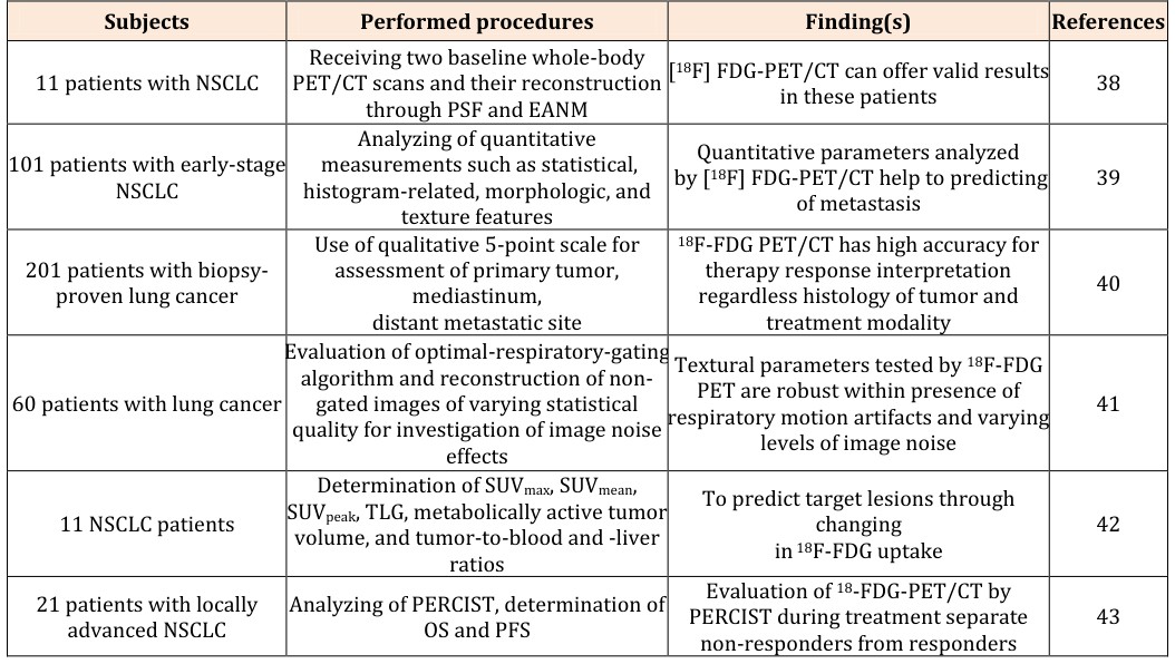
Table 2: Findings of papers published by 2015 in association with role of 18F-FDG PET-CT in assessment of lung cancer Table 2: Findings of papers published by 2015 in association with role of 18F-FDG PET-CT in assessment of lung cancer Study about repeatability features of [18F] FDG-PET/CT in patients with non-small-cell lung cancer were revealed that due to more sensitive of these features than simple standardized uptake value they can offer reliable results in these patients [38]. In addition, [18F] FDG-PET/CT is a good predictor of metastasis in patients suffered from International Journal of Nuclear Medicine & Radioactive Substances
early-stage non-small-cell lung cancer and can be helpful in administration of appropriate remedy in these patients in order to reduce effects of metastasis. Because, evaluation of quantitative parameters analyzed by [18F] FDG-PET/CT can facilitate predicting of metastasis [39]. Use of 18F-FDG PET/CT as an interpretational system to predict therapy response and survival in patients with lung cancer proved by biopsy were determined that 18F- FDG PET/CT regardless histology of tumor and treatment strategy is a reliable tool with high accuracy in order to therapy response interpretation [40]. Given that accurate determination of tumor heterogeneity is pivotal in order to precise characterization of tumor lesions, a research group were examined efficacy of respiratory motion and levels of varying noise on quantification of textural parameters measured by 18F-FDG PET in patients suffered from lung cancer. The results of this study were confirmed that when there are respiratory motion artifacts and varying levels of image noise, textural parameters tested by 18F-FDG PET are robust [41].
Change in 18F-FDG uptake can be one of the way predictions of response to anticancer treatment. To achieve this goal, a study was conducted in association with evaluation of repeatability of quantitative 18F-FDG PET/CT uptake measures in patients with lung cancer. The findings were showed that changing in SUVmean/SUVpeak (15% during 60 min after injection) can be helpful in assessment response in patients with advanced non-small cell lung cancer for up to 5 PERCIST (response criteria in solid tumors) target lesions. In addition, lower thresholds is usable for averaged PERCIST (response criteria in solid tumors) target lesions [42]. Furthermore, evaluation of 18-FDG-PET/CT by PERCIST (response criteria in solid tumors) was revealed that although there are different in parameters affecting 18- FDG-PET/CT uptake, but use of 18-FDG-PET/CT with PERCIST within treatment can be useful to diagnose non- responders from responders because their overall survival and point spread function were statistically difference [43] (table 3).
- Subjects
- Performed procedures
- Finding(s)
- References
- Receiving two baseline whole-body
- PET/CT scans and their reconstruction
- [18F] FDG-PET/CT can offer valid results
- 11 patients with NSCLC in these patients
- 38 through PSF and EANM
- Analyzing of quantitative measurements such as statistical, histogram-related, morphologic, and
- Quantitative parameters analyzed by [18F] FDG-PET/CT help to predicting
- 101 patients with early-stage
- NSCLC of metastasis texture features
- Use of qualitative 5-point scale for
- 18F-FDG PET/CT has high accuracy for
- 201 patients with biopsyassessment of primary tumor, therapy response interpretation regardless histology of tumor and proven lung cancer mediastinum, distant metastatic site treatment modality
- Evaluation of optimal-respiratory-gating
- Textural parameters tested by 18F-FDG algorithm and reconstruction of non-
- PET are robust within presence of respiratory motion artifacts and varying
- 60 patients with lung cancer gated images of varying statistical quality for investigation of image noise levels of image noise effects
- Determination of SUVmax, SUVmean,
- SUVpeak, TLG, metabolically active tumor
- To predict target lesions through
- 11 NSCLC patients changing in 18F-FDG uptake volume, and tumor-to-blood and -liver ratios
- Evaluation of 18-FDG-PET/CT by
- PERCIST during treatment separate
- 21 patients with locally
- Analyzing of PERCIST, determination of advanced NSCLC
- OS and PFS non-responders from responders
Table 3: Findings of papers published by 2016 in association with role of 18F-FDG PET-CT in assessment of lung
International Journal of Nuclear Medicine & Radioactive Substances
staging, but both them results in equal therapeutic decisions in patients with non-small cell lung cancer. Meanwhile, it can be suggest that 18F-FDG PET/MR replace 18F-FDG PET/CT for clinical NSCLC staging [44]. A research group were designed a study in order to development of a framework for segmentation and labeling of homogeneous versus heterogeneous in lung tumor so that areas with high-uptake simply delineate. Their findings were showed that 18F-FDG-PET can offer a suitable automatic framework for segmentation and labeling of homogeneous versus heterogeneous in lung tumor [45].
| Subjects | Performed procedures | Finding(s) | References |
| 77 NSCLC patients | Evaluation of thoracic tumor staging between $ ^{18} $ F-FDG PET/CT and PET/MR | Both lead to similar therapeutic decisions despite of differences in thoracic tumor staging | 44 |
| 70 patients with lung cancer | Analyzing of TF, segmentation accuracy, evaluation of volumetric by DSC and HD | $ ^{18} $ F-FDG-PET can exhibits an automatic framework for segmentation and labeling of homogeneous versus heterogeneous in lung tumor | 45 |
Table 4: Findings of papers published by 2017 in association with role of 18F-FDG PET-CT in assessment of lung Table 4: Findings
In this study, we reviewed important of 18F-FDG-PET- CT in diagnosis of lung cancer. Given that early and reliable detection of cancer leads to suitable treatment strategy to control disease and reduction of mortality thus presence of efficient tools is require to solve this problem. Based on studies, 18F-FDG PET-CT can be helpful to diagnose tumor when it is curable because 18F-FDG PET- CT offer metabolic and anatomic information about tumor It is a valid criteria to prognosis overall survival by evaluation metabolic tumor volume and total lesion glycolysis. Use of 18F-FDG PET/CT is very helpful in order to predict survival within chemoradio therapy. In addition, changing in 18F-FDG PET/CT predict target lesions in patients with lung cancer. Furthermore, it facilitates treatment decision for patients. Finally, we offer that 18F-FDG PET/CT is a suitable tool to control and manage lung cancer and it can reduce mortality rate in patients suffered from lung cancer.
References
-
Almuhaideb A, Papathanasiou N, Bomanji J (2011) 18F-FDG PET/CT imaging in oncology. Annals of Saudi medicine 31(1): 3-13.
-
Kumar R, Xiu Y, Jian QY, Takalkar A, El-Haddad G, Potenta S, et al. (2004)18F-FDG PET in evaluation of adrenal lesions in patients with lung cancer. Journal of Nuclear Medicine 45(12): 2058-2062.
-
Siegel RL, Miller KD, Jemal A (2015) Cancer statistics 2015. CA: a cancer journal for clinicians 65(1): 5-29.
-
Keidar Z, Haim N, Guralnik L, Wollner M, Bar-Shalom R, et al. (2004) PET/CT using 18F-FDG in suspected lung cancer recurrence: diagnostic value and impact on patient management. Journal of Nuclear Medicine 45(10): 1640-1646.
-
Spiro SG, Silvestri GA (2005) One hundred years of lung cancer. American journal of respiratory and critical care medicine 172(5): 523-529.
-
Socinski MA, Morris DE, Masters GA, Lilenbaum R (2003) Chemotherapeutic management of stage IV non-small cell lung cancer. CHEST Journal 123(1): 226S-243S.
-
Nahmias C, Hanna WT, Wahl LM, Long MJ, Hubner KF (2007) Time course of early response to chemotherapy in non–small cell lung cancer patients with 18F-FDG PET/CT. Journal of Nuclear Medicine 48(5): 744-751.
-
Ben-Haim S, Ell P (2009) 18F-FDG PET and PET/CT in the evaluation of cancer treatment response. Journal of Nuclear Medicine 50(1): 88-99.
-
Juweid ME, Cheson BD (2006) Positron-emission tomography and assessment of cancer therapy. New England Journal of Medicine 354(5): 496-507.
-
Hillner BE, Siegel BA, Liu D, Shields AF, Gareen IF, et al. (2008) Impact of positron emission tomography/computed tomography and positron emission tomography (PET) alone on expected management of patients with cancer: initial results International Journal of Nuclear Medicine & Radioactive Substances from the National Oncologic PET Registry. Journal of Clinical Oncology 26(13): 2155-2161.
-
Delbeke D, Coleman RE, Guiberteau MJ, Brown ML, Royal HD, et al. (2006) Procedure guideline for tumor imaging with 18F-FDG PET/CT 1.0. Journal of nuclear Medicine 47(5): 885-895.
-
Stauss J, Franzius C, Pfluger T, Juergens K, Biassoni L, et al. (2008) Guidelines for 18F-FDG PET and PET-CT imaging in paediatric oncology. European journal of nuclear medicine and molecular imaging 35(8): 1581- 1588.
-
Kalff V, Hicks RJ, P. MacManus M, Binns DS, McKenzie AF, et al. (2001) Clinical impact of 18F fluorodeoxyglucose positron emission tomography in patients with non–small-cell lung cancer: a prospective study. Journal of clinical oncology 19(1): 111-118.
-
Mah K, Caldwell CB, Ung YC, Danjoux CE, Balogh JM, et al. (2002) The impact of 18 FDG-PET on target and critical organs in CT-based treatment planning of patients with poorly defined non-small-cell lung carcinoma: a prospective study. International Journal of Radiation Oncology* Biology* Physics 52(2): 339- 350.
-
Vanuytsel LJ, Vansteenkiste JF, Stroobants SG, De Leyn PR, De Wever W, et al. (2000) The impact of 18 F-fluoro-2-deoxy-D-glucose positron emission tomography (FDG-PET) lymph node staging on the radiation treatment volumes in patients with non- small cell lung cancer. Radiotherapy and Oncology 55(3): 317-324.
-
Nestle U, Walter K, Schmidt S, Licht N, Nieder C, et al. (1999)18 F-deoxyglucose positron emission tomography (FDG-PET) for the planning of radiotherapy in lung cancer: high impact in patients with atelectasis. International Journal of Radiation Oncology* Biology* Physics 44(3): 593-597.
-
Paulino AC, Thorstad WL, Fox T (2003) Role of fusion in radiotherapy treatment planning. Seminars in nuclear medicine 33(3): 238–243.
-
Gupta NC, Rogers JS, Graeber GM, Gregory JL, Waheed U, et al. (2002) Clinical role of F-18 fluorodeoxyglucose positron emission tomography imaging in patients with lung cancer and suspected malignant pleural effusion. CHEST Journal 122(6): 1918-1924.
-
Hicks RJ, Kalff V, MacManus MP, Ware RE, Hogg A, et al. (2001) 18F-FDG PET provides high-impact and powerful prognostic stratification in staging newly diagnosed non–small cell lung cancer. Journal of Nuclear Medicine 42(11): 1596-1604.
-
Gould MK, Maclean CC, Kuschner WG, Rydzak CE, Owens DK, et al. (2001) Accuracy of positron emission tomography for diagnosis of pulmonary nodules and mass lesions: a meta-analysis. Jama 285(7): 914-924.
-
Lee P, Weerasuriya DK, Lavori PW, Quon A, Hara W, et al. (2007) Metabolic tumor burden predicts for disease progression and death in lung cancer. International Journal of Radiation Oncology* Biology* Physics 69(2): 328-333.
-
La TH, Filion EJ, Turnbull BB, Chu JN, Lee P, et al. (2009) Metabolic tumor volume predicts for recurrence and death in head-and-neck cancer. International Journal of Radiation Oncology* Biology* Physics 74(5): 1335-1341.
-
Larson SM, Erdi Y, Akhurst T, Mazumdar M, Macapinlac HA, et al. (1999) Tumor treatment response based on visual and quantitative changes in global tumor glycolysis using PET-FDG imaging: the visual response score and the change in total lesion glycolysis. Clinical Positron Imaging 2(3): 159-1571.
-
Antoniou AJ, Marcus C, Tahari AK, Wahl RL, Subramaniam RM (2014) Follow-up or surveillance 18F-FDG PET/CT and survival outcome in lung cancer patients. Journal of Nuclear Medicine 55(7): 1062- 1068.
-
Hyun SH, Ahn HK, Kim H, Ahn MJ, Park K, et al. (2014) Volume-based assessment by 18F-FDG PET/CT predicts survival in patients with stage III non-small- cell lung cancer. European journal of nuclear medicine and molecular imaging 41(1): 50-58.
-
Heusch P, Buchbender C, Köhler J, Nensa F, Gauler T, et al. (2014) Thoracic staging in lung cancer: prospective comparison of 18F-FDG PET/MR imaging and 18F-FDG PET/CT. Journal of Nuclear Medicine 55(3): 373-378.
-
Tixier F, Hatt M, Valla C, Fleury V, Lamour C, et al. (2014) Visual versus quantitative assessment of intratumor 18F-FDG PET uptake heterogeneity: prognostic value in non–small cell lung cancer. Journal of Nuclear Medicine 55(8): 1235-1241. International Journal of Nuclear Medicine & Radioactive Substances
-
Everitt SJ, Ball DL, Hicks RJ, Callahan J, Plumridge N, et al. (2014) Differential 18F-FDG and 18F-FLT uptake on serial PET/CT imaging before and during definitive chemoradiation for non–small cell lung cancer. Journal of Nuclear Medicine 55(7): 1069- 1074.
-
Huang W, Fan M, Liu B, Fu Z, Zhou T, et al. (2014) Value of metabolic tumor volume on repeated 18F- FDG PET/CT for early prediction of survival in locally advanced non–small cell lung cancer treated with concurrent chemoradiotherapy. Journal of Nuclear Medicine 55(10): 1584-1590.
-
Chung HW, Lee KY, Kim HJ, Kim WS, So Y (2014) FDG PET/CT metabolic tumor volume and total lesion glycolysis predict prognosis in patients with advanced lung adenocarcinoma. Journal of cancer research and clinical oncology 140(1): 89-98.
-
Caicedo C, Garcia-Velloso MJ, Lozano MD, Labiano T, Diaz CV, et al. (2014) Role of [18F] FDG PET in prediction of KRAS and EGFR mutation status in patients with advanced non-small-cell lung cancer. European journal of nuclear medicine and molecular imaging 41(11): 2058-2065.
-
Cook GJ, O’Brien ME, Siddique M, Chicklore S, Loi HY, et al. (2015) Non–small cell lung cancer treated with erlotinib: heterogeneity of 18F-FDG uptake at PET— association with treatment response and prognosis. Radiology 276(3): 883-893.
-
Calais J, Thureau S, Dubray B, Modzelewski R, Thiberville L, et al. (2015) Areas of high 18F-FDG uptake on preradiotherapy PET/CT identify preferential sites of local relapse after chemoradiotherapy for non–small cell lung cancer. Journal of Nuclear Medicine 56(2): 196-203.
-
Park SY, Cho A, Yu WS, Lee CY, Lee JG, et al. (2015) Prognostic Value of Total Lesion Glycolysis by 18F- FDG PET/CT in Surgically Resected Stage IA Non– Small Cell Lung Cancer. Journal of Nuclear Medicine 56(1): 45-49.
-
Schaarschmidt BM, Buchbender C, Nensa F, Grueneien J, Gomez B, et al. (2015) Correlation of the apparent diffusion coefficient (ADC) with the standardized uptake value (SUV) in lymph node metastases of non-small cell lung cancer (NSCLC) patients using hybrid 18F-FDG PET/MRI. PLoS One 10(1): e0116277.
-
Yossi S, Krhili S, Muratet JP, Septans AL, Campion L (2015) Early Assessment of Metabolic Response by 18F-FDG PET During Concomitant Radiochemotherapy of Non–Small Cell Lung Carcinoma Is Associated With Survival: A Retrospective Single-Center Study. Clinical nuclear medicine 40(4): e215-e221.
-
Chirindel A, Adebahr S, Schuster D, Schimek-Jasch T, Schanne DH, et al. (2015) Impact of 4D-18 FDG- PET/CT imaging on target volume delineation in SBRT patients with central versus peripheral lung tumors. Multi-reader comparative study. Radiotherapy and Oncology 115(3): 335-341.
-
Van Velden FH, Kramer GM, Frings V, Nissen IA, Mulder ER, et al. (2016) Repeatability of radiomic features in non-small-cell lung cancer [18F] FDG- PET/CT studies: impact of reconstruction and delineation. Molecular imaging and biology 18(5): 788-795.
-
Wu J, Aguilera T, Shultz D, Gudur M, Rubin DL, et al. (2016) Early-Stage Non–Small Cell Lung Cancer: Quantitative Imaging Characteristics of 18F Fluorodeoxyglucose PET/CT Allow Prediction of Distant Metastasis. Radiology 281(1): 270-278.
-
Sheikhbahaei S, Mena E, Marcus C, Wray R, Taghipour M (2016) 18F-FDG PET/CT: therapy response assessment interpretation (Hopkins criteria) and survival outcomes in lung cancer patients. Journal of Nuclear Medicine 57(6): 855-860.
-
Grootjans W, Tixier F, van der Vos CS, Vriens D, Le Rest CC, et al. (2016) The impact of optimal respiratory gating and image noise on evaluation of intratumor heterogeneity on 18F-FDG PET imaging of lung cancer. Journal of nuclear medicine 57(11): 1692-1698.
-
Kramer GM, Frings V, Hoetjes N, Hoekstra OS, Smit EF, et al. (2016) Repeatability of quantitative whole-body 18F-FDG PET/CT uptake measures as function of uptake interval and lesion selection in non–small cell lung cancer patients. Journal of Nuclear Medicine 57(9): 1343-1349.
-
Fledelius J, Khalil AA, Hjorthaug K, Frokiær J (2016) Using positron emission tomography (PET) response criteria in solid tumours (PERCIST) 1.0 for evaluation of 2′‐deoxy‐2′‐[18F] fluoro‐D‐glucose‐PET/CT scans to predict survival early during treatment of locally International Journal of Nuclear Medicine & Radioactive Substances advanced non‐small cell lung cancer (NSCLC). J Med Imaging Radiat Oncol 60(2): 231-238.
-
Schaarschmidt BM, Grueneisen J, Metzenmacher M, Gomez B, Gauler T, et al. (2017) Thoracic staging with 18F-FDG PET/MR in non-small cell lung cancer–does it change therapeutic decisions in comparison to 18F- FDG PET/CT?. European radiology 27(2): 681-688.
-
Soufi M, Kamali-Asl A, Geramifar P, Rahmim A (2017) A Novel Framework for Automated Segmentation and Labeling of Homogeneous Versus Heterogeneous Lung Tumors in [18F] FDG-PET Imaging. Molecular Imaging and Biology 19(3): 456-468.
- Contribution of 18FDG PET in Atypical HORTON Disease
- Living Conditions, Healthy Practice and State of Households of a Town Rural in Colombia
- Background to the Health and Safety Regulation at Work in Colombia
- Risk Factors Psychology Workers Sena (Center for the Petrochemical Industry) Regional Bolívar, Colombia
- Diffuse Intense Pleural FDG Uptake with Smooth Thickening: A MARKER of Tuberculosis in Isolated Pleural Effusion
- Hypermetabolic Splenomegaly with Infarct in FDG PET/CT: A Clue to Scrub Typhus in PUO