To the Question of Cancer Therapy of the Upper Third of the Esophagus
<p>The article proposes a way of cancer therapy of the upper third of the eso-phagus after it, or resection, with subsequent</p> <p>recanalization or alloplastikoj. The ability to use for these purposes has been proved experimentally alloplasty, and</p> <p>recanalization is clinically.</p>
Introduction
Because of the peculiarities of the topography and Anatomy of the esophagus with contact aortas, trachea, major arterial and venous blood vessels and other organs and tissues of the upper part of the mediastinum, a cancer of the upper third of the esophagus was considered inoperable, even without sprouting it into neighboring organs and tissues. The basic method of treatment of this tumor is tele-gammaterapija using betatron and other external sources of radiation. However, such treatment is accompanied by extensive exposure of neighboring tissues, accompanied by their damage, up to the development of radiation sickness. However, with the advent of the latest models and manipulators video scopes, technical possibilities of oncologists have expanded. Relatively easy they can remove the top third of the esophagus from access by the neck. But for that they had to have a method that allows you to simply and reliably restore the patency of the esophagus without opening the chest and abdominal cavities. In the period from 1950 to 1980 years, when there was an intensive introduction into clinical practice techniques esophagoplasty hollow organs of the abdominal cavity, there were sporadic reports of esophageal alloplasthic plastic tubes [1, 2, 3]. For example, the 1975 such operations were made 122. They mainly produced at the tumor in the middle third of this body, with favorable outcome was observed in operation 66.4% of patients. Their life expectancy after the surgery ranged from 1 month to 2.5 years [3]. However, with cancer of the upper third of the esophagus, for the reasons mentioned above, has not been applied, although alloplasty surgical removal of tumors of the esophagus is considered to be the most effective way to treat it. In subsequent years, stopped and alloplasty middle third of this body, which was due to co morbidity and relapses and Metastases of esophageal cancer [4, 5]. Thus, the most promising way to treat cancer of the upper third of the esophagus is alloplasty using fibro video endoskopowa by manipulation through micro accesses on the neck, with consequent postoperative local beam (radio) treatment.
Material and Methods
To develop a methodology of the esophagus have been implemented alloplasty five series of experiments on transplevralnomu replacement rezecirovannogo International Journal of Nuclear Medicine & Radioactive Substances
esophageal fragment lengths from 4 to 14 cm medium- upper thirds using different models of plastic tubes. Stopped on the way, which gave a positive result. As the alloproteza were used two tubes, one of which (internal). Role of protector and it was 8 cm long, with a diameter of 16 mm, thickness 0.6 mm. At both its end dressed muftochka of porous plastics. The ends of the tubes were introduced into the lumen of the esophagus that accompanied its sealing. Before you enter the lower end of the tube into the lumen of the esophagus, it put the second tube, which has already served as the role of the permanent prosthesis. Its length was 12 cm; diameter 20 mm, wall thickness is 0.2 mm. She had suffered for 6 lateral holes with a diameter of 0.5 cm. They were intended for the germination of granulation in the lumen of the outer tube, thereby strengthening the prosthesis in the tissues of the mediastinum.
Results
To confirm these findings, here are two drawings. Figure 1 submitted radiographs of the esophagus of the animal, which was done through the 3 months after surgery. In the lumen of the esophagus is visible plastic pipe. Figure 2 presented makropreparat esophagus dogs, whose 2.5 years ago was performed by esophageal defect length alloplasty 8 cm. The defect appeared stiff muco- muscular rubber reclaim.
Esophagoscopy, which fulfilled through 3 weeks after surgery, showed that through the side slots in the tube in her skylight serve mushroom shaped red granulation. When the protector is already migrated into the stomach, Rentgeno Esophagography, carried out through 3 months after surgery, testified that the tube is still in the tissues of the mediastinum, although animal has eaten in the normal way. The dog was killed through 2, 5 years after surgery.
On the former site of the denture, over 8 cm was found stiff mucous tissue tubular form with elements of smooth muscle fibers in the form of a cord of different lengths and widths.
Discussion
Was made and attempt to develop a method of summing up the esophagus radioactive containers needles placed in the drainage tube through minidostupy on the neck of the animal, but the experiment was prerace for technical reasons. With the advent of video fibro endoskopowa application of this method in the clinic became topical, as it significantly reduces traumatize of operations, as well as radiation load on the patient's body, while significantly increasing ablasticheskij effect from radiation therapy. The joint work of a surgeon and radioterapevta in this situation should bring progress in this very difficult case.
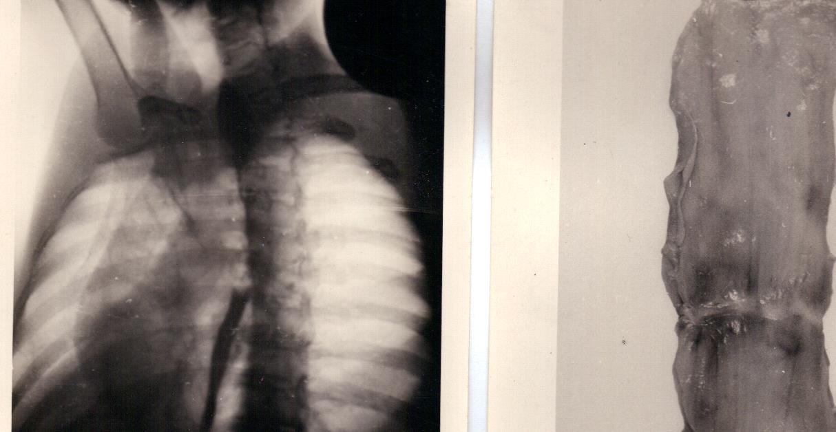
Figures 1 & 2: (1) X-ray Hirsch Esophagus Honed Speiseröhre geschliffe and (2) Eophagus (Makropreparat).
Conclusions
- When the long-term stay in the upper mediastinum tube systems leads to regeneration of removed tissues of the esophagus with the appearance of smooth muscle fibers that accompanied the restoration of patency of the esophagus.
- When the tread inside the esophagus can use medical glue, as well as apply medi-cal prosthesis.
- Operation easily can produce through access to the neck using modern equipment.
International Journal of Nuclear Medicine & Radioactive Substances
4. To suppress the remnants of a malignant growth in tissues of the mediastinum, it is desirable to apply local radiotherapy-by injecting a radioactive substance in rosette drainage tubes, 5. If the tumor turns out to be inoperable, advanced carry out rekanalizaciju of the esophagus.
References
-
Shevchenko IT, Shaposhnikov VI (1972) Esophageal Krebs/ herausgegeben von d.m. Abdurasulova / der "Medizin" in Usbekistan-Taschkent, mit 124.
-
Berrisford RG, Wajed SA, Sanders D, Rucklidge MWM (2008) Short-term otitcomes following total minimally invasive oesophagectomy. BJS 95(5): 602- 610.
-
Das A, Singh V, Fleischer DE, Sharma VK (2008) A Comparison of Endoscopic Treatment and Surgery in Early Esophageal Cancer. An Analysis of Surveillance Epidemiology and End Results Data. Am J Gastroenterol 28: 286-292.
-
Starikov VI (2006) Die esophageal Krebs: Diagnose und Perspektiven der Behan lung. international medizinischen Fachzeitschrift 66-70.
-
Chernousov AF, Bogopolskij MI, Kurbanov FSM (2000) Esophageal Surgery: Medizin, 350.
- Contribution of 18FDG PET in Atypical HORTON Disease
- Living Conditions, Healthy Practice and State of Households of a Town Rural in Colombia
- Background to the Health and Safety Regulation at Work in Colombia
- Risk Factors Psychology Workers Sena (Center for the Petrochemical Industry) Regional Bolívar, Colombia
- Diffuse Intense Pleural FDG Uptake with Smooth Thickening: A MARKER of Tuberculosis in Isolated Pleural Effusion
- Hypermetabolic Splenomegaly with Infarct in FDG PET/CT: A Clue to Scrub Typhus in PUO