Morphometric Study of Nutrient Foramen of Femur in Nepalese Population
Introduction: The femur is the strongest and longest bone that forms the skeleton of the thigh, bears body weight, supports movement of legs and provides attachment to muscles. Nutrient foramen is an opening in the shaft of femur. The nutrient artery enters through nutrient foramen through cortex into the medullary cavity of the femur. The morphometry of nutrient foramina are variable between different individuals with different Nationality. Aims: To study the morphometry of nutrient foramen in femurs of Nepalese subjects. Materials and Methods: The present study was carried out on 80 adult femur bones of both sides. Maximum length of femur, circumference at the level of nutrient foramen, location and position of nutrient foramen was measured and recorded separately for both femur with the help of measuring scale, thread and vernier calliper. All the collected data were represented as mean then analyzed with MS Excel 2007 software and represented graphically. Results: The mean length of femur obtained in the study was 42 cms. The nutrient foramina were found to be present in all femurs and were directed towards the upper end of femur. It has been observed that a total of 47 nutrient foramina were found to be present in 40 right femur and 61 nutrient foramina in 40 left femurs. Conclusion: The parameters measured in the present study are of clinical importance because it will be helpful to orthopaedic surgeons during any surgical procedures such as bone repair, bone grafting and microvascular bone surgery.
Introduction
The femur is the strongest and longest typical long bone that forms the skeleton of the thigh, supports body weight and provides attachment to muscles. It presents upper and lower ends, and an intervening shaft [1]. The femur is a highly vascular bone because of its blood supply via numerous foramina located over its different segments. Among these foramina, nutrient foramen is an important one which gives way to the nutrient artery [2]. The femur is supplied by nutrient artery which enters the bone obliquely through the nutrient foramen, which is directed away from the growing end. Henderson reported that the position of nutrient foramen are variable and may alter during the growth [3]. The number and location of foramina are variable and may alter during the growth of long bones [4]. Knowledge of the number and location of nutrient foramina is useful in certain surgical procedures [5]. The knowledge of variations of nutrient foramina is significantly important for orthopaedic surgeons undertaking an open reduction of a fracture to avoid injuring the nutrient artery and thus lessening the chances of delayed or non-union of the fracture [6]. The morphometry of nutrient foramina are variable between different individuals with different Nationality. Therefore, the aim of this study was to study the morphometry of nutrient foramen of Nepalese subjects which may be useful to curious students, researchers, anatomists, forensic experts, vascular surgeons and also to the orthopaedic surgeons performing vascularised bone microsurgery.
Materials and Methods
After Ethical approval the study was carried out on 80 adult femur bones of both extremities (40 were of right side and 40 were of left side) in the Department of Anatomy, Manipal College of Medical Sciences, Pokhara, from February 2019 to June 2019. Normal femur used in this study was collected from the Osteology laboratory of Anatomy. The femurs were retrieved from cadavers of Nepalese origin aged between 30 to 60 years with irrespective of sex. Femur with any fracture or pathological abnormalities like tumours, deformities, fractures, and trauma was excluded from the study. The side determination was done for the entire femur and following parameters were measured in the study:
- Maximum length of femur.
- Circumference at the level of nutrient foramen
- Position and location of nutrient foramen.
- Distance of nutrient foramen from upper end of femur Location and direction of nutrient foramen in relation with surface and zone were observed and noted.
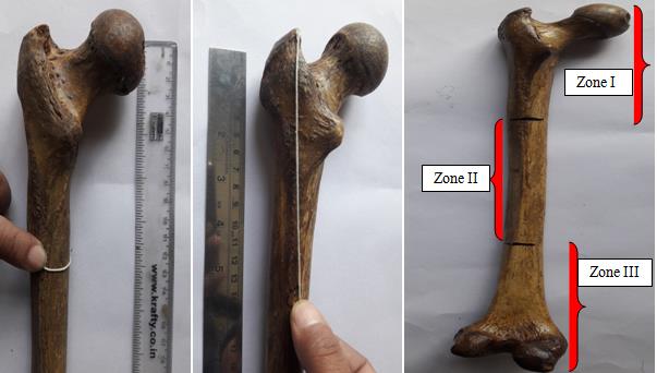
The position of foramina was divided into 3 types of zones as follows (Figure 1). Zone I: from 0% to 33.33%, the foramen was in upper one third of the bone. Zone II: from 33.33% to 66.66%, the foramen was in the middle one third of bone. Zone III: from 66.66% to 100% the foramen was in the lower one third of bone.
All the measurements were measured manually with the help of measuring scale, thread, osteometric board and vernier calliper for right and left femur separately. All the data were represented as mean ± SD then analyzed with MS Excel 2007 software and represented graphically.
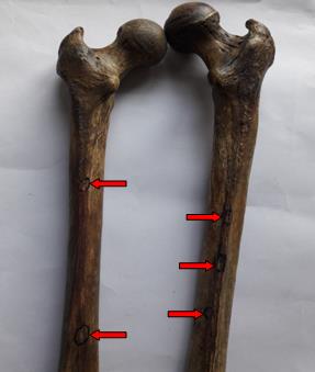
toward the upper end of femur but there was a variation in the number of foramen for both femurs (Figure 2). The single nutrient foramen was present in 85% of right side femur (34) and 47.5% of left side femur (19), double nutrient foramina in 12.5% of right femur (5) and 52.5% of left femur (21). Triple nutrient foramen was seen in 2.5% right femur (1) where as it were not seen in left femur. It has been observed that a total of 47 nutrient foramina were found to be present in right femur and 61 in left femur (table 2). In right femur 38.29% nutrient foramina were present on the posterior surface, 12.76% on medial surface and 6.38% on the lateral surface and 42.55% on the linea aspera. In left femurs, the nutrient foramen were present 37.7% on posterior surface, 13.11% on medial surface, 3.27% on lateral surface and 45.9% on medial lip of linea aspera (Table 3, Figure 3) .
Results
The following observations were found in the present study. The mean results and standard deviation (SD) of different parameters of both right and left femur is summarized in table 1. The nutrient foramina were found to be present in all femurs. The direction of nutrient foramina was not showing any deviation from normal anatomical feature even in single case throughout the study. All the nutrient foramina were directed upwards or
| S.N | Parameters | Right femur | Left femur | ||
| S.N | Parameters | Mean in cm | SD | Mean in cm | SD |
| 1 | Maximum length of femur | 42.70 | 3.09 | 41.30 | 2.54 |
| 2 | Circumference at the level of nutrient foramen | 8.10 | 2.34 | 8.30 | 2.40 |
| 5 | Distance of nutrient foramina from upper end | 14.30 | 2.01 | 16.20 | 2.30 |
Table 1: Showing mean and standard deviation (SD) of different parameters in cm of right and left femur.
| Number of foramen in right femur | Number of foramen in left femur | |
|---|---|---|
| Zone I | 12 | 19 |
| Zone II | 34 | 40 |
| Zone III | 01 | 02 |
| Total | 47 | 61 |
Table 2: Showing position and numbers of nutrient foramina of right and left femur.
| Parameters | Location of nutrient foramen | Total number of foramen | |
| Parameters | Right femur | Total number of foramen | Left femur |
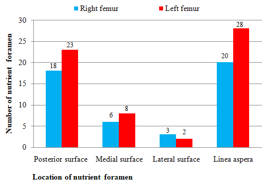
| Posterior surface | 18 | 23 | 41 |
| Medial surface | 6 | 8 | 14 |
| Lateral surface | 3 | 2 | 5 |
| Linea aspera | 20 | 28 | 48 |
| Total | 47 | 61 | 108 |
Table 3: Showing location and numbers of nutrient foramina of right and left femur.
Discussion
Many previous studies on nutrient foramen of femur have been carried out in different countries using different materials and techniques such as dry bones and plain radiographs. The present study was conducted in adult dry femur bones of Nepalese origin. The present study showed a single nutrient foramen in 85% of right side femur and 47.5% of left side femur in Nepalese population and is comparable with Pereira, et al. (2011) studies which was done in South Brazilian populations, who showed nutrient foramen as high as 97.4%, Muhammad Shahwani et al. (2017) studies in 83% Pakistani populations, and 78% in Oladayo Sunday (2014) studies in Nigerian population but less comparable to studies done by Seema, et al. (2015) 45.71% and 52% in north Indian populations, Mysorekar (1967) 50% and Prashanth, et al. (2011) 47.7% in Indian population and Sendemir and Cimen (1991) 46% in Turkish population [7, 8, 9, 10, 11, 12]. In the present study double nutrient foramen was noted in 12.5% of right femur and 52.5% of left femur in Nepalese population. It was more or less comparable with Pereira et al. study in South Brazilian populations who found in 47.71% of femur, 46% in Turkish population done by Sendemir and Cimen studies but it was less than 75% noted by Campos Forrio, et al. (1995) studies in Spanish population [12, 13]. Triple nutrient foramina was seen in 2.5% of right femur in the Nepalese population in the present study which was in agreement with Prashanth, et al. studies in 3.5% in South Indian population and Arvind, et al. (2017) studies in 3.3% of Indian population which shows three nutrient foramina but Campos, et al. studies in Spanish population and Gumusburun, et al. studies in Turkish population showed incidence as high as 10% [11, 14, 15]. The present study did not show any absent nutrient foramen and was in agreement with Sendemir and Cimen studies but Gumusburun, et al. (1994) studies reported it as 1.9% and Forriol F, et al. (1987) studies reported that 10% femur had three nutrient foramina [12, 15, 16].
Most common position of nutrient foramen (76.5%) in femur was in the middle one third of the shaft of femur on the linea aspera as reported by Mysorekar and Sendemir and Cimen and this was in the agreement with the present study which shows 68.5% nutrient foramen was located in middle one third (zone II) of femur [5, 12]. Maulukar O (2011) observed that 48% of the nutrient foramen were located in the proximal third and 52% in the middle third and no foramina in the distal 1/3rd [17]. The present study showed 28.7% nutrient foramen in proximal 1/3rd femur 68.5% nutrient foramen was located in middle 1/3rd and no foramina in the distal 1/3rd. Poornima B (2015) in her study found that nutrient foramina was found more close to medial lip of linea aspera at middle 1/3rd of the shaft of femur and corroborates with the present study [18]. To the best of our knowledge, this study has attempted to completely investigate the morphometry of nutrient foramen in dry femur bone of Nepalese population. The study was done on the dry bones so right and left femurs did not belong to the same individual and is the limitations of the study. Hence, further large scale study with larger sample size should be done to define the parameters in broad perspective.
Conclusion
The mean length of femur obtained in the study was 42 cms. There were a variable number of nutrient foramina, ranging from 1 to 3 on a single bone. The presence of double nutrient foramen was seen more in left femur. The morphometry of nutrient foramen of femur might be helpful for the orthopaedic surgeons during surgical procedures such as bone repair, bone graft, microvascular bone surgery with a possible reduction in post-operative complications. In addition, the knowledge about the variation in the location of nutrient foramina is important for the surgeons because of the increased chances of rupture to the nutrient artery during open or close procedures.
References
-
Standring S (2008) Gray’s Anatomy 40th ed. Edinburgh: Churchill Livingstone, Elsiever, 798.
-
Al-Motabagani MAH (2002) The Arterial Architecture of the Human Femoral Diaphysis. J Anat Soc India_._ 51(1): 27-31.
-
Henderson RG (1978) The position of nutrient foramen in the growing tibia and femur of rat. J Anat. 125(3): 593-599.
-
Bokariya P, Gudadhe D, Kothari R, Murkey PN, Shende MR (2012) Comparison of humerus and femur with respect to location and number of nutrient foramina. Indian J Forensic Med Pathol 5(2): 79-81.
-
Mysorekar VR (1967) Diaphysial nutrient foramina in human long bones. J Anat 101(4): 813-822.
-
Joshi H, Doshi B, Malukar O (2011) A study of the nutrient foramina of the humeral diaphysis. NJIRM 2: 4-17.
-
Pereira GAM, Lopes PTC, Santos AMPY, Silveira FHS (2011) Nutrient foramina in the upper and lower limb Bones: Morphometric Study in Bones of South Brazilian Adults. Int J Morpho 29(2): 514-520.
-
Muhammad BS, Mian AA, Lal MK, Nawab MK (2017) Significance of Morphometric Anatomy of Diaphyseal Nutrient Foramen in dried Pakistani Femurs and its Clinical Applications in Microvascular Bone Graft. PJMHS 11: 352-355.
-
Oladayo Sunday Oyedun (2014) Morphometric Study of Diaphyseal Nutrient Foramen in dried Nigerian femurs: Implications for Microvascular bone graft. Advances in Life Sci Technol 23: 91-96
-
Seema, Poonam Verma, Anupama Mahajan, Deepinder Gandhi (2015) Variation In The Number And Position Of Nutrient Foramina Of Long Bones Of Lower Limb In North Indians. Int J Anat Res 3(4): 1505-1509.
-
Prashanth KU, Murlimanju BV, Prabhu LV, Kumar CJ, Mangala MP, et al. (2011) Anatomy of Nutrient Foramina in the Lower Limb Long Bones. Australasian Med J 4(10): 530-537.
-
Sendemir E, Cimen A (1991) Nutrient foramina in the shafts of lower limb long bones: situation and number. Surg Radiol Anat 13(2): 105-108.
-
Campos FF, Pellico LG, Alias MG, Valencia FR (1995) A study of nutrient foramina of lower limb long bones. Surg Radiol Anat 16(4): 409-412.
-
Arvind D, Srivastava SK, Amit S, Anju B (2017) Morphometric Study of Nutrient Foramina In Human Femur Bone. Global J Res Analysis 6: 4-6.
-
Gumusburun E, Yucel E, OzkanY, Akgun Z (1994) A study of the nutrient foramina of lower limb long bones. Surg Radiol Anat 16(4): 409-412.
-
Forriol F, Gomez L, Gianonatti M,Fernandez R (1987) A study of the nutrient foramina in human long bones. Surg Radiol Anat 9(3): 251-255.
-
Malukar O, Joshi H (2011) Diaphysial Nutrient Foramina In Long Bones And Miniature Long Bones. Natl J Integr Res Med 2(2): 23-26.
-
B Poornima, Angadi AV (2015) A study of nutrient foramina of the dry adult human femur bones. Int J Biomed Res 6(6): 370-373.
- Pattern of Breast Lesions in Ovu Inland, Delta State, South Southern Nigeria
- Morphometric Analysis of the Human Femur: Exploring Platymetric and Robusticity Indices Among the Nigerian Population
- Anatomical Variation of Arteria Lusoria: Clinical Implications for Dysphagia Lusoria and Surgical Risk
- Morphometric Study of the Vertebral Body and Pedicle of Typical Cervical Vertebrae Using Radiological Image
- Epigenetic Mechanisms Driving Human Evolutionary Changes
- Neuroprotective Effects of Ginkgo Biloba Extract on Bilateral Common Carotid Artery Ischaemic Stroke Induced in Wistar Rat