Computerized Tomographic Study of Normal Evans Index in Adults at University of Gondar Comprehensive Specialized Hospital, North West Ethiopia, Gondar 2019
Introduction: Evans indexis one of the most important parameter in diagnosis of hydrocephalus, follow up cases of ventriculoperitoneal shunt, dementia and numerous other pathologies. In this situation, having a standard reference value of the Evan’s index will be supportive in a wide extent of clinical pathologies. The main objective of this study was to establish normal values for Evans index in a population of North West Ethiopia as there is no study found in Ethiopian medical literature. Methods: Computerized tomographic brain scans of 169 normal subjects were reviewed. Evan’s index was measured as the direct proportion of the most extensive frontal horn tips diameter of the cerebral lateral ventricles to the most extensive internal distance across the cranium. Results: In this study, 123(72.78) of the patients were males and 46 (27.22%) were females; their ages ranged from 18 to 79 years with a mean age of 40 years. The mean value for Evans index for the studied population was 0.262 ± 0.03. The Evan’s index increased with age and it was slightly higher among males. The difference in Evans value in males and females was not statistically significant. People over 60 years old had the highest Evans values in both genders. Conclusion: This study found the ranges of normal value for Evans index in north west Ethiopian population. It agrees with the diagnostic cut-off value of > 0.3 for hydrocephalus.
Introduction
Ventricles constitute 2 percent of brain volume.82 percent of the ventricular volume is contributed by lateral ventricles [1]. Ventricular extension caused due to imbalance in the production and absorption of cerebrospinal fluid is called hydrocephalus [2]. For the exact and early diagnosis of sort of hydrocephalus, information of ventricular size is pivotal. Computed Tomography (CT) is an acknowledged strategy in the identification of wide range of pathologic abnormalities and measuring the ventricular size precisely. In Ethiopian situation, CT still remains an effort lessly accessible, reasonable and quicker mode of brain imaging. Ventricular measure can be gotten by straight or volumetric estimations, out of which the direct proportions of the width of ventricles to the width of brain or cranium is easiest reproducible strategy.
Evan’s Index is the ratio which compares the greatest frontal horn tips diameter sidelong ventricles to the most extreme transverse distance across of inward table of the skull [3]. For the diagnosis of Normal Pressure Hydrocephalus (NPH), follow-up cases of ventriculoperitoneal shunt (VPN) [4], alcoholism, dementia, it is an important parameter. In this way, creating a standard reference value of Evan’s index will be valuable in a wide range of clinical conditions for this population where such information is inaccessible. The main objective of this study was to establish normal values for Evans index in a North West Ethiopian population as none has been found in the Ethiopian medical literature.
Material and Methods
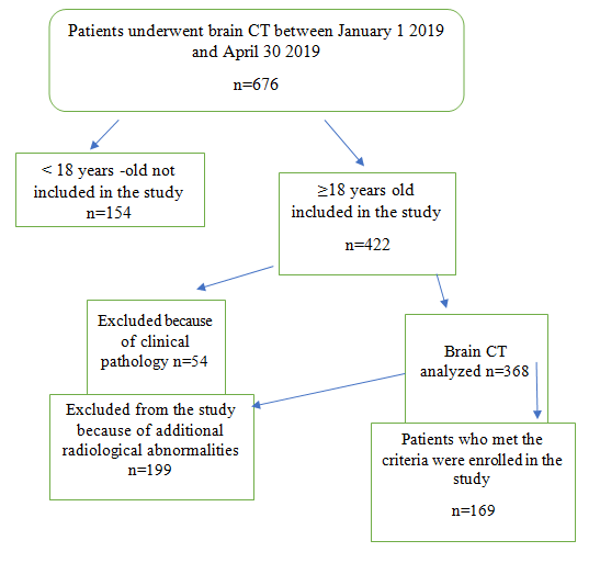
This prospective study was conducted in the Department of Radiology, university of Gondar comprehensive specialized hospital during the period between January 1, 2019 and April 30, 2019. From 675 cranial CT scans performed between January 1 2019 and April 30 2019 in the study area 169 (123 males and 46 females) were analyzed for this study. Of all the patients referred for CT brain to Department of Radiology, between the study period with neurological complaints, only those patients whose CT were reported to be normal were included in this study. Patients with intracranial and intraventricular pathology were avoided from the study. The flow chart diagram of the study is given in figure 1.
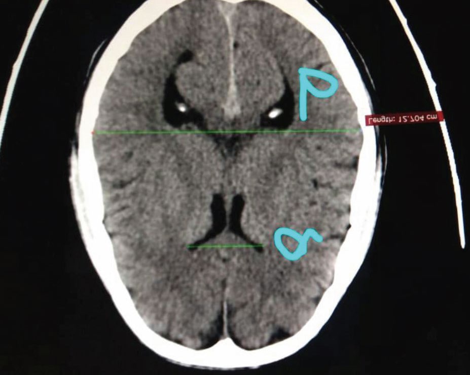
The CT scanner used in this study was the “General Electric GE (Bright speed 4 slices) with scanning time of 12-15 seconds and slice thickness of 5 mm. Measurement was taken on the axial section of CT images using Radi Anti Diacom Works Software. Frontal horn tips diameter (FHTD) of the cerebral lateral ventricles and widest Inner Diameter of the Skull (IDS) were taken as shown in Figure 2. Following measurements were made as seen in Figure 2. EI = a/b.
Statistical Analysis
Statistical analysis was done using SPSS software version 22.0. Descriptive statistics like mean and percentages were used for the analysis.
Results
The difference in Evans value in males and females was not statistically significant. Individuals above 60 years old had the highest Evans values in both sexes. There were a total of 169 healthy individuals between 18 and 79 years, with a mean of 40. Of them, 123 (72.78) of the patients were males and 46 (27.22) were females. The mean value for Evans index for the studied population was 0.262 ± 0.03. The EI increased with age and it was slightly higher among males. There was no much variation in calculation of Evan’s index from CT scan. Evan’s index can be calculated fairly accurately by multiple observers, which can be appreciated in the form of very small standard deviations. We found that less than 5% of patients were having ventriculomegaly but there were no clinical symptoms related to ventriculomegaly (Table 1).
- Mean ± SD p-value
- Males
- Females
- Males + Females
- FHW (mm)
- 31.95 ± 3.73
- 30.23 ± 2.82
- 31.08 ± 3.15
- ˂ 0.05
- IDS (mm)
- 123.85 ± 6.75
- 118.95 ± 5.84
- 121.98 ± 6.37
- ˂ 0.05
- Evans index
- 0.266± 0.03
- 0.264± 0.03
- 0.262 ± 0.03
- 0.06
Table 1: Descriptive statistics of ventricular parameters versus sex, computerized tomographic study of brain.
- Age group (years) sex n
- Ventricular parameters Mean ± SD
- FHTD
- IDS
- EI
- 18-39
- Male
- 123
- 31.84 ± 2.57
- 121.29 ± 4.21
- 0.25 ±0.36
- Female
- 46
- 29.87 ± 1.98
- 118.36 ± 3.74
- 0.24 ±0.17
- 40-59
- Male
- 123
- 32.84 ± 2.76
- 124.62 ± 5.42
- 0.27 ±0.15
- Female
- 46
- 31.43 ± 2.74
- 119.97± 6.12
- 0.25 ±0.14
- 60-79
- Male
- 123
- 34.34 ± 3.23
- 126.32± 6.54
- 0.28 ± 0.22
- Female
- 46
- 32.85 ± 1.34
- 121.85± 7.23
- 0.27 ± 0.18
Table 2: Mean and standard deviations of Evans Index of the adult age groups of 169 subjects, computerized tomographic study
Comparison between mean Evans Index between male and female showed no statistical significance differences (P>0.05) (Table 3).
| Variable | Male | Female | Independent sample test | P- value | 95% confidence interval of the difference | |
|---|---|---|---|---|---|---|
| Mean ± SD | Mean ± SD | Lower | upper | |||
| Evans Index | 0.266 ± 0.03 | 0.264 ± 0.03 | 0.27 | 0.06 | -0.002 | 0.214 |
Table 3: Independent sample test for mean difference of Evans Index by gender computerized study of brain.
Pearson`s correlation finding indicated a weak positive statistically significant correlation (P<0.05) between Evans index and age (r= 0.152) (Table 4).
| Evans index | ||
|---|---|---|
| Age in years | Pearson Correlation | 0.152 |
| Age in years | Sig (2 tailed) | 0.003 |
| Age in years | N | 169 |
Table 4: Pearson`s correlation (r) of Evans Index with age of the subjects computerized study of brain.
Discussion
EI is quantitative criterion has been used extensively in assessment of ventriculomegaly [5, 6, 7, 8, 9]. We also found that EI is increased with advancing age as reported by the other authors [8, 9]. The reason could be brain parenchyma shrinks with age, while cerebrospinal fluid spaces which also include the ventricles increase in size, to compensate for the atrophying brain substance. However, this physiologic ventricular enlargement does not cause Evans ratio to exceed 0.3. No statistically significant difference in the Evans ratio between males and females was found in our study. Haug has reported that females as compared to males has a smaller ventricular system above 15 years of age [10].
Idiopathic normal pressure hydrocephalus consists of triad of dementia, urinary incontinence and gait disturbance and is potentially a reversible cause of dementia in the elderly. It responds well to CSF shunting procedures. The disruptive effects of severe cerebral atrophy on memory, autonomic function and cognitive ability and could then be averted [5]. The ventricular enlargement could be quantitatively assessed by Evans index, with diagnostic cut off value of >0.3 based on international guidelines [7]. In presence of the clinical symptoms EI could be adequate for the diagnosis.
Our finding also supports that this defining criterion (EI > 0.3) could be used in the diagnosis of hydrocephalus in our own environment. EI is also an acceptable predictive index in post-traumatic ventriculomegaly as stated by Poca, et al. while Odebode, et al. [10] used it in determining the inter relation of visual function and ventricular size in children suffering from hydrocephalus. The following is the comparison of different studies measuring EI in different populations (Table 4).
Limitation of the Study
Relatively small Female sample size is one of the limitations of our study. Isolated use of Evan’s Index can lead to missing of ventriculomegaly in cases where occipital horn of lateral ventricle expands earlier than the frontal horns is considered as a limitation.
Conclusions
This study has established the normal range of Evan’s index in North West Ethiopia with respect to age and sex. Mean EI of 0.27 ± 0.03 in our study supports the adaptation of international guideline cut-off value of EI > 0.30 in the diagnosis of hydrocephalus in our North West Ethiopian population as well. EI can be used routinely due to less technically, easily reproducible, not time consuming.
Acknowledgement
We would like to express our deep appreciationto the staff members of Department of Radiology at University of Gondar Comprehensive Specialized Hospital. We wish to acknowledge the medical director/manager of the Universitty of Gondar Comprehensive hospital for his support and permission.
Availability of Data and Materials
The data set supporting this study are available in the manuscript.
Funding
This study received money from the University of Gondar for data collection only. The authors declare that they have received no funds for the publication of this manuscript and that they have no external source of fund for both data collection and publication.
Authors’ Contributions
Conceived the idea: AA. Designed the study methodology: AA, BT, MJ. Conducted the study: AA, BT, MJ. Analyzed the data: AA, BT, MJ. Interpreted the results: AA, BT, MJ. Wrote the draft manuscript: AA. Revised and edited the final manuscript: AA, BT, MJ. Approved the manuscript: AA, BT, MJ.
Ethical Approval and Consent to Participate
This study obtained ethical approval from College of Medicine and Health Sciences Institutional Review Board of the University of Gondar. The participants gave written informed consent prior to data collection.
Competing interests
The authors declare that they have no conflict of interests.
References
-
Akdogan I, Kiroglu Y, Onur S, Karabulut N (2010) The volume fraction of brain ventricles to total brain volume: a computed tomography stereological study. Folia morphologica 69(4): 193-200.
-
Hamidu AU, Olarinoye-Akorede SA, Ekott DS, Danborno B, Mahmud MR, et al. (2015) Computerized tomographic study of normal Evans index in adult Nigerians. Journal of neurosciences in rural practice. 6(1): 55-58.
-
Haslam R (1992) The nervous system. Behrman RE, Kliegman RM, Nelson WE (Eds.), Textbook of pediatrics. Philadelphia: WB Saunders.
-
Hashimoto M, Ishikawa M, Mori E, Kuwana N, SoIo N (2010) Diagnosis of idiopathic normal pressure hydrocephalus is supported by MRI-based scheme: a prospective cohort study. Cerebrospinal Fluid Research 7(1): 18.
-
Skullerud K (1985) Variations in the size of the human brain. Influence of age, sex, body length, body mass index, alcoholism, Alzheimer changes, and cerebral atherosclerosis. Acta neurologica Scandinavica Supplementum 102: 1-94.
-
Patnaik P, Singh V, Singh D, Singh S (2016) Age and gender related variations in lateral ventricle brain ratios. Int J Health Sci Res 6(5): 78-84.
-
Patnaik P, Singh V, Singh S, Singh D (2014) Lateral Ventricle Ratios Correlated to diameters of cerebrum-a study on CT scans of head. J Anat 22(2): 5-11.
-
Toma AK, Holl E, Kitchen ND, Watkins LD (2011) Evans’ index revisited: the need for an alternative in normal pressure hydrocephalus. Neurosurgery 68(4): 939-944.
-
Tullberg M, Jensen C, Ekholm S, Wikkelso C (2001) Normal pressure hydrocephalus: vascular white matter changes on MR images must not exclude patients from shunt surgery. American Journal of Neuroradiology 22(9): 1665-1673.
-
Kosourov A, Gaĭvoronskiĭ I, Rokhlin G, Blagova I, Panfilenko A (2002) In vivo assessment of various parameters of the brain ventricles with magnetic resonance tomography. Morfologiia 122(4): 71-73.
- Pattern of Breast Lesions in Ovu Inland, Delta State, South Southern Nigeria
- Morphometric Analysis of the Human Femur: Exploring Platymetric and Robusticity Indices Among the Nigerian Population
- Anatomical Variation of Arteria Lusoria: Clinical Implications for Dysphagia Lusoria and Surgical Risk
- Morphometric Study of the Vertebral Body and Pedicle of Typical Cervical Vertebrae Using Radiological Image
- Epigenetic Mechanisms Driving Human Evolutionary Changes
- Neuroprotective Effects of Ginkgo Biloba Extract on Bilateral Common Carotid Artery Ischaemic Stroke Induced in Wistar Rat