History of the Evolution of Human Anatomy Textbooks and Atlases, from Antiquity to the Present Day
This article presents information, accompanied by many illustrations, thematically about the history and evolution of human anatomy textbooks and atlases, from antiquity to the present day. The textual information of the materials of the study conducted by the author of this article is rich and abundant, with scans-illustrations of many anatomy textbooks and atlases, from different historical periods and by different authors, from many countries of the world - both in black and white and in color. This research work will be the first of a series of similar works of the author, devoted to the history of development and formation of the science of both normal and pathological human anatomy.
Introduction
The study of the history of medicine and, in particular, the history of human anatomy, at all stages of its formation, is a very relevant and demanded issue for many scientists and researchers. It is impossible to imagine a doctor of any medical specialty who would not study, and not apply in practice his anatomical knowledge! Since the first days of medical education, at any time and in any country, it has been mandatory for all students to study the subject of normal anatomy. Anatomy textbooks, anatomic atlases, anatomical plaster casts and charts are an essential element of anatomy training, not to mention the manuals for practical training and improvement of practical skills on a dead human body, next to which there are always anatomy textbooks! Since the first days of medical education, at any time and in any country, it has been mandatory for all students to study the subject of normal anatomy. Anatomy textbooks, anatomic atlases, anatomical plaster casts and charts are an essential element of anatomy training, not to mention the manuals for practical training and improvement of practical skills on a dead human body, next to which there are always anatomy textbooks [1]. The author of this article also studied normal anatomy at one time, first as a medical student and then as a university student. I remember with gratitude my anatomy teachers, as well as the anatomy textbooks and atlases that helped me to learn the mysteries of this great science. In most countries, these textbooks and atlases are different, belonging to both domestic and foreign authors. It is very difficult to imagine a time when there were no such textbooks and anatomical atlases at all! Anatomy teachers and their pupils had to find a suitable corpse, dissect it, and explain to their pupils everything that was going on and what they had seen! And, of course, this whole process, and individual body parts, had to be recorded in the form of a drawing and, preferably, in color in order to preserve and present the anatomical features of this or that body part. To accomplish this, the anatomist needed an assistant, a talented artist, because, unfortunately, many of the first anatomists did not always have the talent for drawing! Well, in later times, with the advent and perfection of the printing process and the creation of lithography and prints, both black and white and color, an entire team of specialists worked to perfect the process, creating the first anatomy textbooks and anatomical atlases. And it took many years, and sometimes decades or centuries, for these special books to appear, on which thousands and hundreds of thousands of doctors were educated, and to which we are so accustomed today, and take the process of learning from them as a natural process. In this, the first, research article, we will talk about such authors, the first textbooks on anatomy and anatomical drawings and anatomical atlases, as Avicenna, Andrew Vesalius, Ambroise Paré, Paolo Mascagni, Peter Camper [1].
Aim
The purpose of this article is to present the materials of the author’s study, thematically devoted to the search and presentation of textbooks and atlases on human anatomy, both by different authors and from different periods of time, using as illustrations, scans of these books.
Well, in later times, with the advent and perfection of the printing process and the creation of lithography and prints, both black and white and color, an entire team of specialists worked to perfect the process, creating the first anatomy textbooks and anatomical atlases. And it took many years and sometimes decades or centuries, for these special books to appear, on which thousands and hundreds of thousands of doctors were educated, and to which we are so accustomed
today, and take the process of learning from them as a natural process.
Naturally, in this one research article, it is very difficult to present all the material devoted to the question under study. Its presentation will require a large series of articles and new research materials. Therefore, the materials of his initial research, the author tried to present in his first article, which he presents to the court, the interested reader.
Material and Methods
In carrying out this research work, such means were used as the method of literary-critical analysis of available sources of information on the issue related to the ongoing research, using such available media as reference books, encyclopedias, catalogs of various collectibles, specialized periodicals, the Internet -resources on the subject under study.
Result and Discussion
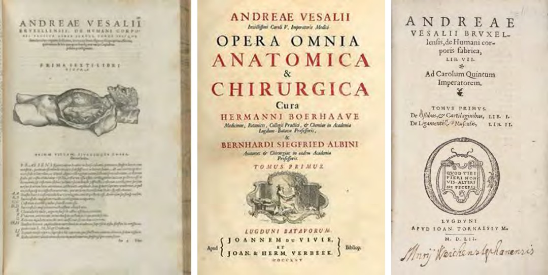
I would like to begin my story by presenting the earliest textbooks and atlases on human anatomy, belonging to the first doctors and anatomists of antiquity. In particular, in (Figure 1), we have a small collection of books by Andreus Vesalius, which are dedicated to human anatomy, which can be called the first textbooks on anatomy and anatomical atlases of the structure of the human body. These books were used for many decades by the first doctors, anatomists, and surgeons [1, 2, 3].
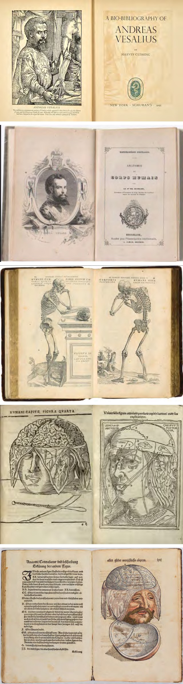
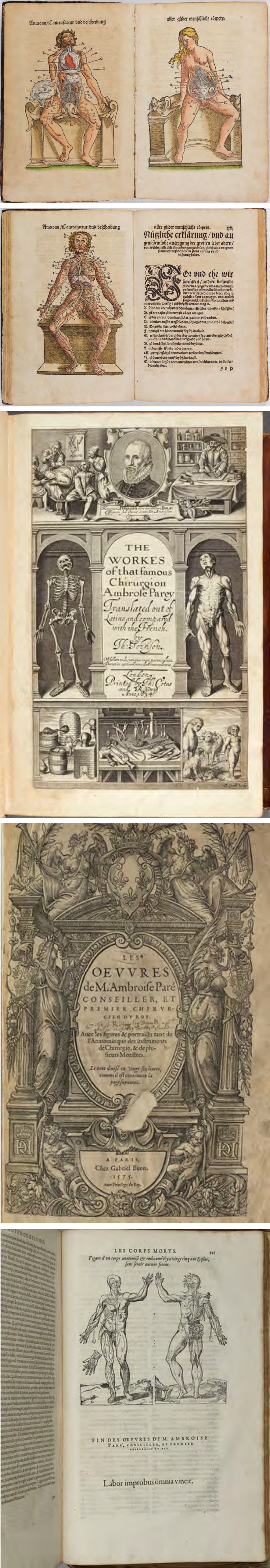
Figure 2 shows a small selection of books on anatomy by the world-renowned anatomist and surgeon Ambroise Paré [1, 4, 5, 6].

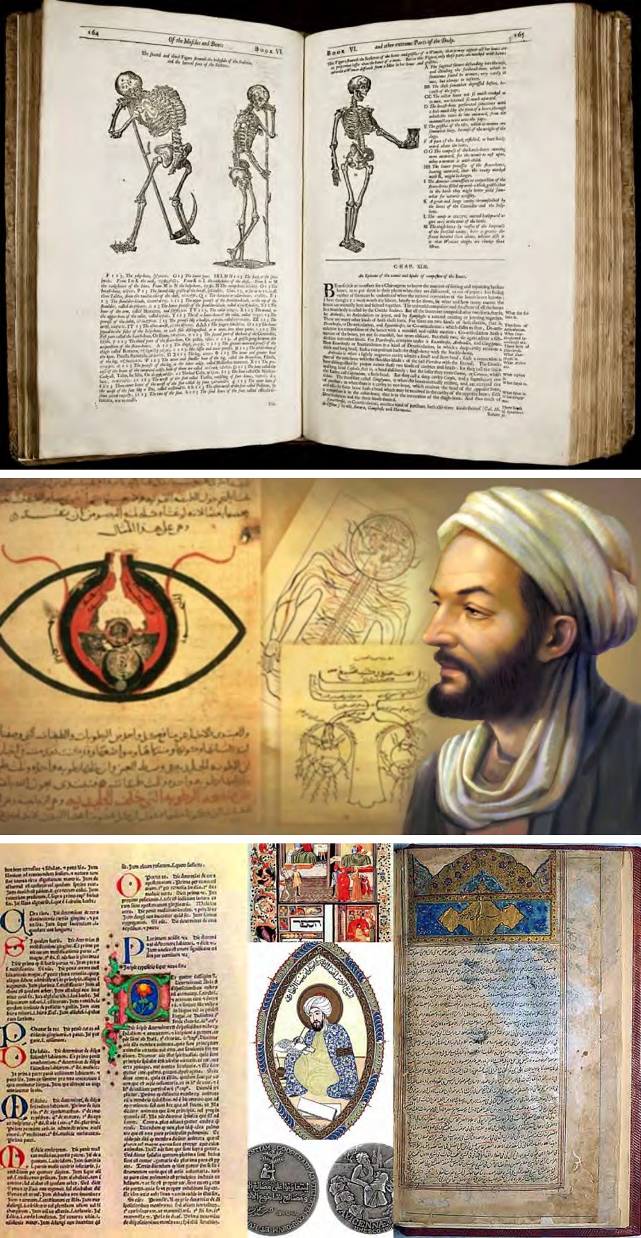
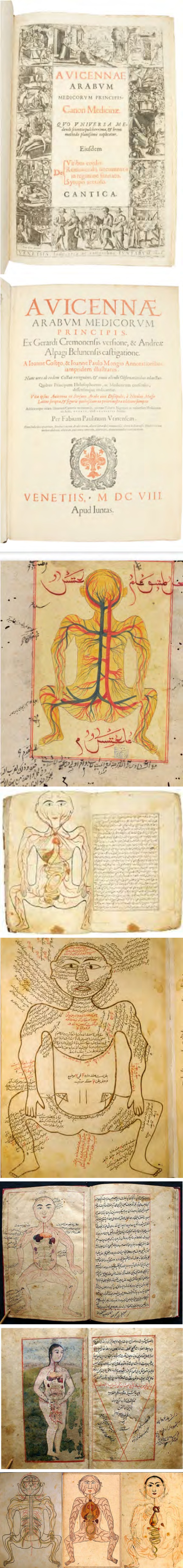
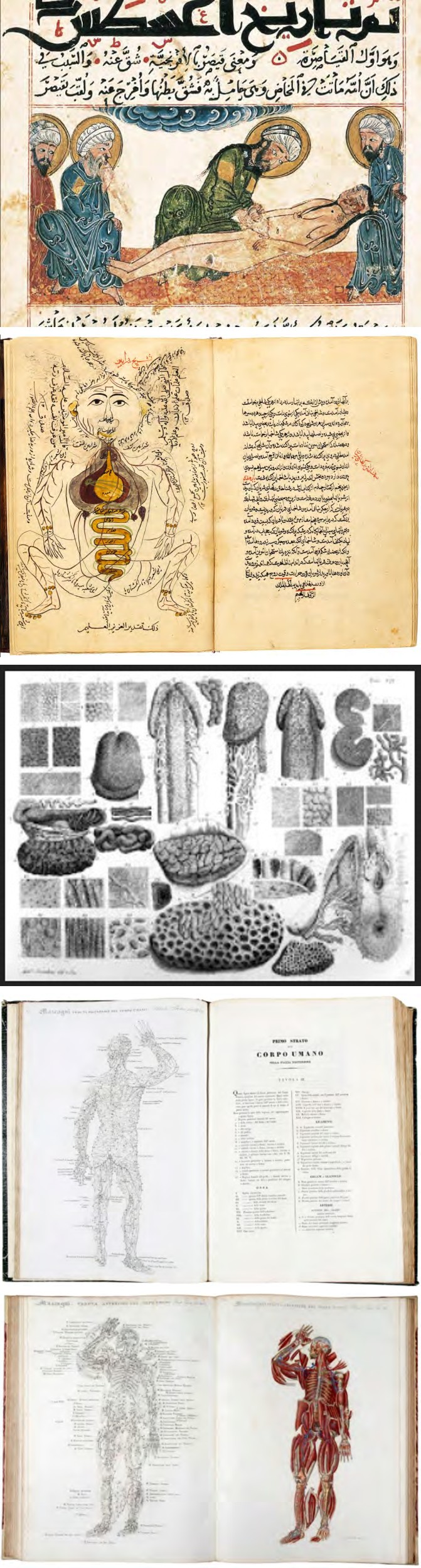
Figure 3, presents a small selection of ancient books on anatomy and medicine, with illustrations on anatomical subjects [7, 8, 9, 10, 11]. We are talking primarily about the books by Avicenna and a number of Arab authors - doctors who were actively engaged in anatomical research. – MANSUR BIN MUHAMMAD BIN AHMAD BIN YUSUF BIN FAQIR ILYAS (FL. 15TH CENTURY); AL-TASHRIH BI’L-TASWIR; ZAYN AL-DIN JURJANI (D.1136 AD), TASHRIH ZAKHIRAH-I KHWARAZMSHAHI; Tasrîh-i Mansûrî [7, 8, 9, 10, 11].
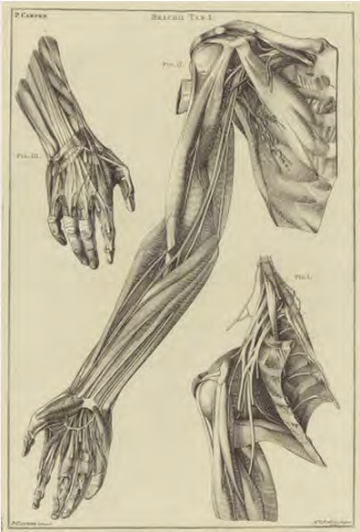
A small collection selection, which is shown in (Figure 4) (portraits of the scientist, commemorative bronze medal, obverse and reverse, his anatomical books and illustrations), dedicated to the famous Italian anatomist and illustrator of anatomical atlases and textbooks of that time, on human anatomy - Paolo Mascagni (1755-1815) [12, 13, 14]. From 1801
he was professor of anatomy and taught anatomy at the University of Pisa, anatomy, physiology and chemistry, at the Santa Maria Nuova hospital in Florence. It was he who studied the anatomy and functioning of the spongy body in the male penis [12, 13, 14]. Also, he created a large collection of wax bodies, in the anatomical museum, in Florence [12, 13, 14].
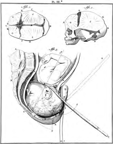
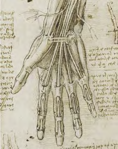
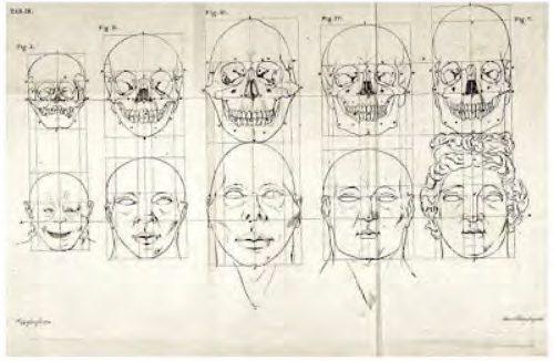
A selection of collection materials (lifetime books of the scientist, illustrations-drawings), shown in (Figure 5), tell about the famous scientist anatomist and anthropologist - Peter Camper (1722-1789) [14, 15, 16].
Conclusion
- This article presents materials devoted to the reflection in philately and numismatics, the anatomy of different parts of the human body.
- The presented illustrative materials can be used as an auxiliary informative tool in the study of such disciplines as “Anatomy”, “Operative surgery and topographical anatomy” and “History of Medicine” in the specialized universities and departments.
- This research work will be the first of a series of similar works of the author, devoted to the history of development and formation of the science of both normal and pathological human anatomy.
- The article presents new materials of the conducted research, devoted to the reflection in the means of collecting, the memory of a number of world famous scientists-anatomists, their scientific contribution to the world anatomical and medical science, as well as the formation and formation of the foundations of modern anatomy.
- The presented text and illustrative materials, taking into account the limited volume of the article, fully enough correspond to the purposes and tasks of writing this article, presenting the information in a reader-friendly format.
- Materials of article can be interesting to scientists, teachers and students of medical educational institutions, practical doctors and other categories of the medical workers of different orientation, interested in anatomy, medicine.
- The presented illustrative materials can be used as an auxiliary information tool in teaching such disciplines as “Anatomy”, “Operative surgery and topographical anatomy”, and “History of medicine” in profile institutes of higher education and departments.
- Modern means of collecting, in all their variety, quite fully, brightly and creatively, reflect the information about any medical discipline and its heroes.
References
-
Human Anatomy on Stamps - Stamp Community Forum.
-
Andreas V (2023) History of Medicine.
-
Andreas V (2023) Books, Health and History.
-
Bugaevsky KA, Bugaevskaya NA (2017) Ambroise Paré: life history and service to medicine in the reflection of collection tools Bulletin of SMUS74. 4 (19): 22-26.
-
The Apologie and Treatise of Ambroise Paré, CURL.
-
Ambroise Pare, Works, Second English edition.
-
Ambroise Paré | Books.
-
Ibn Sina (Avicenna).
-
Ibn Sina’s The Canon of Medicine URL: https:// (Can Muslim Heritage - M.
-
ZAYN AL-DIN JURJANI (D.1136 AD), TASHRI National Library of Medicine.
-
Islamic Medical Manuscript.
-
Islamic Medical Manuscript/ Islamic Medical Illustrations - CLAS 3239.
-
Figure Drawing: How to Draw the Head.
-
Petrus Camper: A history and overview of the clinical importance of Camper’s fascia in surgica.
-
Frank FA IJpma, Robert C van de Graaf, Thomas M van Gulik (2010) Petrus Camper’s Work on the Anatomy and Pathology of the Arm and Hand in the 18th Century. J Hand Surg 35(8): 1382-1387.
- Pattern of Breast Lesions in Ovu Inland, Delta State, South Southern Nigeria
- Morphometric Analysis of the Human Femur: Exploring Platymetric and Robusticity Indices Among the Nigerian Population
- Anatomical Variation of Arteria Lusoria: Clinical Implications for Dysphagia Lusoria and Surgical Risk
- Morphometric Study of the Vertebral Body and Pedicle of Typical Cervical Vertebrae Using Radiological Image
- Epigenetic Mechanisms Driving Human Evolutionary Changes
- Neuroprotective Effects of Ginkgo Biloba Extract on Bilateral Common Carotid Artery Ischaemic Stroke Induced in Wistar Rat