Sciatic Nerve Morphometry in the Gluteal Region in a Nigerian Population: An Anatomical Study
The sciatic nerve is frequently used in anesthesia, neurology, orthopedics, and rehabilitative medicine procedures. Due to its girth, the sciatic nerve is one of the human body's most often injured nerves. Unawareness of the structural differences of this nerve may be a contributing factor in damage. The purpose of the study is to evaluate the usual morphomety of the sciatic nerve and piriformis muscle. Lower limbs (N = 10) from Nigerian cadavers were chosen for this study's morphological examination and dissection. There were 8 males and 2 females in total. Using a computerized sliding calliper, the piriformis and sciatic nerve characteristics were examined and statistically analyzed.The right medial width (32.20±1.45mm) of the piriformis muscle was larger compared to the left (31.91±1.65mm). However, it was not so for lateral width of the piriformis muscle as the left was larger (17.35±0.25mm) compared to the right (17.09±0.17mm).The right length of superior piriformis border was larger (54.57±1.58mm) compared to the left (52.19±2.87mm). In comparison to the female specimens, the mean length of the sciatic nerve was substantially longer in the male specimens (p 0.05).
Abbreviations
LSBP: Length of the Superior Border of the Piriformis; LIBP: Length of the Inferior Border of the Piriformis; SNW: Width of the Sciatic Nerve; SNL: Length of the Sciatic Nerve; TL: Length of the Thigh; SPSS: Statistical Package for Social Sciences
Introduction
The sciatic nerve is constantly involved in the daily medical practices of anaesthesia, neurology, orthopaedics, and rehabilitative medicine [1]. The sciatic nerve, and its branches, are also the most frequently injured nerves within the human body [2, 3]. This nerve is unique because of its extensive course throughout the gluteal region, and lower limbs [4].The nerve is susceptible to damage from a variety of medical conditions because of its very long route [3, 4, 5]. A lack of understanding of the nerve’s structural differences may also contribute to injury [3, 6]. Because of this, understanding the nerve and its variations is crucial for clinical science [5, 7].
Numerous books have been written that detail the anatomy of the sciatic nerve [8]. This is not the case, though, for the variations of the sciatic nerve and the ensuing variations of the structures in the gluteal region that are intimately connected to and innervated by this nerve [3]. The intimate connection between the piriformis and the sciatic nerve is one such correlation that is causing growing alarm [1].The possibility that it contributes to the development of sciatic-related pain and discomfort is the main reason for the interest in this link. Variations in the sciatic nerve’s architecture make it more likely that this nerve will sustain damage during treatments and operations [9].
Different population groups around the world have variable prevalences of variations of the sciatic nerve and its bifurcation [10]. While the sciatic nerve is the subject of in-depth research in the Northern and Western parts of the world, this is not the case for Africa. Only six academic publications from African medical universities were discovered after a review of the literature, and they came from Kenya, Ethiopia, Uganda, and Nigeria [6, 7]. By examining the prevalence of sciatic nerve variations and associated variations of the piriformis muscle in a South African population, this research seeks to address some of the issues raised by the paucity of research on variations of the sciatic nerve for the African population [7]. The project will make use of lower limbs from dissected cadavers. Even with advancements in medical technology, according to Prakash, et al. [5], the cadaver is still the greatest way to study anatomy at this level [1, 9].Therefore, the purpose of this study was to assess the typical anatomical characteristics of the sciatic nerve and piriformis muscle as well as to define the frequency of anatomical alterations along the sciatic nerve’s course in relation to the piriformis muscle.
Materials and Methods
The Department of Human Anatomy at Achievers University in Owo’s Research and Ethics Committee provided their clearance with reference number: ANA18/023/032. Cadavers for this study came from Achievers University in Owo and Delta State University in Abraka, both in Nigeria. The departments and divisions generally used two techniques to procure cadavers. First, informed consent was used to obtain cadavers.A person gives permission for the donation of their body for use in medical research and education before they pass away. Second, cadavers that are not claimed by family members from the state within 30 days are automatically given to a local institution for the sake of medical study and instruction. The latter arrangement does not require formal consent; rather, the inspector of anatomy for Nigeria grants consent on behalf of the individual, their family, and the government. A thorough record of the cadaver’s whereabouts is also kept. No specific information on the deceased is made accessible in any case. Cadavers’ fundamental cause of death, date of death, sex, and age will all be included in a university-specific identification number that will be used for storage and identification. When conducting the current investigation, no cadaver’s personal or medical information was used. The study was conducted with complete anonymity for the cadavers.
Ten lower limbs from embalmed cadavers were used in the current study. At the two medical universities already stated, the researcher conducted this investigation while medical students regularly dissected the lower limb. All the cadavers utilized for the medical dissection programs at the universities were included in the study, therefore the specimens were obtained at random. All ten cadavers’ lower limbs, both right and left, were utilized. There is a corresponding left lower limb for each right lower limb as a result. There were 8 male and 2 female specimens among the 10 cadavers.
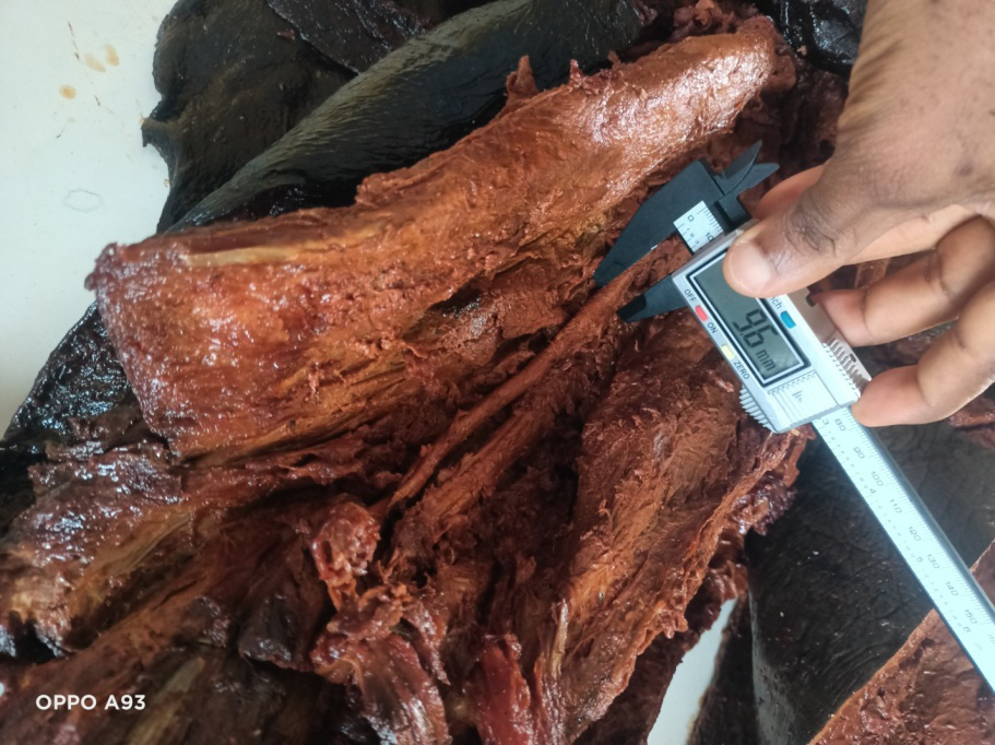
The length of the superior border of the piriformis (LSBP), the length of the inferior border of the piriformis (LIBP), the width of the sciatic nerve as it emerges below the inferior border of the piriformis (SNW), the length of the sciatic nerve (SNL), and the length of the thigh (TL) are among the parameters measured.
The study’s quantitative and descriptive statistics were gathered. The Statistical Package for Social Sciences (SPSS), version 25.1, was used for the statistical analysis. Comparisons were done between the lower limbs’ left and right sides, as well as between sexes and demographic groupings.
Results
From the study, it showed that the right medial width (32.20±1.45mm) of the piriformis muscle was larger compared to the left (31.91±1.65mm). However, it was not so for lateral width of the piriformis muscle as the left was larger (17.35±0.25mm) compared to the right (17.09±0.17mm). The right length of superior piriformis border was larger (54.57±1.58mm) compared to the left (52.19±2.87mm).
Similar trend was seen for the length of inferior piriformis. For the sciatic width, the left width was larger (16.24±0.84mm) compared to the right width; 14.76±0.73mm. Similar trend was also seen for the sciatic length, as the left was larger compared to the right side (Table 1).
| Measurement | Abv | Mean±SD |
|---|---|---|
| Right medial width of piriformis | RMVP | 32.20±1.45 |
| Left medial width of priformis | LMVP | 31.91±1.65 |
| Right lateral width of piriformis border | RLWP | 17.09±0.17 |
| Left lateral width piriformis | LLWP | 17.35±0.25 |
| Right length of superior piriformis border | RPSBL | 54.57±1.58 |
| Left length of superior piriformis border | LPSBL | 52.19±2.87 |
| Right length of inferior piriformis | RPIBL | 54.57±1.58 |
| Left length of inferior piriformis | LPIBL | 52.19±2.87 |
| Right width of sciatic nerve | RSNW | 14.76±0.73 |
| Left width of sciatic nerve | LSNW | 16.24±0.84 |
| Right length of sciatic nerve | RSNL | 390.25±0.17 |
| Left length of sciatic nerve | LSNL | 390.62±0.24 |
Table 1: Summary of the side morphometric measurements conducted in present study (measured in mm).
From the study, it showed that the male medial width (33.87±0.95mm) of the piriformis muscle was larger compared to the female (32.31±0.24mm). A similar trend was also seen for male lateral width of the piriformis muscle as it was larger (17.80±0.14mm) compared to the left (15.59±0.28mm). The male length of superior piriformis border was larger (55.78±0.18mm) compared to the female (55.28±0.23mm). Female length of inferior piriformis was larger compared to the male. The female sciatic width (15.24±0.02mm) was larger compared to the male (15.19±0.02mm) but the male sciatic length was larger compared to that of the female (Table 2).
| Measurement | Abv | Mean±SD |
|---|---|---|
| Male medial width of piriformis | MMVP | 33.87±0.95 |
| Female medial width of priformis | FMVP | 32.31±0.24 |
| Male lateral width of piriformis border | MLWP | 17.80±0.14 |
| Female lateral width piriformis | FLWP | 15.59±0.28 |
| Male length of superior piriformis border | MPSBL | 55.78±0.18 |
| Female length of superior piriformis border | FPSBL | 55.28±0.23 |
| Male length of inferior piriformis | MPIBL | 59.26±0.33 |
| Female length of inferior piriformis | FLPIBL | 60.21±0.06 |
| Male width of sciatic nerve | MSNW | 15.19±0.02 |
| Female width of sciatic nerve | FSNW | 15.24±0.02 |
| Male length of sciatic nerve | MSNL | 396.09±0.01 |
| Female length of sciatic nerve | FSNL | 384.51±1.92 |
Table 2: Summary of the morphometric measurements based on gender conducted in present study (measured in mm).
Discussion
The most frequent cause of nerve injury in children is damage brought on by injections into the sciatic nerve or structures closely connected to the nerve [11, 12]. Even though intramuscular injection is regarded as a fundamental skill, it needs to be handled carefully. Medical professionals must have a thorough understanding of regional anatomy to prevent issues [13, 14, 15]. The many injection sites, as well as the benefits and drawbacks of each site, must also be understood by practitioners [16].
There is a dearth of thorough anatomical information in the literature about the morphology of the piriformis itself [17]. The anatomical and radiological literature lacks information on variations of the piriformis. In particular, if ultrasound is being utilized to assist in the administration of an injection, this is crucial for the interpretation of magnetic and ultrasound pictures [17]. In order to better understand the sciatic nerve, piriformis, and other components in the gluteal region, researchers must do morphometric analyses. As far as we are aware, this is the third study of its kind to morphometrically quantify the variations seen in the gluteal region between sides and sex in a Nigerian population.
Vicente, et al. [18] found that the average sciatic nerve width was 18.85 mm for the 17 non-variant right lower limbs and 22.34 mm for the left lower limbs. Furthermore, the scientists went on to say that, when comparing the right and left groups, there was a statistically significant difference in the sciatic nerve width [18]. The mean sciatic nerve width in the 18 non-variant right lower limbs was 19.45 mm, while the mean sciatic nerve width in the 18 left lower limbs was 19.46 mm. Brooks, et al. [1] also found similar results in their specimens. But since the values of these figures were nearly similar, no statistical significance was determined when contrasting the two groups. In addition, Brooks, et al. [1] claimed that due to the study sample’s small sample sizes, statistical comparisons and conclusions were not possible. Given that the sample sizes in both Brazilian investigations were the same at 40 samples, it is unclear whether Vicente, et al. [18] were able to draw meaningful inferences from their sample. However, the reason for this gender differences may be attributed to ethnic variability and sciatic morphology.
All 297 specimens used in the computation for the current study’s sciatic nerve have a mean width of 15.19 mm, with a standard deviation of 3.44 mm. This width is comparable to that determined by Tomaszewski, et al. [10], and it’s plausible that this is because the sample sizes used in the two studies were similar. Although Vicente, et al. [18, 19] and Brooks, et al. [1] have slightly wider widths, this might be because of the significantly smaller sample sizes they employed.
Conclusion
From this study, it can be concluded that majority of the parameters measured were larger on the right compared to that of the left with an exception of the sciatic width and lateral width of piriformis muscle. Also the male had larger measurements compared to that of the females with an exception of the inferior piriformis border, and sciatic width. In order to strengthen comparisons and findings of reported research, there is a need for a rise in published literature for the Nigerian subpopulation groups. Researchers must also examine differences among bigger ethnic groups within the Southern and Western Nigerian population. It is strongly recommended that further study be done on the alterations along the sciatic nerve’s course in relation to the piriformis muscle.
Source of Fund: Nil
Conflicts of Interest: Nil
References
-
Brooks JBB, Silva CAC, Soares SA, Kai MR, Cabral RH, et al. (2011) Anatomical variations of the sciatic nerve in a group of Brazilian Cadavers. Revista Dor São Paulo 12(4): 332-336.
-
Kumar MT, Srimathi, Rani A, Latha S (2011) Cadaveric Study of Sciatic Nerve and its Level of Bifurcation. Journal of Clinical and Diagnostic Research 5(8): 1502-1504.
-
Budhiraja V, Rastogi R, Jain SK, Sharma N, Garg R, et al. (2016) Variation Relation of the Sciatic Nerve to the Piriformis Muscle: A Cadaveric Study from North India. Argentine Journal of Clinical Anatomy 8(1): 38-42.
-
Kotian SR, Sinha A, Souza ASD, Sumalatha S (2015) Variations of the sciatic nerve and its relation with the piriformis muscle in South Indian Population. Journal of Experimental and Integrative Medicine 5(3): 144-158.
-
Prakash BAK, Devi MN, Sridevi NS, Rao PK, Singh G (2010) Sciatic nerve division: a cadaver study in the Indian population and review of the literature. Singapore Medical Journal 51(9): 721-723.
-
Kiros MD, Woldeyes DH (2015) Anatomical variations in the level of bifurcation of the sciatic nerve in Ethiopia. Journal of Experimental and Clinical Anatomy 14(1): 1-4.
-
Ogeng’o JA, El-Busaidy H, Mwika PM, et al. (2011) Variant anatomy of sciatic nerve in a black Kenyan population. Folia Morphol 70(3): 175-79.
-
Kanawati AJ (2014) Variations of the sciatic nerve anatomy and blood supply in the gluteal region: a review of the literature. ANZ Journal of Surgery 84(11): 816- 819.
-
Smoll NR (2010) Variations of the Piriformis and Sciatic Nerve with Clinical Consequence: A Review. Journal of Clinical Anatomy 23(1): 8-17.
-
Tomaszewski KA, Graves MJ, Henry BM, Popieluszko P, Roy J, et al. (2016) Surgical Anatomy of the Sciatic Nerve: A MetaAnalysis. Journal of Orthopaedic Research 34(10): 1820-1827.
-
Sen A, Rajesh S (2011) Accessory piriformis muscle: an easily identifiable cause of piriformis syndrome on magnetic resonance imaging. Neurol India 59(5): 769- 71.
-
Enaohwo TM, Godswill OG (2018) Anthropometric study of the frontal sinus on plain radiographs in Delta State University Teaching Hospital. Journal of Experimental and Clinical Anatomy 17(2): 49-49.
-
Oladunni AE, Ogheneyebrorue GO, Joyce EI (2021) Radiological assessment of age from epiphyseal fusion at the wrist and ankle in Southern Nigeria. Forensic Science International Reports 3: 100164.
-
Mamerhim ET, Godswill OO (2020) Morphometric study of hypoglossal canal of occipital bone in dry skulls of two states in southern nigeria. Bangladesh Journal of Medical Science 19(4): 670-672.
-
Godswill OO, Nwaokoro IC, Owhefere OG, Felicia AT, Ebeye OA (2023) Stages of Epiphyseal Fusion at the Distal End of Radius and Ulna in Nigeria; A Radiological study. International Research in Medical and Health Sciences 5(5): 1-6.
-
Small SP (2004) Preventing sciatic nerve injury from intramuscular injections: literature review. Journal of Advanced Nursing 47(3): 287-296.
-
Windisch G, Braun EM, Anderhuber F (2007) Piriformis Muscle: Clinical Anatomy and Consideration of the Piriformis Syndrome. Surgical and Radiologic Anatomy 29(1): 37-45.
-
Vicente EJ, Viotto MJ, Barbosa CA, Vicente PC (2007) Study on Anatomical Relationships and Variations between the Sciatic Nerve and Piriformis Muscle. Revista Brasileira de Fisioterapia 11(3): 227-232.
-
Mbaka G, Osinubi A (2022) Morphological study of sciatic nerve and its topographic anatomical variations in relation to landmark structures around the pelvis: a Nigerian population study. Folia Morphol (Warsz) 81(1): 44-51.
- Pattern of Breast Lesions in Ovu Inland, Delta State, South Southern Nigeria
- Morphometric Analysis of the Human Femur: Exploring Platymetric and Robusticity Indices Among the Nigerian Population
- Anatomical Variation of Arteria Lusoria: Clinical Implications for Dysphagia Lusoria and Surgical Risk
- Morphometric Study of the Vertebral Body and Pedicle of Typical Cervical Vertebrae Using Radiological Image
- Epigenetic Mechanisms Driving Human Evolutionary Changes
- Neuroprotective Effects of Ginkgo Biloba Extract on Bilateral Common Carotid Artery Ischaemic Stroke Induced in Wistar Rat