Analyzing Foramen Ovale Morphometrically in Adult Dry Skulls of Nigerian-Based Population
Introduction: The foramen ovale is one of the major foramina on the greater wing of the sphenoid that is used for advanced surgical therapeutic and diagnostic procedures pertaining to the middle cranial fossa. The study was aimed at determining the length and width of foramen ovale in a Nigerian population. Materials and Methods: Seven dehydrated adult skulls, all the same gender, were acquired from the anatomy departments of Achievers University in Owo and Delta State University in Abraka, Nigeria. The length and the width of the foramen ovale were measured using digital sliding calipers. The paired sample t-test was used for the analysis. Results: The mean length of the foramen ovale was 0.611±0.21cm on the right side, and 0.42±0.12cm on the left side. The mean width was 0.5±0.15cm on the right side, and 0.43±0.12cm on the left side. Conclusion: Understanding the dimensions of the foramen ovale vary is crucial for neurosurgery during invasive surgical procedures like percutaneous trigeminal rhizotomy, as well as for biopsying Meckel cave lesions and cavernous sinus tumors.
Introduction
The foramen ovale, located in the greater wing of the sphenoid bone, is significant foramina that facilitate communication between the middle cranial fossa and the infra-temporal fossa. It can be found medial to the foramen spinosum and lateral to the foramen lacerum. It conveys the emissary vein that joins the cavernous sinus to the pterygoid venous plexus, the accessory meningeal artery, the lesser petrosal nerve, and the mandibular division of the trigeminal nerve [1].
Numerous invasive surgical and diagnostic procedures make use of the foramen ovale [2]. One of the main entrances into the cranial cavity via which nasopharyngeal carcinomas spread is the foramen ovale [3].
The foramen ovale varies in size, shape, and other morphological characteristics, just like other foramina of the skull. An osseous ligament that runs from the lateral pterygoid plate to the sphenoid bone’s spine may occasionally cover it. The foramen ovale is divided into four compartments by osseous pterygospinous and pterygoalar ligaments [4, 5]. They have the ability to compress the structures that go through it or obstruct the needle’s passage through the foramen ovale [6, 7]. The anteromedial side of the foramen ovale is divided in half by a bony spur [8].
The foramen ovale on the right side (RS) is smaller than the one on the left side (LS) [9]. The literature that is currently available shows that there are many different variations in the foramen ovale. The goal of the current study is to measure the foramen ovale’s length and width because this information can help to minimize complications during neurological disease surgeries.
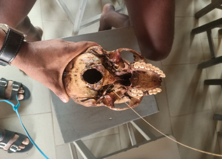
Materials and Methods
The present study was conducted on 7 dried adult skulls of unknown gender and age obtained from the Department of Anatomy of the Achievers University, Owo and Delta State University Abraka, Nigeria. Ethical clearance was obtained from the Institutional ethical committees to conduct this study. The measurement of the length (Anteroposterior diameter) and width (trans- verse diameter) of the foramen ovale was taken using sliding digital caliper. The paired sample t-test was used for the analysis (Figure 1).
Results
The present study was conducted on a total of 14 sides of 7 dry adult skulls. The mean length of the foramen ovale was 0.11±0.21cm on the RS, and 0.42±0.12cm on the LS. The mean width was 0.5±0.15cm on the RS, and 0.43±0.12cm on the LS (Table 1).
The length and the width were longer on the RS when compared with the LS. The difference between the length of the RS and of the LS was not statistically significant (p>0.05). Similarly, there was no statistically significant difference in the widths of the RS and LS.
| Parameters | Mean and SD |
|---|---|
| Right side length | 0.611 ± 0.21cm |
| Left side length | 0.42±0.12cm |
| P-value | 0.24 |
| Right side width | 0.5± 0.15cm |
| Left side width | 0.43±0.12cm |
| P-value | 0.25 |
Table 1: Descriptive statistics of right and left foramen ovale length and width.
Discussion
In the present study, the mean length of the foramen ovale was 0.611±0.21cm on the RS, and 0.42±0.12cm on the LS. The mean width was 0.5±0.15cm on the RS, and 0.43±0.12cm on the LS. As a result, in comparison to earlier research, the RS has longer length and width.
Yanagi [10] stated in his developmental study that the foramen ovale measures 3.85 mm in length in newborns and 7.2 mm in adults. The foramen ovale first appears as a ring-shaped region in the seventh month of intrauterine life and continues for three years after delivery [8]. According to Lang, et al. [11] adults’ foramen ovales typically measure 7.48 mm at their maximum length and 4.17 mm at their minimum. From 1.81mm in newborns to 3.7mm in adults, the width varies [12]. In a study involving 35 dry skulls, Ray, et al. [2] found that the foramen ovale’s mean length was 7.46±1.41 mm on the RS and 7.01±1.41 mm on the LS. On the RS, the mean width was 3.21±1.02 mm, while on the LS, it was 3.29±0.85 mm.
According to the current study, the RS has longer length and width than the LS, which is consistent with research done by Daimi, et al. [6] and Patel, et al. [12]. Many invasive surgeries and diagnostic procedures are carried out through the foramen ovale [2]. Through the foramen ovale, percutaneous trigeminal rhizotomy is also carried out in cases of trigeminal neuralgia [13, 14]. The foramen ovale is used for the percutaneous biopsy of cavernous sinus tumors and the biopsy of deep brain lesions, such as Meckel cave lesions, which lowers patient morbidity and is economical [15, 16]. Moreover, meningiomas and squamous cell carcinomas can be identified through the foramen ovale using transfacial fine needle aspiration guided by computed tomography (CT) [17, 18].
Conclusion
The foramen ovale’s morphometric significance is demonstrated in this study, which may benefit neurosurgeons and radiologists. The present study’s findings regarding variations in foramen ovale measurements may prove beneficial in the treatment of trigeminal neuralgia, the removal of abnormal tumors, and the acquisition of biopsies from deeper regions of the brain.
References
-
Standring S (2008) The Anatomical Basis of clinical practice. In: Standring S, et al. (Eds.), Gray’s Anatomy. 40th(Edn.), Churchill Livingstone Elsevier, London, pp: 415-416.
-
Ray B, Gupta N, Ghose S (2005) Anatomic variations of foramen ovale. Kathmandu Univ Med J 3(1): 64-68.
-
Chong VF, Fan YF, Khoo JB (1996) Nasopharyngeal carcinoma with intracranial spread: CT and MR characteristics. J Comput Assist Tomogr 20(4): 563-569.
-
Błaszczyk B, Kaszuba A, Kochanowski J (1980) Atypical foramina of the base of the skull. Folia Morphol (Warsz) 39(2): 201-209.
-
Tubbs RS, May WR, Apaydin N, et al. (2009) Ossification of ligaments near the foramen ovale: an anatomic study with potential clinical significance regarding transcutaneous approaches to the skull base. Neurosurgery 65(6): 60- 64.
-
Daimi SR, Siddiqui AU, Gill SS (2011) Analysis of foramen ovale with special emphasis on pterygoalar bar and pterygoalar foramen. Folia Morphol (Warsz) 70(3): 149- 153.
-
Enaohwo M, Godswill O (2018) Anthropometric study of the frontal sinus on plain radiographs in Delta State University Teaching Hospital. Journal of Experimental and Clinical Anatomy 17(2): 49.
-
Reymond J, Charuta A, Wysocki J (2005) The morphology and morphometry of the foramina of the greater wing of the human sphenoid bone. Folia Morphol (Warsz) 64(3): 188-193.
-
Neto HS, Camilli JA, Marques MJ (2005) Trigeminal neuralgia is caused by maxillary and mandibular nerve entrapment: greater incidence of right-sided facial symptoms is due to the foramen rotundum and foramen ovale being narrower on the right side of the cranium. Med Hypotheses 65(6): 1179-1182.
-
Yanagi S (1987) Developmental studies on the foramen rotundum, foramen ovale and foramen spinosum of the human sphenoid bone. Hokkaido Igaku Zasshi 62(3): 485-496.
-
Lang J, Maier R, Schafhauser O (1984) Postnatal enlargement of the foramina rotundum, ovale et spinosum and their topographical changes. Anat Anz 156(5): 351-387.
-
Patel R, Mehta CD (2014) Morphometry of foramen ovale at base of skull in Gujarat. J Dent Med Sci 13(6): 26-30.
-
Philips CX, Bilodi AKS (2013) A study on foramen ovale in human skulls. Indian J Med Case Rep 2(4): 65-76.
-
Wieser HG, Siegel AM (1991) Analysis of foramen ovale electroderecorded seizures and correlation with outcome following amygdalohippocampectomy. Epilepsia 32(6): 838-850.
-
Sindou M, Chavez JM, Pierre GS, Jouvet A (1997) Percutaneous biopsy of cavernous sinus tumors through the foramen ovale. Neurosurgery 40(1): 106-110.
-
Dresel SH, Mackey JK, Lufkin RB, Desalles AA, Layfield LJ, et al. (1991) Meckel cave lesions: percutaneous fine- needle-aspiration biopsy cytology. Radiology 179(2): 579-582.
-
Barakos JA, Dillon WP (1992) Lesions of the foramen ovale: CTguided fine-needle aspiration. Radiology 182(2): 573-575.
-
Enaohwo TM, Okoro OG (2020) Morphometric study of hypoglossal canal of occipital bone in dry skulls of two states in southern nigeria. Bangladesh Journal of Medical Science 19(4): 670-672.
- Pattern of Breast Lesions in Ovu Inland, Delta State, South Southern Nigeria
- Morphometric Analysis of the Human Femur: Exploring Platymetric and Robusticity Indices Among the Nigerian Population
- Anatomical Variation of Arteria Lusoria: Clinical Implications for Dysphagia Lusoria and Surgical Risk
- Morphometric Study of the Vertebral Body and Pedicle of Typical Cervical Vertebrae Using Radiological Image
- Epigenetic Mechanisms Driving Human Evolutionary Changes
- Neuroprotective Effects of Ginkgo Biloba Extract on Bilateral Common Carotid Artery Ischaemic Stroke Induced in Wistar Rat