Investigation of in Vitro Immunostimulating Effects of Freeze-Thaw Antigens against Visceral Leishmaniasis Caused by L. Infantum
Background: Visceral leishmaniasis, also known as Kala-Azar among the public, is among the notifiable infectious diseases. The fact that an effective and safe drug or vaccine formulation suitable for use on humans has not been developed causes the disease threat to continue. The aim of this study is to determine the immune system stimulating properties and cytotoxicity of the formulations obtained by combining the antigen obtained from Leishmania infantum parasites with different adjuvants. Methods: L. infantum (EP 126) antigens were obtained by the freeze-thaw method. Freund's adjuvant (Complete and Incomplete), a classical adjuvant, was used in the studies. Mouse macrophage (J774) and mouse fibroblast (L929) cell lines were used for cytotoxicity studies. Additionally, the responses at both 10ug/ml and 40ug/ml were investigated to ensure that the developed formulations were easily adaptable to in vivo. While the cytotoxicity of the formulations was determined by the MTT cell viability analysis method, the antileishmanial property was determined by the Nitric Oxide (NO) determination.method. Results: It was determined that all the developed formulations did not have a toxic effect on the host cell, the immunostimulating activity increased with increasing concentration, and the highest value was obtained at a concentration of 200 µg/ml. Cell viability at the maximum concentration was found to be between 78% and 89% for both J774 and L929 cells. The highest cell viability values and NO production abilities (37% increase compared to the control group) were obtained with Incomplete Freund's adjuvant (40 ug/ml formulation). Conclusion: It has been understood that the combination of antigens obtained by the freeze-thaw method with Freunds adjuvant (incomplete) supports NO production, thus having an immunostimulating effect.
Introduction
Leishmaniasis, caused by protozoans of the genus Leishmania, is a parasitic disease that is endemic in approximately 98 countries including Turkey [1]. There are three clinical forms caused by different types of leishmania: Cutaneous Leishmaniasis (CL), Mucocutaneous Leishmaniasis (MCL), which causes deep lesions on the skin, and Visceral Leishmaniasis (VL), which causes death if left untreated. L. major and L. tropica are among the most common CL agents, while VL agents are L. infantum and L. donovani.
VL, one of the notifiable communicable diseases, poses a serious threat to public health worldwide. According to the report published by the World Health Organization in 2019, it is known that more than 20,000 deaths and 300,000 new cases occur worldwide every year. It is known that most of the cases are in developing countries. Weak immunity, problems in the diagnosis of the disease, climate changes, wars, migrations, and most importantly, drug resistance of the active substances are the factors that increase the incidence of the disease [2]. It is known that if the disease is not treated, it has a mortality rate of up to 100% in a short period of two years in developing countries.
Although limited therapeutic agents, including paromomycin, amphotericin B, fluconazole, pentamidine, and antimony-containing compounds are used as drug therapy in VL [3], the options in vaccine form are limited due to the need for long-term regimens, drug-related side effects, and limited efficacy research is important [4, 5]. It is known that there is no effective and safe antileishmanial vaccine developed against human VL to date [6]. Although many vaccines against Leishmaniasis have been formulated in the last few decades, none of the strategies developed have provided complete protection. Vaccines to be developed for leishmaniasis, as in other infections, are required to provide reliable, stable, long-term protection, and to be easy to manufacture as well as low cost [2]. Therefore, there is sufficient scientific and epidemiological rationale for studies to develop antileishmanial vaccines.
In addition to the appropriate vaccine candidate selection, the selection of the right adjuvant or carrier is also important in developing an effective vaccine. It is seen in the literature that adjuvants such as Freund’s, Monophosphoryl lipid A, alum, saponin and BCG have been investigated against VL [7, 8]. Freund’s adjuvant, which is a water/oil emulsion, has two forms as complete and incomplete. Complete Freund’s adjuvant (CFA) contains heat-killed Mycobacterium, while incomplete Freund’s adjuvant (IFA) does not contains Mycobacteria. The most important advantages of emulsion- based adjuvants are their association with antigen and easy distribution in lymph nodes. Thus, they help interact with immune system cells. It also induces stimulation of antibody production due to non-specific immunopotentiation of macrophages [9]. It is known that Freund’s adjuvants are currently used in many vaccine formulations [10]. When the literature is examined, it is known that different antigens are formulated with Freund’s adjuvants and vaccine candidate researches for leishmaniasis are carried out [11, 12].
It is believed that in the fight against leishmaniasis, vaccines can be a much more economical and safer option than treatment methods that usually use chemotherapeutic drugs. For this purpose, in our study, as a vaccine candidate against VL, the immunostimulatory activities of soluble Leishmania antigens prepared by us were combined with different adjuvants and evaluated under in vitro conditions. Freund’s species (CFA, IFA) were used as adjuvant in the study, and the efficacy of vaccine formulations was evaluated in J774 macrophage and L929 fibroblast cells.
Materials and Methods
RPMI-1640, Fetal bovine serum (FBS), Dimethylsulfoxide (DMSO), Trypsin-EDTA, Dulbecco’s Modified Eagle Medium (DMEM), gentamicin, Hoffmann’s filter paper, 0.45 and 0.22 µm filters used to perform Leishmania and cell cultures were purchased from Sigma-Aldrich. Media were prepared with milli-Q water. leishmania parasites and morphological changes in cells were monitored using an inverted microscope Olympus CKX 41.
Cell Culture
Mouse fibroblast cell line -L929 and mouse macrophage cell line -J774 in our laboratory cryobank were used to detect immunostimulant activity. Cells were cultured in 75cm2 (Polystyrene surface, NEST) flasks using appropriate media. Stock media were prepared by adding 1% penicillin- streptomycin and 1% L-glutamine. For the growth and proliferation of cells, the culture of L929 fibroblast cell line was carried out in DMEM medium containing 10% FBS, and the culture of J774 macrophage cell line was carried out in RPMI- 1640 medium containing 10% FBS. The passage numbers of the cell lines used in the study were kept between 10 and 15.
During the experimental period, cells were incubated at 37°C, 95% humidity and 5% CO2 incubation conditions required for culture. Cell growth was monitored daily with an inverted microscope. After the cells reached 80-90% confluency, L929 cells were collected enzymatically and J774 macrophage cell line was collected by physical means and centrifuged at 25°C, 1000 rpm for 5 minutes. After centrifugation, the supernatant was discarded. On the other hand, 1 ml of medium was added on the J774 macrophage cells and L929 fibroblast cells remaining in the pellet part and cell count was performed on the thoma slide. In 96-well cell culture plates, cells were seeded at 1x10⁵ cells/mL per well and incubated for 24 hours.
Soluble Leishmania Antigen Preparation
The VL agent L. infantum (EP 126), which was present in the laboratory cryobank, was incubated in RPMI 1640 medium in a 27°C cooled oven. 7 ml of medium was added to 25 cm2 flasks and parasites were inoculated from the culture at a rate of 3x10⁵ parasites/ml. The growth of promastigotes was monitored daily using an inverted microscope. When the number of 107 parasites/ml reached in the 25 cm2 flask, the L. infantum promostigos were transferred to the 75 cm2 flask and incubated for 48 hours by adding 30 ml of RPMI 1640 medium with 5% FBS. After incubation, it was centrifuged at 4000 rpm at + 4°C for 25 minutes, and the supernatant was discarded, and the pellet was washed with 1 ml of PBS. The supernatant was removed by centrifugation again at +4°C at 4000 rpm for 15 minutes. The resulting pellet was brought to -20°C for later use.
Freeze-thaw method was used for the preparation of soluble Leishmania antigens (SLA). In the freeze-thaw method, parasites removed from -20°C were dissolved in a water bath at 37°C by adding 1 ml of PBS in a 15 ml falcon. The thawed cells were then placed in a liquid nitrogen container to be covered with liquid nitrogen, and the parasites were allowed to freeze for 15 minutes. Frozen parasites were kept in a 37°C water bath for 15 minutes and thawed. The parasites suspended in PBS in 15mL falcons were frozen with liquid nitrogen and thawed in a water bath 5 times, then centrifuged at 10,000 rpm for 3 minutes and the supernatant was taken. The determination of the protein amount in the lysate was carried out using the Warburg-Christian method in UV spectrometry at 280 and 260 nm wavelengths.
Cytotoxicity Analysis
To determine the percentage of cell viability, cells containing 3-(4,5-dimethylthiazol-2-yl)-2,5- Diphenyltatrazilium bromide were placed on cells incubated for 2 days under certain conditions (37°C temperature, 95% humidity and 5% CO2). MTT cell viability analysis was performed. 10 µl of MTT solution was added to the microplates and incubated at 37°C for 3 hours in a dark environment. At the end of the 3rd hour, liquids containing MTT solution were aspirated. After adding 100 µl of DMSO to each well, the microplates were incubated for half an hour in a dark environment. To detect cell viability, the microplates were placed in the ELISA Reader and the optical density was analyzed at 570 nm wavelength. The percentage of cell viability was determined using equation 1 [13].
Cell viability ( ) 0 *
100 0 sampleabsorbancevalue control absorbancevalue = (1)
Nitric Oxide (NO) Analysis
The amount of NO produced by the L929 fibroblast and J774 macrophage cell lines was determined by the Griess method. In this way, different concentrations of immunostimulatory activity of SLA antigens, alone or in combination with different adjuvants, were detected. After incubating the J774 and L929 cells in a 37⁰C oven containing 5% CO2 for 1 day, antigen and adjuvant combinations (CFA and IFA) prepared at six different concentrations (25-200 µg/ml) were added by adjusting with the medium.
Supernatants obtained after 2 days of incubation were reacted with Griess reagent. To prepare Griess reagent, 0.1 g of N-(1-Naphthyl) Ethylenediamine, 2.5 ml of phosphoric acid and 1 g of Sulfanilamide were added into 100 ml of distilled water. 50 µl of the culture medium to be analyzed was added to the microplates for NO measurement. Then, 50 µl of Griess reagent was added and incubated at room temperature for 10 minutes. Absorbance values were determined using an ELISA Reader at a wavelength of 540 nm. All studies were performed in 3 replicates.
Statistical Analysis
The experiments were repeated three times and the results were averaged. Fisher Behran’s d-test was used for statistical analysis of the data.
Results
SLA was prepared from L. infantum parasites by freeze- thaw methods. The immunostimulatory efficacy of vaccine formulations developed with SLAs alone or in combination with different adjuvants was determined in vitro in J774 macrophage and L929 fibroblast cell lines, and their cytotoxicity was determined.
The percentage of viability of the positive control group was accepted as 100%, and the percentage of viability of other cells was calculated with a simple ratio- proportionality. At the end of the 2 days, cell viability was calculated for all groups. In the first stage of the study, the immunostimulatory activity of both 10ug/ml and 40ug/ml CFA and their combinations with IFA, SLA was investigated in the L929 fibroblast cell culture system.
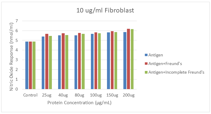
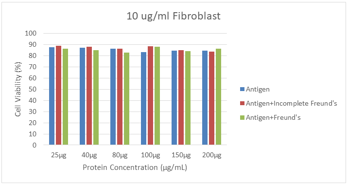
Immunostimulatory activity and cytotoxicity of SLA and 10 µg/ml adjuvants obtained by freeze-thaw method from L. infantum parasites in L929 fibroblast cell culture are shown in Figure 1 and Figure 2, respectively. In the study, it was seen that the immunostimulatory activity increased with increasing concentration and the highest value was obtained at 200 µg/ml concentrations. The highest immunostimulatory activity obtained from SLA treatment with fibroblasts at a concentration of 200 µg/ml appears to be 5,853 nmol/ml. In the cytotoxicity analysis, 84.42% viability was determined.
The maximum efficacy value of the combination of the lysate with CFA in combination with fibroblasts at the maximum concentration was 6.202 nmol/ml, and the viability rate of 83.45% in the cytotoxicity experiments was determined. In addition, it was determined that the maximum activity in fibroblast cells in combination with IFA was 6,163 nmol/ml and the viability rate was 86.43% in cytotoxicity analyses.
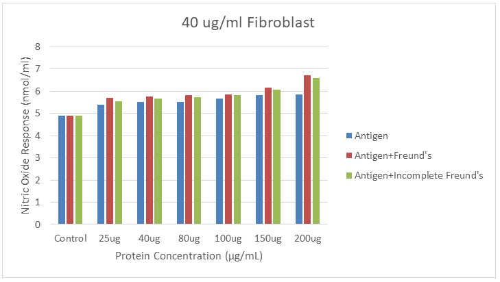
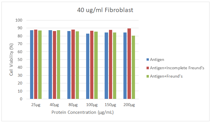
The immunostimulant and cytotoxic activity of SLA and 40 µg/ml Freund’s adjuvants in L929 fibroblast cell culture are shown in Figure 3 and Figure 4. It was determined that the highest efficiency obtained in fibroblast cells due to the combination of lysate with 40 ug/ml CFA at the maximum concentration was 6,705 nmol/ml and the viability rate was 80.47%. In combination with IFA, the maximum efficiency was found to be 6.589 nmol/ml, and 89.41% viability according to MTT analysis results.
In the second phase of the study, we determined the immunostimulatory activity of both 10 ug/ml and 40 ug/ml CFA and IFA in combination with SLA in J774 macrophage cell culture. In Figure 5 and Figure 6, immunostimulant and cytotoxic values of vaccine formulations obtained from the combination of 10 µg/ml Freund’s adjuvants with SLA are given. It was determined that the immunostimulatory activity at the maximum concentration was 6.24 nmol/ ml. Depending on the MTT analysis results at the same concentration, the viability rate is 84.25%.
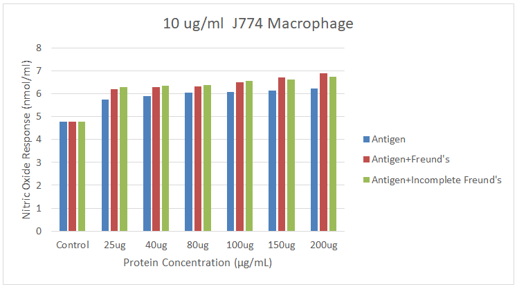
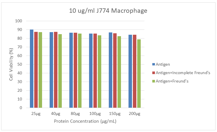
It is seen that the formulations of SLAs with 10 ug/ml CFA provided 6.899 nmol/ml activity in J774 cells, and 78.83% viability was detected. In addition, it was determined that the efficacy value was 6,744 nmol/ml and the viability rate was 84,06% in their combination with IFA.
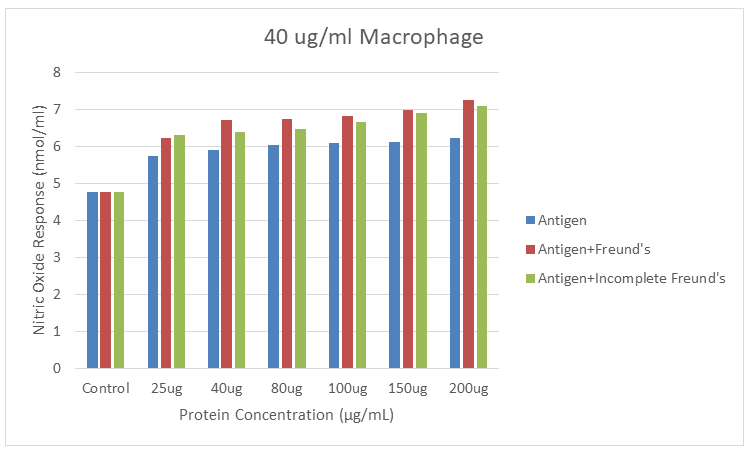
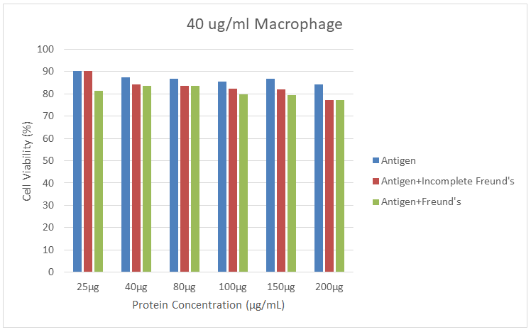
In the study, the results of immunostimulatory activity of combinations of 40 ug/ml CFA and IFA antigens in the J774 macrophage cell culture system are given in Figure 7 and Figure 8. The formulation of the lysate with 40 µg/ ml CFA at a concentration of 200 µg/ml achieved the maximum efficiency of 7.248 nmol/ml in J774 cells. In MTT analysis, 77.32% viability was detected. The highest efficacy value obtained from the treatment of SLAs with IFA in combination with macrophages was 7,093 nmol/ml. In the cytotoxicity analysis, the viability rate was found to be 77.07%.
Conclusion
It is known that VL is a tropical disease that puts an average of 350 million people at risk [14]. Although drug formulations have been developed for the treatment of this neglected disease, it is likely that the disease will be eradicated by vaccination. Prophylactic vaccination seems to be one of the most effective strategies in the control of VL.
In order to develop an effective vaccine, it has been shown in animal model studies of leishmaniasis that the selection of an appropriate adjuvant, which can elicit a long- lasting immune response against VL, is also necessary [9]. In general, the addition of the appropriate adjuvant will increase the immunogenicity of the vaccine candidate by inducing a cell-mediated immune response against disease. Adjuvants are important as they can stimulate and direct adaptive and innate and immune responses. Although there are many studies on the development and research of many vaccines against VL, data on the use of freeze-thawed parasite antigens as vaccine candidates against VL are very limited. There are some studies in the literature using freeze- thawed parasites as vaccine candidates against VL [15, 16, 17]. When compared with the literature, the results obtained were found to be satisfactory.
In this study, which can be seen as a preliminary study, the anti-leishmanial activity and cell cytotoxicity of the vaccine formulations developed in both mouse macrophage cells, and mouse fibroblast cells were evaluated under in vitro conditions. In the in vitro studies, both nitroxide assays and MTT cell viability analyses were performed. The results of the present study demonstrate the antileishmanial potential of soluble leishmania antigens combined with CFA and IFA adjuvants against VL. In the next stages, it is planned to evaluate the results under in vivo conditions with animal experiments.
Conflict of Interest Statement
Confirms that there is no conflict of interest between the authors.
Ethics Approval
None
Research Involving Human Participants and/or Animals
None
Informed Consent
None
References
-
Braga MS, Neves LX, Campos JM, Roatt BM, Braga SL, et al. (2014) Shotgun proteomics to unravel the complexity of the Leishmania infantum exoproteome and the relative abundance of its constituents. Molecular and biochemical parasitology 195(1): 43-53.
-
Kelleci K (2022) Determination of Immunostimulatory Efficacy of Benzimidazole Derivatives against Leishmania infantum. Systematic Reviews in Pharmacy 13(12).
-
Zorbozan O, Harman M, Evren V, Erdoğan MA, Kilavuz A, et al. (2018) Glia hücrelerinin antimona dirençli Leishmania tropica ile enfekte edilmesi: yeni bir ex-vivo modeli. Mikrobiyol Bul 52(1): 49-55.
-
Kelleci K, Gölebatmaz E (2023) In Vitro Determination of Antileshmanial Activities of Benzimidazolium Derivatives on L. major Promastigotes and Amastigotes. Acta Parasitologica 68(1): 51-55.
-
Roatt BM, Cardoso ODJM, Brito RCF, Coura-Vital W, Aguiar-Soares RDO, et al. (2020) Recent advances and new strategies on leishmaniasis treatment. Applied Microbiology and Biotechnology 104(21): 8965-8977.
-
Abdellahi L, Iraji F, Mahmoudabadi A, Hejazi SH (2022) Vaccination in Leishmaniasis: A Review Article. Iranian Biomedical Journal 26(1): 1-35.
-
Kelleci K, Allahverdiyev A, Bağirova M, Ihlamur M, Abamor EŞ, et al. (2023) Particulate and non-particle adjuvants in Leishmaniasis vaccine designs: A review. Journal of vector borne diseases 60(2): 125-141.
-
Chatzikleanthous D, O’Hagan DT, Adamo R (2021) Lipid- based nanoparticles for delivery of vaccine adjuvants and antigens: toward multicomponent vaccines. Molecular Pharmaceutics 18(8): 2867-2888.
-
Mutiso JM, Macharia JC, Gicheru MM (2010) A review of adjuvants for Leishmania vaccine candidates. Journal of biomedical research 24(1): 16-25.
-
Salk J, Salk D (1977) Control of influenza and poliomyelitis with killed virus vaccines. Science 195(4281): 834-847.
-
Pinto AR, Beyrodt CGP, Lopes RAM, Barbiéri CL (2000) Identification of a 30 kDa antigen from Leishmania (L.) chagasi amastigotes implicated in protective cellular reponses in a murine model. International journal for parasitology 30(5): 599-607.
-
Dole VS, Raj VS, Ghosh A, Madhubala R, Myler PJ, et al. (2000) Immunization with recombinant LD1 antigens protects against experimental leishmaniasis. Vaccine 19(4-5): 423-430.
-
Kelleci K, Ihlamur M, Ozkan M, Abamor E (2024) Cytotoxic Effects of Momordica Charantina Extract with Amphotericin B and Miltefosine Lac Combinations on Leishmania Parasites. Journal of Faculty of Engineering 39(1):1-7.
-
Kushawaha PK, Tripathi CDP, Dube A (2022) Leishmania donovani secretory protein nucleoside diphosphate kinase b localizes in its nucleus and prevents ATP mediated cytolysis of macrophages. Microbial Pathogenesis 166: 105457.
-
Vilela MC, Gomes DCO, Marques-da-Silva EA, Serafim TD, Afonso LCC, et al. (2007) Successful vaccination against Leishmania chagasi infection in BALB/c mice with freeze-thawed Leishmania antigen and Corynebacterium parvum. Acta Tropica 104(2-3): 133-139.
-
Grenfell RF, Marques-da-Silva EA, Souza-Testasicca MC, Coelho EA, Fernandes AP, et al. (2010) Antigenic extracts of Leishmania braziliensis and Leishmania amazonensis associated with saponin partially protects BALB/c mice against Leishmania chagasi infection by suppressing IL- 10 and IL-4 production. Memórias do Instituto Oswaldo Cruz 105(6): 818-822.
-
Thakur A, Kaur H, Kaur S (2015) Studies on the protective efficacy of freeze thawed promastigote antigen of Leishmania donovani along with various adjuvants against visceral leishmaniasis infection in mice. Immunobiology 220(9): 1031-1038.
- Epidemiological and Clinical Aspects of Intestinal Parasitoses Among Students in the City of Bocaranga, Central African Republic
- Artificial Intelligence Empowers Global Infectious Disease Prevention and Control: Opportunities and Challenges
- Factors that Affect the Incidence of Babesia and Blood Donor Testing in Select States: A Regression Analysis
- Neuro-TB: The Battle between Tuberculosis and the Nervous System
- The Biological and Health Implications of Cat Fleas (Ctenocephalides felis): Assessing Zoonotic Risks and Hygiene Strategies
- Biostatistical Analysis of Medicinal Plants for Treating Schizophrenia