Histophatological Damage to Heart and Lungs on Canine Dirofilariosis in Panama
Latin America and countries with hot climates have long suffered with vector-borne diseases, be they fleas, mosquitoes or ticks, among others. Canine Dirofilariasis is a vector disease whose etiological agent in America is Dirofilaria immitis, however, with little information on its real damage to canines and no information on its pathogenesis in humans in this country. Five anatomical pieces made up of heart and lungs, preserved in 10% formaldehyde, were evaluated histopathologically to define the damage caused by the nematode. The most common lesion found in the lungs was pulmonary emphysema and lymphocytic interstitial pneumonitis. Pseudo papillary hyperplasia of the endothelium was observed in one of the cases. The most common lesions found in the heart were endocarditis and pericarditis. We can conclude that the damage caused by D. immitis to the heart and lungs tissues of dogs is evident and that further studies are necessary to better understand this disease.
Introduction
Latin America and countries with hot climates have long suffered with vector-borne diseases, be they fleas, mosquitoes or ticks, among others. This situation has been aggravated in recent decades due to the strong climatic changes that the planet has been undergoing, and which have led to an increase in vector populations and therefore in the diseases transmitted by them. The Panamanian isthmus does not escape this phenomenon, showing an increasing incidence of this type of disease, a large part of these zoonoses being transmitted by mosquitoes to humans, some re-ermerging others of an emerging nature and many of this little studied, mainly in developing countries.
Canine Dirofilariasis is a vector disease whose etiological agent in America is a nematode of the Onchocercidae family, Dirofilaria immitis, reported in Panama causing disease in dogs in the Chiriquí region [1], however, with little information about its real damage to canines. No information on its pathogenesis to humans in this country. Part of the life cycle of this parasite has an obligatory passage within mosquitoes of various species that function as vectors, where biological changes occur that transform this nematode into an infectant through the insect bite, causing damage to various tissues of its definitive and accidental hosts. In the definitive hosts, which are normally canines, the cycle is completed, developing adults that invade the pulmonary artery where reproduction occurs and the release of microfilariae into the circulation, which are ingested by the vector mosquitoes during their feeding on the vertebrate. The human being is an accidental host where the cycle is not completed; however, larval migration can cause granulomatous lesions on lungs.
The evaluation of histopathological damage is essential to trace the pathogenesis of an etiological agent and then be able to measure the real impact on animal health, trying to elucidate the physiopathological mechanisms that lead to tissue damage. The objective of this work is to report the histopathological damage caused by this nematode to canine heart and lungs in Panama.
Methods
Five anatomical pieces made up of the heart and lungs, preserved in 10% formaldehyde and obtained from the autopsy of patients who died of severe heart failure or who were euthanized at the request of their owners due to lack of quality of life and suffering, using Xylazina 4.4 mg/Kg, Ketamine 20 mg/Kg and Potassium Chloride 100 mg/Kg [2], all with a positive diagnosis for Dirofilaria immitis performed through rapid tests, immunochromatography tests Uranotest Quattro® Diagnostic Kit from Urano Vet for detection of Ehrlichia canis, Anaplasma plays and Leishmania infantum antibodies, and Dirofilaria Immitis antigens; and Elisa test Snap 4Dx Plus® Diagnostic Kit from Idexx for detection of Ehrlichia canis / Ehrlichia ewingii, Anaplasma platys / Anaplasma phagocytophilum and Borrelia burgdorferi antibodies, and Dirofilaria Immitis antigens. The nematodes were removed from hearts and lungs and these organs were washed and fixed in 10% formaldehyde with buffered pH for 96 hours and made the histological sections that were stained with hematoxylin – eosin for microscopic evaluation of the histopathological lesions and visualized in an optical microscope with a 200x lens.
Results
The canines from which the organs for the study were obtained came from two regions, one from El Manzal beach (PM) in San Lorenzo to the east and four from Puerto Armuelles (PA) to the west, both from the province of Chiriquí. This is the first record of D. immitis at El Manzal beach. It should be noted that the five animals also had antibodies against E. canis and one of them against A. platys / A. phagocytophilum. Nematodes were found in the right ventricle and atrium, along the entire length of the pulmonary artery, and within medium-caliber vessels of the lung parenchyma. Nematodes were also removed from the left atrium and pulmonary vein. The number of nematodes and the histopathological observations in heart and lungs are listed in Table 1 & Figures 1a-11b.
Both organs were affected by the action of the parasitism, being the lungs the ones that suffered more tissue damage since they presented a hepatic aspect without alveolar aeration. The most common lesion found in the lungs was pulmonary emphysema and lymphocytic interstitial pneumonitis, which were observed in all five cases; however, chronic vasculitis and chronic non-granulomatous bronchiolitis were also observed. A pseudopapillary hyperplasia of the vascular endothelium or intravascular papillae in pulmonary parenchyma arterioles endothelium was observed in one of the cases. The heart and its structures presented less severity in relation to histopathological damage. The most common lesions were endocarditis and pericarditis, observed in three and two cases, respectively. Chronic myocarditis, pericarditis, hypertrophy of both right cardiac chambers, and coronary arteriosclerosis were also observed.
Discussion
The damage caused to host tissues due to parasitism by D. immitis is evident, with lesions with inflammatory infiltrates in the lungs and hearts; however, the pathophysiological mechanism that causes the appearance of pseudopapillary hyperplasia of the vascular endothelium is still under discussion.
In studying the pathogenesis of filariasis in vascular endothelial tissue, it was reported that the reaction of the pulmonary artery wall of dogs infected with D. immitis, previously attributed to endothelial tissue damage and/or vascular smooth muscle hyperplasia, appeared to be more likely caused by changes in endothelial cell behavior, and that pulmonary artery obstruction by adult parasites or intimal litter proliferation can lead to pulmonary hypertension and right ventricular failure [3]. Parasites cause mechanical damage to the pulmonary artery wall, causing inflammation of endothelial cells and the growth of structures such as villi made of endothelial smooth muscle and endothelium [4]. The author comments that this growth can be stimulated by platelet growth factor and that it contributes to vessel obstruction due to lumen reduction, causing embolism and pulmonary hypertension, and dilatation and hypertrophy of the right ventricle, congestive heart failure, Valvular damage and endocarditis. Lesions indicative of right-sided heart failure, consisting of right ventricular dilatation, were also observed in necropsied infected dogs; other lesions were observed in the interalveolar septum.
| AGE | OrigIn | N° Nem | Heart | Lungs |
|---|---|---|---|---|
| 3 years 5 months | PA | 54 | Mononuclear infiltrate in myocardium (Figure 2a) Chronic myocarditis. Mesothelial cell proliferation (Figure 2b) and mononuclear infiltrate in pericardium (Figure 5b) Pericarditis. Mononuclear infiltrate in epicardium (Figure 3a) Epicarditis. Focal endothelial proliferation in endocardium (Figure 3b) Focal endocarditis. | Hepatized lung (Figure 1b), without evidence of alveolar aeration, but consolidated. Vascular congestion and mononuclear infiltrate in small caliber capillaries towards pulmonary interstitium (Figure 6a). Diffuse alveolar damage and expansion of the interstitium with mononuclear infiltrate (Figure 6b) Pulmonary emphysema, lymphocytic interstitial pneumonitis. |
| 1 year 9 months | PA | 75 | No anatomical or microscopic pathological changes of the heart or great vessels were found. Moderate dilatation of the right chambers Right atrioventricular hypertrophy. No infarcts or vegetations were found. In the right ventricle there was a focus of mononuclear inflammatory infiltrate in the endocardium (Figure 4a) Focal endocarditis. | Both lungs were hepatized (Figure 1b), with marked vascular congestion. Atelectasis with marked thickening of the interstitium at the expense of mononuclear inflammatory infiltrate (lymphocytes and macrophages) and vascular congestion with endothelial edema and presence of lymphocytes, monocytes and plasma cells in the vascular lumen of medium and small caliber vessels (Figure 7a). Marked expansion of pulmonary interstitium with mononuclear infiltrate. Reactive hyperplastic proliferation of mesothelial cells in visceral pleura (Figure 7b) Pulmonary emphysema, chronic lymphocytic interstitial pneumonitis. |
| 1 year 6 months | PM | 20 | Mild enlargement and dilatation of the right ventricle and thickening of its wall. No vegetations or infarcts were identified. Right ventricular hypertrophy. | Both lungs were congestive (Figure 9a) and hepatized (Figure 1b). Presence of intravascular parasites in medium caliber vessels (Figure 8a). Marked expansion of the interstitium due to inflammatory infiltrate of mononuclear cells (lymphocytes and macrophages) (Figure 8b). Marked vascular congestion with abundant lymphocytes and monocytes in the lumen (Figure 9a). Pulmonary emphysema, chronic lymphocytic interstitial pneumonitis. |
| 6 years | PA | 27 | Lymphocytic infiltrate at the left atrium endocardium level (Figure 4b), as well as nodular lymphocytic and histiocytic infiltrate in the soft tissues of pericardium (Figure 5a). Nematodes were observed in the left atrium (Figure 1a). No valvular lesions, infarcts, thrombi, emboli or vegetations were observed. Pericardium presented foci of hemorrhage. Nodular inflammatory infiltrate composed predominantly of mature lymphocytes in pericardium and endocardium. Chronic endocarditis, chronic non- granulomatous pericarditis. | Peribronchiolar lymphocytic infiltrate, without alterations of the airway or respiratory epithelium (Figure 9b). Pseudopapillary hyperplasia of the vascular endothelium or intravascular papillae in pulmonary parenchyma arterioles endothelium and nodular inflammatory infiltrate predominantly lymphocytic and histiocytic, endothelial and interstitial, adjacent to the tunica adventisia in arterioles (Figure 10 a) and predominantly lymphocytic in venules (Figure 10b), and in pulmonary parenchyma (Figure 11a). Endovascular parasites were observed in lung parenchyma (Figure 11b). No thrombi were identified in the main blood vessels but marked vascular congestion and bee- panel appearance in the airways of the pulmonary parenchyma, with marked dilatation. Chronic lymphocytic interstitial pneumonitis, chronic vasculitis in medium caliber vessels (arterioles and venules), pulmonary emphysema, chronic non-granulomatous bronchiolitis. |
| 6 Years | PA | 9 | Calcified coronary arteries at ventricular level with arteriosclerotic plaques. No vegetations were observed inside the cardiac chambers nor thrombi or emboli in the coronary arteries. No histological alterations were found in large caliber vessels. Arteriosclerosis in coronary arteries. | Lungs were well aerated without thrombi or emboli. An interstitial lymphocytic infiltrate of nodular configuration around small vessels was identified in the right lung. Destruction of interalveolar septa without fibrosis or other changes was observed in both lungs. Interstitial lymphocytic pneumonitis, pulmonary emphysema. |
Table 1: Histopathological observations on hearts and lungs with canine Dirofilariosis from two localities in Chiriquí, Panamá.
Figure 1a: Canine heart whole preserved in formaldehyde with adults of D. immitis emerging in the left atrium and pulmonary veins.
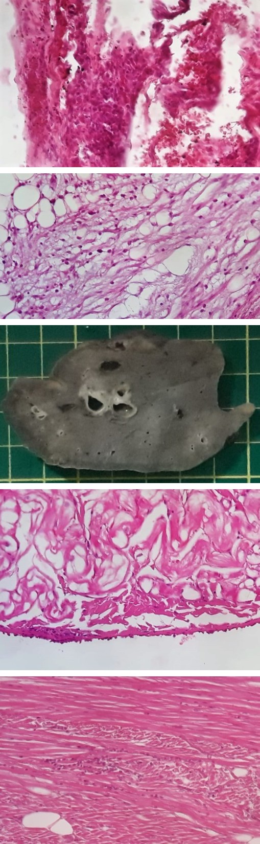
Figure 1b: Hepatized lung without evidence of alveolar aeration. Photo MDofChiriqui.
Figure 2a: Chronic myocarditis with mononuclear infiltrate.
Figure 2b: Mesothelial cell proliferation and mononuclear infiltrate in pericardium. Micrograph 200x, Hematoxylin- Eosin. Photo MDofChiriqui.
Figure 3a: Mononuclear infiltrate in epicardium.
Figure 3b: Focal endothelial proliferation in the endocardium. Micrograph 200x, Hematoxylin-Eosin. Photo MDofChiriqui.
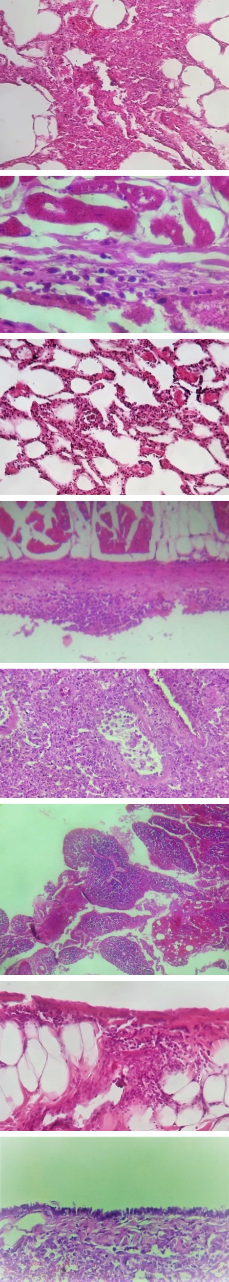
Figure 4a: Mononuclear inflammatory infiltrate in the endocardium.
Figure 4b: Lymphocytic infiltrate at the level of left atrium endocardium. Micrograph 200x, Hematoxylin-Eosin. Photo MD of Chiriqui.
Figure 5a: Nodular lymphocytic and histiocytic infiltrate in pericardial soft tissues.
Figure 5b: Mononuclear infiltrate in pericardium. Micrograph 200x, Hematoxylin-Eosin. Photo MDofChiriqui.
Figure 6a: Pulmonary emphysema, vascular congestion and mononuclear infiltrate in small caliber capillaries to pulmonary interstitium.
Figure 6b: Diffuse alveolar damage and expansion of the pulmonary interstitium with mononuclear infiltrate. Micrograph 200x, Hematoxylin-Eosin. Photo MD of Chiriqui.
Figure 7a: Atelectasis with marked thickening of the pulmonary interstitium at the expense of mononuclear inflammatory infiltrate (left) and vascular congestion with endothelial edema and presence of lymphocytes and monocytes in the vascular lumen of medium and small caliber vessels (center).
Figure 7b: Mesothelial cell proliferation in pulmonary visceral pleura. Micrograph 200x, Hematoxylin-Eosin. Photo MD of Chiriqui.
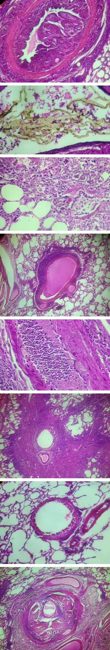
Figure 8a: Presence of poorly preserved intravascular nematodes in the pulmonary circulation.
Figure 8b: Marked expansion of the pulmonary interstitium due to inflammatory infiltrate of mononuclear cells. Micrograph 200x, Hematoxylin-Eosin. Photo MD of Chiriqui.
Figure 9a: Marked pulmonary vascular congestion with abundant lymphocytes and monocytes in the lumen.
Figure 9b: Peribronchiolar lymphocytic infiltrate, without airway or respiratory epithelium alterations.. Micrograph 200x, Hematoxylin-Eosin. Photo MD of Chiriqui.
Figure 10a: Pseudopapillary hyperplasia of the vascular endothelium or intravascular papillae in endothelium, and lymphocytic and histiocytic endothelial and interstitial nodular infiltrate adjacent to the tunica adventitia at the level of arterioles in the pulmonary parenchyma.
Figure 10b: Predominantly lymphocytic infiltrate around venules at the pulmonary parenchyma level. Micrograph 200x, Hematoxylin-Eosin. Photo MD of Chiriqui.
Figure 11a: Predominantly interstitial lymphocytic infiltrate in the lung parenchyma.
Figure 11b: Endovascular parasites in lung parenchyma. Micrograph 200x, Hematoxylin-Eosin. Photo MD of Chiriqui.
Dogs with chronic Dirofilariosis including endothelial degeneration and hypertrophy, perivascular aggregation of plasma cells, hyperplasia and hypertrophy of endothelial smooth muscle cells, and interstitial fibrosis [5]. In Turkey they reported finding adult parasites in the right ventricle and atrium with enlargement of both and found microfilariae in vessels of various organs including lungs, liver, kidneys, heart, brain and spleen. They also reported that the most significant lesion observed was obstructive pulmonary artery fibrosis and villous endarteritis [6]. Lesions in all organs caused by circulatory disorders due to severe parasitism and by the toxic action of D immitis were observed in dogs, where erosion of the vascular endothelium of the pulmonary artery and proliferation of pseudovillous-like structures were observed because of the mechanical action of the parasites in the vascular lumen [7]. No relationship was observed between the number of nematodes and the appearance of pseudopapillary hyperplasia. In our opinion, the hyperplasia in the vascular endothelium of arterioles of the pulmonary parenchyma observed is related to the time of parasitism and the continuous mechanical action of the nematode body on the vascular endothelium.
The study of some cardiopulmonary and inflammatory biomarkers in patients positive for D. immitis proved myocardial damage and heart failure, being able to evidence that the inflammatory process may contribute to disease progression [8]. The authors also conclude that the possibility of detecting whether the patient has pulmonary hypertension and endothelial damage through blood tests opens a new window of opportunity for Veterinary Medicine. More recently, chronic myocarditis, chronic pericarditis and mild right ventricular hypertrophy were confirmed in Panama in an animal from Boca Chica, district of San Lorenzo in Chiriqui [1].
D. immitis maintains a symbiotic relationship with bacteria of the genus Wolbachia sp. of the Rickettsia group [9]. The involvement of the bacterium in the pathogenesis of the disease is questioned and it is suspected that animals diagnosed with Dirofilariasis, which had a bacterial load of Wolbachia sp. high enough to be detected in lung tissues by immunohistochemistry and/or polymerase chain reaction (PCR), probably have a more severe pulmonary disease than those with low levels of bacteria detected at the same time it is thought that changes in the pulmonary artery may be produced by the bacteria [6, 10]. Clearly, the relationship of these bacteria with the nematode and with the disease must be studied. It is very difficult to define the damage caused solely by the nematode, especially when it is common to find simultaneous positive diagnoses for E. canis/E. ewingii, although the cardiorespiratory symptoms were very evident, for which reason genetic studies must be performed to confirm which bacteria are involved in the disease and more histopathological studies are necessary to discover what is the real responsibility of each one of these bacteria in the pathogenesis of the damages caused to the dog tissues.
Conclusion
From the observations presented we can conclude that the damage caused by D. immitis to the heart and lung tissues of its definitive host is evident, being the lungs the most affected. In our opinion and due to our investigations, we can speculate that the damage caused to the vascular endothelium is directly related to the time of parasitism. Further studies are needed to define the real prevalence and distribution of this disease and other vector-borne diseases in Panama, the study of its pathogenesis in tissues, its real zoonotic impact in this country, as well as seroprevalence studies in inhabitants of endemic areas, and to define the participation of Wolbachia sp. in the course of the disease, among others. The study and identification of its vectors is also of vital importance, as well as a more active participation of the government in this regard, through epidemiological surveys, education of the population and prevention tools for dogs.
Competing Interests
The authors declare that they have no competing interests.
Author´s Contributions
Both authors made substantial contributions to the manuscript. SCCh and PGCC performed the laboratory tests and necropsy. SCCh wrote the manuscript. PGCC made corrections to the text. Both authors read and approved the final manuscript.
Acknowledgement
To my patients for offering me the casuistry and experience necessary for the correct understanding and exercise of Veterinary Medicine. To our friends from Medical Diagnostics of Chiriquí (MDofChiriquí), Pathology Laboratory, Dr. Rolando Alvarado Anchisi, Pathologist, Hospital Chiriquí, Chiriquí, Panamá, for the anatomy histopathological evaluation and pictures.
References
-
Chacón SC, Candanedo PGC (2021) Canine Dirofilariasis in Panama First Cases Report Immunochromatography and Necropsy Diagnosis. J Appl Microb Res 4(1): 7-17.
-
Leary SL, Underwood W, Anthony R, Cartner S, Corey D, et al. (2013) AVMA Guidelines for the euthanasia of animals.
-
Mupanomunda M, Williams JF, Mackenzie CD, Kaiser L (1997) Dirofilaria immitis: heartworm infection alters pulmonary artery endotelial cell behavior. J Appl Physiol 82(2): 389-398.
-
Guerrero J (2005) Heartworm Pathophysiology in Dogs and Cats. World Small Animal Veterinary Association World Congress Proceedings.
-
Ranjbar Bahadori S, Mohri M, Helan JA, Jamshidi K, Kashefinejad M (2010) Clinico-Pathologic Evaluation of the Heartworm Infestation. Res Jour Parasitol 5(2): 90- 98.
-
Ceribasi AO, Simsek S (2012) Histophatology Effects of Dirofilaria immitis Microfilaria on Internal Organs of Dog Confirming by PCR Technique. Iranian J Parasitol 7(2): 103-107.
-
Pasca SA, Acatrinei D, Oprean OZ, Lazar M (2012) Vascular, hepatic and renal lesions by Dirofilaria immitis invasión in dogs. Arq Bras Med Vet Zootec 64(4): 841- 846.
-
Carretón E, Morchón R, Montoya Alonso JA (2017) Cardiopulmonary and inflamatory biomarkers in heartworm disease. Parasit Vectors 10(2): 534.
-
Rossi MID, Paiva J, Bendas A, Almeida FM, Knackfuss F, et al. (2010) Effects of doxycycline on the endosymbiont Wolbachia in Dirofilaria immitis (Leydi, 1856)–Naturally infected dogs. Vet Parasitol 174(1-2): 119-123.
-
Dingman P, Levy JK, Kramer LH, Johnson CM, Lappin MR, et al. (2010) Association of Wolbachia with heartworm disease in cats and dogs. Vet Parasitol. 170(1-2): 50-60.
- Anomalous Origin of the Left Coronary System from the Right Coronary Cusp: A Rare Coronary Anomaly in a Patient Undergoing Aortic Stenosis Workup
- Optimizing the Therapeutic Potential of Sacubitril/Valsartan: The Promise of Co-Crystal Engineering
- Association between Mortality and Topography of Peripheral Arterial Disease due to Atherosclerosis Obliterans
- Mitral Valve Replacement vs Mitral Valve Repair
- Characteristics and Evolution of Patients with Heart Failure Hospitalized in the Cardiology Department of Dalal Jamm Hospital
- Distribution of Association between Basic Knowledge of Chest Pain and Myocardial Infarction (Heart Attack) and Demographic Variables: A Survey-Based Study