Nematicidal Activity of Lactic Acid Bacteria against Root-Knot Nematodes
Root-knot nematodes are economically important obligate parasites of plant root that parasitize more than 3000 species of plant. Lactic Acid Bacteria (LAB) are well known as strong producers for wide range of organic acids that have been suggested recently to possess a remarkable lethal effect on root-knot nematodes. In the present ‘study’ 37 LAB strains were isolated from soil and agricultural wastes by using MRS medium and screened for their nematicidal activity under lab conditions. Four bacterial isolates, recorded lethal effect on second stage juveniles by more than 94%, were selected and identified by molecular method. Further, the nematicidal activity at two concentrations (i.e 50 and 10%) of the most promising ten LAB cultures was screened. The nematicidal activity of LAB was positively correlated to the concentration of their suspension cultures.2S4 (Pediococcus pentosaceus MW558270), 2S5 (Pediococcus pentosaceus MW558883), 3S1 (Pediococcus pentosaceusMW558885) and 1A3 (Pediococcus pentosaceus MW558152) isolates showed significant mortality effect by 88.42, 87.37, 81.05 and 85.96%at the low concentration (10%), respectively. In order to confirm the relationship between organic acid production by LAB and their nematicidal activity, an artificial succinic acid, lactic acid, malic acid, acetic acid, and their mixtures were tested at concentration of 1% for their ability to inhibit juvenilesvitality. The mixture of acids induced the maximum mortality effect by 100% followed by lactic acid which recorded 99.66% mortality effect. The microscopic studies and the malformation pattern of the juveniles indicating that the nematicidal activity of LAB may be derived mainly from their natural organic acids mainly lactic acid.
Introduction
Plant-parasitic nematodes are considered one of the most important pests that infect many economic crops, causing many heavy losses. The losses were estimated at more than $77 billion a year worldwide [1, 2]. The seriousness of these pests lies in the fact that the family range has a wide range [3, 4, 5]. These pests cause many problems in the cultivated fields, and the most important symptoms are wilting, stunting and yellowing, the appearance of scattered areas in the field devoid of plants [6]. Some symptoms also appear on the roots, which are ulcers, scaling of the roots and the appearance of some knots on the roots as a result of infection with the root- knot nematode [7]. All this leads to weak plants and reduced production [8]. One of the most common nematodes is the root-knot nematode, which includes more than 100 species, the most common of which are Meloidogyne incognita, M. arenaria, M. javanica and M. hapla [3, 9, 10]. Infection with root-knot nematodes causes knots on the roots, in addition, it allows the entry of other pathogens present in the soil [11]. All of this is reflected on the plant and the quality of the crop, which necessitates management against this pest. Control of RKNs (Root-knot nematodes) is difficult because they have short generation times and high reproduction rates [12]. As a result of the various harms of chemical control on human and animal health and the disruption of the ecosystem, concerns about public health and environmental safety are pushing for reducing chemical pesticides used in controlling plant diseases [13].
So scientists were forced to find safe alternative methods. Biological control strategies have been proposed as a safer and highly practical alternative to the management of phytonematodes. The term biological control is defined as the use of microorganisms for the purpose of reducing the population density of harmful pests [14]. These microorganisms can interact with ‘pathogen’ whether directly by antibiosis and competition for nutrients or indirectly by inducing plant resistance [15, 16, 17]. Biological control is one of the effective and safe ways to control many species of plant parasitic nematodeas using some species of fungi and bacteria has led to an anti-nematode effect [18, 19, 20]. Lactic acid bacteria (LAB) are heterogeneous group of Gram-positive bacteria, non-respiring non-spore-forming, cocci or rods, and their importance is associated mainly with their safe metabolic activity [21]. LABS have been reported recently as an agricultural input for improvement of crop yields and protection of agriculturally important species against certain phytopathogens [22]. LAB has long been appreciated by proponents of holistic, ecologically oriented agricultural systems. The ease of growing wild LAB without the use of laboratory facilities or microbiological knowledge has led to the widespread use of these microbes by farmers [23].
For ‘example’ LAB showed significant antagonistic potential against phytopathogenic bacteria and is widely accepted by the United States Food and Drug Administration (FDA) as healthy (GRAS), rendering it suitable for the production of bioprotective agents in fresh fruits and vegetables [24]. LAB produces a number of antimicrobial substances, including antifungal diketopiperazines, derivatives of fatty acid hydroxide, 3-phenylactate, antibacterial bacteriocins and bacteriocin-like compounds, and general antimicrobials such as organic acids, hydrogen peroxide, diacetyl and reuterine (b-OH-propionic aldehyde) pyrrolidone-5-carboxylic acid [25]. Further, Seo, et al. [26] confirmed the vital role of natural organic acids produced by LAB in controlling root-knot nematodes. The main objective of the current study was to isolate novel LAB from different environmental sources and evaluate the antagonistic and suppression effects of their culture broths and culture filtrates on juveniles (J2s) of M.incognita. under laboratory conditions.
Material and Methods
Samples Collection
Four samples (250 g) from each environmental source soil (citrus, grapes and peanuts) and agricultural wastes (beet, dill, cabbage and lettuce) were collected. Samples were transported in an ice-box and directly tested in our microbiology lab upon arrival.
Isolation and Characterization of LAB
One gram of each sample (was suspended in 9ml of sterile distilled saline (0.9% NaCl).The isolates were obtained by plating a serial of nine-fold dilutions of each sample suspension on tode Man Rogosa and Sharpe (MRS) agar plates by pour plate method [27]. Nine- fold serial dilutions then made from the mixed solution and 1ml from the last 3 dilutions (10-7, 10-8, and 10-9) pipetted and plated on (MRS) agar plates by pour plate method [27]. LAB were isolated on de Man Rogosa and Sharpe (MRS) agar (LAB-M, Bury, UK) after an incubation with MRS broth supplemented with 2% NaCl for 12 h at 37°C. Plates were incubated at 37°C for 24 h aerobically. The Gram-staining and catalase tests were performed for pure colonies with morphological characters. Only the isolates that were Gram-positive, catalase-negative, positive growth at 15, 45°C and pH (5-7-9-11) were sub- cultured in MRS broth. Glycerol stocks (60% v/v) were used for preservation of each selected isolate and stored at − 80°C.
Safety Assessment
Susceptibility to Antibiotics: The antibiotic susceptibility of the selected isolates was determined against six antibiotics, namely: ampicillin, vancomycin, kanamycin, tetracycline, erythromycin and fusidic acid. Antibiotic discs were obtained from bioMerieux (Marcy l’Etoile, France). Antibiotic susceptibility was determined semi-quantitatively using the agar overlay diffusion methods of the National Committee for Clinical Laboratory Standards [28]. The susceptibility to antibiotic was graded as either resistant or susceptible according to Kim, et al. [29]. Hemolytic Activity: In order to perform the hemolytic activity tests, the LAB strains were streaked on Columbia blood agar plates containing 5% of sheep blood. The plates were incubated for 48 h at 37°C. As suggested by Maragkoudakis, et al. [30], the strains that produced green- hued zones around the colonies or did not produce any effect on the blood plates were considered non hemolytic. The strains showed blood lysis zones around the colonies were classified as hemolytic (β-hemolysis). Molecular Identification of Selected Isolates: Genomic DNA was extracted from pure cultures of the bacterial isolates using the DNeasy mini kit (Qiagen, CA, USA) according to the manufacturer’s instructions. The primers used to amplify the 16S ribosomal RNA gene were 8F 5′AGT TGA TCC TGG CTC AG 3′ and 1492R 5′TAC CTTGTT ACG ACT T 3′, Rochelle, et al. [31]. DNA amplification was performed using 20ng of DNA template 250 mM each dNTPs [dATP, dCTP, dGTP, dTTP], 25 pmol each primer, 2.5mM MgCl2 10 μl of 5x PCR buffer and 2.5 U Taq polymerase (promega) and added up to total volume of 50 μl ddH2O. The reactions were performed in an automatic thermal cycler (GeneAmp1 PCR System 9700, Perkin-Elmer) under the following conditions: initial denaturation at 94°C for 3 min; 35 cycles of 94°C for 50 s, 55°C for 1 min, 72°C for 1.30 min; final extension at 72°C for 7 min. An aliquot of 10 μl PCR product were analyzed on 1% agarose gel. The purification of PCR product was done using high pure PCR purification kit (Qiagen). Purified PCR products were sent for sequencing at Macrogen (South Korea). The deduced sequences were submitted to the GenBank database and accession numbers were obtained. Further, identification of phylogenetic neighbours and calculation of pairwise similarity of these sequences were done by using the National Center for Biotechnology Information (NCBI) taxonomy tree building service, ClustalW and MEGA 5.2 software. Extraction of Nematode Juveniles: Eggs were extracted from galled tomato roots infected with M. incognita using 1 % sodium hypochlorite solution [32]. Eggs were collected on 500 Mesh sieve and transferred to a beaker containing tap water. Hatching of second stage juveniles was stimulated by aerating the egg suspension with oxygen in the dark for 7 days as described by Naserinasab, et al. [33]. Freshly hatched second-stage juveniles were separated from the un-hatched eggs using a modified Baermanntechnique [34] and used to evaluate the nematicidal potentiality of LAB. Juveniles (J2) Mortality Bioassay: Thirty seven bacterial isolates and their culture filtrates were screened for their antagonistic activity against second stage juveniles (J2) of M. incognita. One ml from each bacterial culture and culture filtrate was transferred separately to wells of a 96- well Microtest™ Tissue Culture Plate, thenone ml of water suspension containing 100 of juveniles was added to each well and incubated at 28-30ºC for 24 hours. The same number of juveniles received sterilized distilled water (SDW) and was served as control. Control and each treatment were replicated thrice. After 24 hours, the dead juveniles were counted in each well depending on their mobility [35]. Morphological changes and distortions noticed in treated juveniles were also studies by aid of compound microscope [36]. The mortality percentage was calculated according to Schneider- Orelli formula [37]: Mortality (%) = [(mortality percentage in treatment – mortality percentage in the negative control)/ (100 – mortality percentage in the negative control)] × 100. In vitro Nematicidal Activity of Two Concentrations of the Most Prominent LAB: Ten promising LAB isolates which showed distinct morphological and recorded significant mortality effect on J2 were selected, grown and adjusted to contain 108 CFU. Two concentrations (50 and 10%) of bacterial suspension culture soft he selected bacterial isolates were tested for their namaticidal activity at lower concentrations. In vitro Nematicidal Activity of Some Artificial Organic Acids: According to Seo, et al. [26], in order to investigate the relationship between organic acid production potentiality of LAB and their nematicidal activity, four artificial edible organic acids, which are known to be produced naturally by LAB i. e.: succinic, lactic, malic, acetic acid, and their mixtures were screened at concentration of 1% for their nematicidal activity against M. incognita. The organic acid solutions were transferred to wells of a 96-well Microtest™ Tissue. Culture Plate with three replications, into which about 100 J2 of M. incognita were placed. After 24 hours, the dead juveniles were counted in each well and morphological changes were studies by aid of compound microscope. Statistical analysis: Experiments were carried out in a completely randomized design. Data were subjected to analysis of variance (ANOVA). Means were compared by Duncan’s multiple range tests [38].
Results
Isolation and Presumptive characterization of LAB
In total, 40 LAB strains were isolated from the selected environmental sources (soil and agricultural crop wastes), based on their typical morphological appearance (small pinpointed and creamy white colonies), catalase and coagulase-negative and non-motile, coccus and rod-shaped characteristics (Table1).
Furthermore, the selected isolates were able to grow at different pH concentrations as shown at (Table 2). Most of the strains were able to survive in pH 11. The study of bacteria growth according to parameters by Bergey’s
manual 9th edition was pH, temperature and salinity. For temperature, corresponds to the favorable temperature for optimal growth of lactic acid bacteria; so all strains are mix between mesophilic and thermophilic, also, 17 soil isolate and 15 agriculture waste isolates were gram positive (Table 3).
| Samples | Elevation | Colony texture and Edge | Colony color |
|---|---|---|---|
| 1S 1 | Raised | Coarse surface and rough | Creamy |
| 1S 2 | Raised | Coarse surface and rough | Creamy |
| 1S 3 | Convex | Shiny surface and rough | Whitish |
| 1S 4 | Raised | Shiny surface and rough | Creamy |
| 1S 5 | Convex | Shiny surface and smooth | Whitish |
| 2S1 | Raised | Shiny surface and smooth | Creamy |
| 2S 2 | Raised | Shiny surface and rough | Creamy |
| 2S 3 | Raised | Coarse surface and rough | Creamy |
| 2S 4 | Convex | Shiny surface and smooth | Whitish |
| 2S 5 | Raised | Coarse surface and rough | Creamy |
| 3S1 | Raised | Coarse surface and rough | Creamy |
| 3S2 | Raised | Shiny surface and smooth | Creamy |
| 3S3 | Raised | Shiny surface and rough | Creamy |
| 3S4 | Convex | Shiny surface and smooth | Whitish |
| 3S5 | Raised | Shiny surface and smooth | Creamy |
| 4S1 | Raised | Shiny surface and rough | Creamy |
| 4S2 | Raised | Shiny surface and rough | Creamy |
| 4S3 | Raised | Coarse surface and rough | Creamy |
| 4S4 | Convex | Shiny surface and smooth | Whitish |
| 4S5 | Convex | Shiny surface and smooth | Whitish |
| 1A1 | Raised | Shiny surface and rough | Creamy |
| 1A2 | Convex | Shiny surface and smooth | Whitish |
| 1A3 | Convex | Shiny surface and smooth | Whitish |
| 1A4 | Convex | Shiny surface and smooth | Whitish |
| 1A5 | Convex | Shiny surface and smooth | Whitish |
| 2A1 | Raised | Shiny surface and rough | Creamy |
| 2A2 | Convex | Shiny surface and smooth | Whitish |
| 2A3 | Convex | Shiny surface and smooth | Whitish |
| 2A4 | Convex | Shiny surface and smooth | Whitish |
| 2A5 | Convex | Shiny surface and smooth | Whitish |
| 3A1 | Raised | Shiny surface and rough | Creamy |
| 3A2 | Convex | Shiny surface and smooth | Whitish |
| 3A3 | Convex | Shiny surface and smooth | Whitish |
| 3A4 | Convex | Shiny surface and smooth | Whitish |
| 3A5 | Convex | Shiny surface and smooth | Whitish |
| 4A1 | Raised | Shiny surface and rough | Creamy |
| 4A2 | Convex | Shiny surface and smooth | Whitish |
| 4A3 | Convex | Shiny surface and smooth | Whitish |
| 4A4 | Convex | Shiny surface and smooth | Whitish |
| 4A5 | Convex | Shiny surface and smooth | Whitish |
Table 1: Phenotypic characteristics of lactic acid bacteria (LAB) isolates.
| Isolates No. | Control pH 7/37ºC cfu/ml10-7 | pH 5 | pH 9 | pH 11 | 15ºC | 30ºC | 45ºC |
|---|---|---|---|---|---|---|---|
| 5-Oct | 7-Oct | 7-Oct | 5-Oct | 7-Oct | 5-Oct | ||
| 1S 1 | 132±0.3 | ++ | ++ | ++ | ++ | ++ | + |
| 1S 2 | 112±0.2 | +++ | +++ | +++ | +++ | +++ | ++ |
| 1S 3 | 123±0.1 | +++ | +++ | +++ | +++ | +++ | ++ |
| 1S 4 | 130±0.2 | +++ | +++ | +++ | +++ | +++ | ++ |
| 1S 5 | 43±0.3 | +++ | +++ | +++ | +++ | +++ | ++ |
| 2S1 | 132±0.2 | ++ | ++ | ++ | ++ | ++ | + |
| 2S 2 | 45±0.3 | ++ | ++ | ++ | ++ | ++ | + |
| 2S 3 | 137±0.2 | + | ++ | ++ | + | ++ | + |
| 2S 4 | 51±0.3 | +++ | +++ | +++ | +++ | +++ | ++ |
| 2S 5 | 129±0.2 | ++ | ++ | ++ | ++ | ++ | + |
| 3S1 | 58±0.3 | +++ | +++ | +++ | +++ | +++ | ++ |
| 3S2 | 67±0.3 | ++ | ++ | ++ | ++ | ++ | + |
| 3S3 | 129±0.2 | ++ | ++ | ++ | ++ | ++ | + |
| 3S4 | 188±0.3 | +++ | +++ | +++ | +++ | +++ | ++ |
| 3S5 | 120±0.3 | ++ | ++ | ++ | ++ | ++ | ++ |
| 4S1 | 138±0.2 | + | + | ++ | + | + | + |
| 4S2 | 144±2.0 | ++ | ++ | ++ | ++ | ++ | + |
| 4S3 | 98±0.3 | ++ | ++ | ++ | ++ | ++ | + |
| 4S4 | 121±0.2 | +++ | +++ | +++ | +++ | +++ | ++ |
| 4S5 | 130±0.1 | +++ | +++ | +++ | +++ | +++ | ++ |
| 1A1 | 121±0.1 | +++ | ++ | ++ | +++ | ++ | ++ |
| 1A2 | 180±0.2 | +++ | +++ | +++ | +++ | +++ | ++ |
| 1A3 | 123±0.3 | ++ | ++ | ++ | ++ | ++ | + |
| 1A4 | 170±0.2 | +++ | +++ | +++ | +++ | +++ | ++ |
| 1A5 | 121±0.2 | +++ | ++ | ++ | +++ | ++ | ++ |
| 2A1 | 145±0.3 | ++ | ++ | ++ | ++ | ++ | ++ |
| 2A2 | 137±0.2 | + | ++ | ++ | + | ++ | + |
| 2A3 | 145±0.2 | ++ | ++ | ++ | ++ | ++ | + |
| 2A4 | 137±0.1 | ++ | ++ | ++ | ++ | ++ | + |
| 2A5 | 121±0.2 | +++ | + | ++ | +++ | + | ++ |
| 3A1 | 180±0.2 | +++ | +++ | +++ | +++ | +++ | ++ |
| 3A2 | 123±0.1 | ++ | ++ | ++ | ++ | ++ | + |
| 3A3 | 170±0.2 | +++ | +++ | +++ | +++ | +++ | ++ |
| 3A4 | 180±0.2 | +++ | +++ | +++ | +++ | +++ | ++ |
| 3A5 | 123±0.3 | ++ | ++ | ++ | ++ | ++ | + |
| 4A1 | 170±0.1 | +++ | +++ | +++ | +++ | +++ | ++ |
| 4A2 | 121±0.2 | +++ | ++ | ++ | +++ | ++ | ++ |
| 4A3 | 180±0.2 | +++ | +++ | +++ | +++ | +++ | ++ |
| 4A4 | 123±0.1 | ++ | ++ | ++ | ++ | ++ | + |
| 4A5 | 170±0.1 | +++ | +++ | +++ | +++ | +++ | ++ |
| Isolate code | (VA 30µg) | (FA 10µg) | (E 15µg) | (TE 30µg) | (K 30µg) | (AMP 10µg) | |
| 1S2 | R | S | R | S | I | R | |
| 2S4 | R | I | R | S | R | R | |
| 2S5 | R | I | R | S | R | I | |
| 3S1 | R | I | R | R | R | I | |
| 4S4 | R | I | R | S | R | I | |
| 1A2 | R | I | R | S | R | S | |
| 1A3 | R | R | R | S | R | I | |
| 2A5 | R | I | R | S | I | S | |
| 3A2 | R | R | R | I | R | S | |
| 4A5 | R | I | R | S | R | I |
Table 2: Growth of lactic acid bacteria (LAB) at different pH concentrations and different temperatures. Isolates.
VA: Vancomycin; FA: Fusidic Acid; E.: Erythromycine; TE: Tetracycline; K: Kanamycin; and; Amp: Ampicillin; (Inhibition zone with mm, resistant (R), intermediate (I), and susceptible (S), S: ≥ 21 mm, R:≤ 15 mm; I: 16-20 mm. Table 3: Antibiotic susceptibility profiles of the selected LAB isolates.
Nematicidal Activity of LAB Isolated from Soil and Agricultural Waste In Vitro
Four soil samples were used to isolate LAB. According to the morphological characters and growth pattern, seventeen bacterial isolates were selected for the current assay. It was noticed that all different isolations gave suppressive effect on juveniles activity. The maximum percentage of juveniles mortality was recorded with bacterial culture of isolate 2S4,
2S5, 3S1with percentage of 100% mortality followed by 4S4 (98.63%) then 1S2 (95.9%). However the bacterial culture of isolate 1S1 gave the lowest percentage of juveniles mortality (72.01%). As for culture filtrate, isolates 2S4, 2S5 and 3S1 also showed100 %of juveniles mortality followed by 4S4, 2S1, and 1S2 with percentages of (97.95, 95.22 and 81.57%), respectively (Table 4). In most isolates, we can conduct that bacterial culture showed best result than culture filtrate.
| Isolate code | Juveniles Mortality % | |||
|---|---|---|---|---|
| Treatment type | ||||
| Bacterial Culture | Culture Filtrate | |||
| No. of dead juveniles | Mortality % | No. of dead juveniles | Mortality % | |
| 1S1 | 72.66i | 72.01 | 65.33ef | 64.5 |
| 1S2 | 96.00abc | 95.9 | 82.00bc | 81.57 |
| 1S3 | 87.33efgh | 87.03 | 78.66cd | 78.15 |
| 1S4 | 90.00defg | 89.76 | 70.66ef | 69.96 |
| 2S1 | 95.33abcd | 95.22 | 95.33ab | 95.22 |
| 2S2 | 93.33bcd | 93.17 | 82.00bc | 81.57 |
| 2S3 | 91.33cdef | 91.1 | 82.66bc | 82.25 |
| 2S4 | 100.00a | 100 | 100.00a | 100 |
| 2S5 | 100.00a | 100 | 100.00a | 100 |
| 3S1 | 100.00a | 100 | 100.00a | 100 |
| 3S4 | 82.66i | 82.25 | 78.33cd | 77.81 |
| 3S5 | 85.33gh | 84.98 | 84.00bc | 83.62 |
| 4S1 | 86.00fgh | 85.67 | 81.00bcd | 80.55 |
| 4S2 | 92.00cde | 91.81 | 84.66bc | 84.29 |
| 4S3 | 91.00cdef | 90.78 | 82.00bc | 81.57 |
| 4S4 | 98.66ab | 98.63 | 98.00a | 97.95 |
| 4S5 | 82.66h | 82.25 | 74.00de | 73.38 |
| Control | 2.33j | -- | 2.33f | -- |
| L.S.D at 0.05 | 5.01 | - | 6.54 | - |
| Treatment type | ||||
| Bacterial Culture | Culture Filtrate | |||
| No. of dead juveniles | Mortality % | No. of dead juveniles | Mortality % | |
| 1A1 | 100.00a | 100 | 98.00a | 97.95 |
| 1A2 | 100.00a | 100 | 98.66a | 98.63 |
| 1A3 | 100.00a | 100 | 100.00a | 100 |
| 1A4 | 100.00a | 100 | 99.33a | 99.31 |
| 1A5 | 100.00a | 100 | 92.66b | 92.48 |
| 2A1 | 99.33a | 99.31 | 98.66a | 98.63 |
| 2A2 | 100.00a | 100 | 99.33a | 99.31 |
| 2A3 | 99.33a | 99.31 | 99.33a | 99.31 |
| 2A4 | 100.00a | 100 | 100.00a | 100 |
| 2A5 | 100.00a | 100 | 100.00a | 100 |
| 3A1 | 100.00a | 100 | 99.33a | 99.31 |
| 3A2 | 100.00a | 100 | 100.00a | 100 |
| 3A3 | 99.33a | 99.31 | 98.00a | 97.95 |
| 4A2 | 100.00a | 100 | 100.00a | 100 |
| 4A5 | 100.00a | 100 | 96.66a | 96.58 |
| Control | 2.30b | -- | 2.33c | -- |
| L.S.D at 0.05 | 1.46 | - | 3.08 | - |
Table 3: Nematicidalactivity of the tested lactic acid bacteria (LAB) isolated from soil against root-knot nematode (Meloidogyne
Means in each column followed by the same letter(s) did not differ at P ≤ 0.05 according to Duncan`s multiple range test. Table 4: Nematicidalactivity of the tested lactic acid bacteria (LAB) isolated from soil against root-knot nematode (Meloidogyne incognita) under laboratory conditions.
Data in (Table 5) illustrate the nematicidal activity of fifteen bacterial isolates from agricultural waste on juveniles mortality of M.incognita. It was obvious that all different bacterial culture isolates showed approximately 100 % of nematode mortality. Similar trend was noticed with culture filtrate except the isolate 1A5. Five bacterial isolates encoded, 1A2, 1A3, 2A5, 3A2 and 4A5 recorded the maximum mortality effect on J2of M.incognita. Were selected for further study.
Means in each column followed by the same letter(s) did not differ at P ≤ 0.05 according to Duncan`s multiple range test. Table 5: Nematicidal activity of the tested lactic acid bacteria (LAB) isolated from agricultural waste against root-knot nematode (Meloidogyne spp.) under laboratory conditions.
| Isolate code | Juveniles Mortality % | |||
|---|---|---|---|---|
| Concentration (%) of bacterial culture | ||||
| 50% | 10% | |||
| No. of dead juveniles | Mortality % | No. of dead juveniles | Mortality % | |
| 1S2 | 91.66b | 91.22 | 67.66b | 65.96 |
| 2S4 | 100.00a | 100 | 89.00a | 88.42 |
| 2S5 | 100.00a | 100 | 88.00a | 87.37 |
| 3S1 | 100.00a | 100 | 82.00a | 81.05 |
| 4S4 | 86.33b | 85.61 | 61.00bc | 58.95 |
| 1A2 | 87.00b | 86.32 | 64.66b | 62.8 |
| 1A3 | 100.00a | 100 | 86.66a | 85.96 |
| 2A5 | 87.00b | 86.32 | 67.00b | 65.26 |
| 3A2 | 87.66b | 87.01 | 46.33d | 43.51 |
| 4A5 | 86.66b | 85.96 | 54.66cd | 52.27 |
| Control | 5.00c | -- | 5.00e | -- |
| L.S.D at 0.05 | 4.79 | - | 8.83 | - |
Table 4: Comparative study of two bacterial culture concentrations of the most promising lactic acid bacteria (LAB) against root-
Means in each column followed by the same letter(s) did not differ at P ≤ 0.05 according to Duncan`s multiple range test. Table 6: Comparative study of two bacterial culture concentrations of the most promising lactic acid bacteria (LAB) against root- knot juveniles vitality.
Through the results shown in (Tables 4 & 5), isolates (1S2, 2S4, 2S5,3S1 and 4S4) from soil and isolates (1A2, 1A3,2A5, 3A2 and 4A5) showed the best results in increasing the mortality rate of nematodes, so we compared two bacterial culture concentrations (10 and 50%) of these isolates on survival of nematodes under laboratory conditions (Table 6). Either at a concentration of 50 or 10%, it was found that the best results were obtained by using isolates 2S4, 2S5, 3S1 and 1A3which gave a mortality percentage 100% at a concentration of 50% and >80% at concentration of 10%.
Nematicidal Activity of Different Artificial Organic Acids
Through some previous studies, some scientists have shown that lactic acid bacteria secrete some organic acids such as Succinic, lactic, Malic and Acetic acid. Therefore, in this study, we evaluated the effect of these acids artificially either individually or in a mixture on the death of nematode juveniles to examine the potential of organic acids as main products of LAB as nematicidal compounds. In general all acids effect significantly on survival of juveniles. Acids mixture gave (100 %) of nematode mortality followed by lactic acid (99.66%) and acetic acid (98.33%) (Table 7).
| Artificial acids (1%) | No. of dead juveniles | Mortality % |
|---|---|---|
| Succinic acid | 65.33c | 65.33 |
| Lactic acid | 99.66a | 99.66 |
| Maleic acid | 92.66b | 92.66 |
| Acetic acid | 98.33ab | 98.33 |
| Acids mixture | 100.0a | 100 |
| Control | 0.0d | 0 |
| L.S.D | 5.88 | ---- |
Table 5: ** Nematicidal activity of succinic, lactic, malic, and acetic acid and their mixture on the mortality of juveniles of M
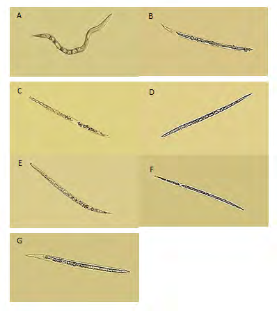
Means in each column followed by the same letter(s) did not differ at P ≤ 0.05 according to Duncan`s multiple range test. Table 7: Nematicidal activity of succinic, lactic, malic, and acetic acid and their mixture on the mortality of juveniles of Meloidogyneincognita under laboratory conditions Treatment with artificial acids at concentration 1% and their mixture look like that juveniles of M.incognita were shown to be dead with rigid and straight body. Similar trend was noticed with treatment by lactic acid bacteria (LAB) isolates. However the juveniles in the untreated control were vital and still moving. The intestinal tract of juveniles treated whether with LAB isolates or their artificial acids showed pathological changes such as vamp and the structures were clearly visible, after those destroying internal organs which are followed by creating cavities comparing to the control (Figure 1).
Safety Assessment of LAB
Susceptibility to Antibiotics: Antibiotic susceptibility profiles of the ten tested isolates were shown at Table 3. There was resistance of 2 antibiotics used (Vancomycin 30µg and Erythromycin 15 µg) for all isolates but eight isolates recorded high sensitivity for Tetracycline 30 µg. Also the strains showed variance effects against Ampicillin, Kanamycin and Fusidic acid respectively. The results of strains showed safe properties for human use and susceptible to all of the tested antibiotics showing clear inhibition zones of diameters ranging between 9 and 30 mm.
Hemolytic Activity: There was no change in the color surrounding colonies of the selected isolates when tested on bloodagar. The absence of β-hemolysis in any of the tested strains is an indication of their safe and GRAS properties. Historically, LAB associated with food has been generally recognized as safe, GRAS [39].
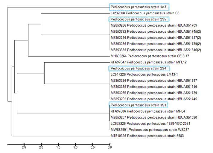
Molecular Identification of the Most Potent Strains and Phylogenetic Tree: PCR products of approximately 1500 bp amplified with the 8F/ 1498R primers and corresponding to the 16SrDNA region were obtained from all bacterial isolates. Sequence alignments of the four bacterial isolates showed identities over 99% with Pediococcus pentosaceus strains. Alignment analysis using 1500 bp of each identified fragment sequence was displayed against the corresponding top five homologous sequences that were determined from the NCBI database. Bacterial isolates sequences have been submitted to GenBank database and accession numbers were obtained (2S4 (MW558270), 2S5 (MW558883), 3S1 (MW558885) and 1A3 (MW558152). Mega-alignment and the phylogenetic tree were generated (Figure 2) among the four nucleotide sequences that were indicated as Pediococcus pentosaceus.
Discussion
Lactic acid bacteria are considered one of the most important types of bacteria that improve the nutritional value of many foods, as well as resist some intestinal diseases for humans [40, 41]. It is also considered one of the bacterial types used in the manufacture of many medicines because of its anti-effect to some fungi and pathogenic bacteria. For this it became possible to use it against some pathogens of the plant [42, 22]. There is a basic group consisting of four genera, which are Lactobacillus, Streptococcus, Pediococcus and Leuconostoc. The modern methods of examination and classification have proven the existence of other genera which are Tetragenococcus, Aerococcus, Carnobacterium, Alloiococcus, Dolosigranulum, Enterococcus, Lactococcus, Vagococcus, Globicatella, Oenococcus, and Weissella [43]. Bourdichon, et al. [44] explained that this group of bacteria is important due to its metabolic activity during growth, as it uses available sugar to produce organic acids, and its safe presence in many foods has led to it being considered suitable as GRAS (Generally Recognized as Safe) for human use. Kim, et al. [45] reported that LAB isolates induce such morphological modifications in several pathogenic microbes.
These morphological modifications may be due to deviations in membrane lipogenesis, altered membrane fluidity and/or membrane integrity resulting in cell wall lysis and loss of dense intracellular substance [46]. Bajpai, et al. [47] studied the antibacterial potential of lactic acid bacteria Pediococcus pentosaceus against Staphylococcus aureus and Escherichia coli. The cell free supernatant of P. pentosaceus revealed its antibacterial action against S. aureus and E. coli on membrane integrity as established by increased potassium ion release, reduced absorption at 260-nm, reduction in cell viability, and increased relative electrical conductivity. GC- mass analysis for this bacteria identified major compounds included organic acid, amino acid and fatty acid. Anas, et al. [48] showed that the antimicrobial activities may be related to the various compounds, such as H2O2, organic acids and bacteriocin-like substances. From this standpoint, the lactic acid bacteria drew attention to their use in the agricultural field as alternative method to chemical pesticides to reduce the harmful effect caused by them, in addition to the percentage of pesticide residues in the fruits.
In this study, some types of bacteria producing lactic acid were isolated from soil (17 isolates) and agricultural waste (15isolates) and their effect against root-knot nematodes were studied under laboratory conditions. Most of isolates were able to survive in pH 11. This result is disagreed with Trias, et al. [22] who reported that lactic acid bacteria can growth well and survives at low pH such as fruit where it is a good source for sugar and nutrients needed for bacterial growth. In our study, we noticed that the nematicidal activity of lactic acid bacteria isolates showed remarkable death at root-knot nematode juveniles. The findings of Borgo, et al. [49] reported that the ability of Enterococcus italicus and Lactococcus garvieae to kill the nematode Caenorhabditis elegans and they suggested that this was due to the production of hydrogen peroxide (H2O2) by these bacteria. In another study on the same species of nematode Caenorhabditis elegans by Zanni, et al. [50], Leuconostoc lactis, Lactobacillus fermentum and L. delbrueckii, were found as the main species and shown to display different colonization capacity at worm gut. Moreover, they noticed that lactic acid bacteria supplementation decreased nematode lifespan. Also Fasseas, et al. [51] reported that Lactobacillus reuteri and Pediococcusacidilactici reduced the lifespan of wild- type and short-lived daf-16 worms of C. elegans. Gustin, et al. [52] conducted that some bacteria produced H2O2 could kill nematodes due to its toxicity. Ralmi, et al. [53] studied the effect of H2O2 against root-knot nematode infected Ficus deltoidea and they reported that no root gall formation.
There is little information on the use of lactic acid as a biological control against plant parasitic nematode. Takei, et al. [54] explained the possibility of using some capsules that contain lactic acid bacteria against root-knot nematodes. Seo, et al. [55] also showed that the use of lactic acid bacteria (Lactobacillus farraginis) with concomitant with Bacillus
cereus and B. thuringiensis can reduce the number of egg masses on the roots of Cucumis melo without phytotoxicity. The impact of four artificial organic acids (succinic, lactic, acetic and maleic acid) and their mixture was determined at 1% concentration on the survival of root-knot nematode juveniles. All treatments showed significant influence on juveniles mortality. Treatment with organic acids displayed the appearance of some gaps in the bodies of dead juveniles. This result is supported by the findings of Seo and Kim [56] who reported that incidence 100% mortality to juveniles at all concentrations of acetic Acid and mixture of acetic and lactic acid and only at 1.0% and 0.5% of Lactic Acid. Also Yeon, et al. [26] reported that Maleic acid showed 100% second stage juveniles mortality. Seo, et al. [26] conducted that the nematicidal activity of the culture filtrate of L. brevis WiKim0069 referred to organic acids such as acetic acid, lactic acid, malic acid, and succinic acid produced by bacteria [57, 58, 59, 60, 61, 62].
Conclusion
Based on this discussion, it can be summarized that using lactic acid bacteria can be used as ecofriendly method to control root-knot nematode. Nevertheless, greenhouse and field experiments are needed on a large scale to confirm the results. Besides, isolation and identification of secondary metabolites compounds from these bacteria are required for further studies under laboratory and greenhouse conditions to understand their mode of action and nematicidal activities and to approve their effectiveness under field conditions.
References
-
Ralmi NHAA, Khandaker MM, Mat N (2016) Occurrence and control of root-knot nematode in crops: a review. Aust J Crop Sci 10(12): 1649-1654.
-
Yadav U (2017) Recent trends in nematode management practices: the Indian context. Int Res J Eng Technol 4(12): 482-489.
-
Jones JT, Haegeman A, Danchin EG, Gaur HS, Helder J, et al. (2013) Top 10 plant-parasitic nematodes in molecular plant pathology. Mol Plant Pathol 14(9): 946-961.
-
Ramadan M (2015) Checklist of Host Range of Root-Knot Nematodes (Meloidogyne species and races) in Jordan. Jordan Journal of Agricultural Sciences 11(3) Article ID 761-769.
-
Bernard GC, Egnin M, Bonsi C (2017) The Impact of Plant- Parasitic Nematodes on Agriculture and Methods of Control. In: Shah MM, Mahamood M (Eds.), Nematology - Concepts, Diagnosis and Control. IntechOpen.
-
Mitiku M (2018) Plant-parasitic nematodes and their management: A review. Agric Res Technol open Access J 16(2): 30-38.
-
Ziems TAJ (2016) Diseases Caused by Parasitic Nematodes. Papers in Plant Pathology, pp: 525.
-
Rius JEP, Escobar C, Cabrera J, Vovlas A, Castillo P (2017) Anatomical alterations in plant tissues induced by plant- parasitic nematodes. Front Plant Sci 8: 1987.
-
Elling AA (2013) Major emerging problems with minor Meloidogyne species. Phytopathology 103(11): 1092- 1102.
-
Molinari S (2009) Antioxidant enzymes in (a) virulent populations of root-knot nematodes. Nematology 11: 689-697.
-
Molinari S, Fanelli E, Leonetti P (2014) Expression of tomato salicylic acid (SA)-responsive pathogenesis- related genes in Mi-1-mediated and SA-induced resistance to rootknot nematodes. Mol Plant Pathol 15(3): 255-264.
-
Moens M, Perry RN, Starr JL (2009) Meloidogynespecies–a diverse group of novel and important plant parasites. Root-Knot Nematodes, CABI Publishing, pp: 1-17.
-
Mantelin S, Bellafiore S, Kyndt T (2017) Meloidogyne graminicola: a major threat to rice agriculture. Mol Plant Pathol 18(1): 3-15.
-
Poveda J, Urias PA, Escobar C (2020) Biological control of plant-parasitic nematodes by filamentous fungi inducers of resistance: Trichoderma, mycorrhizal and endophytic fungi. Front Microb 11: 992.
-
Pal KK, Gardener BMS (2006) Biological control of plant pathogens. Plant Health Instr 2: 1-25.
-
Stirling GR (2018) Biological control of plant-parasitic nematodes in Diseases of Nematodes. In: Poinar GO, Jansson HB, (Eds.). CRC Press, Boca Raton, Florida, pp: 103-150.
-
Xiang N, Lawrence KS, Donald PA (2018) Biological control potential of plant growth-promoting rhizobacteria suppression of Meloidogyne incognita on cotton and Heteroderaglycines on soybean: a review. J Phytopathol 166(7-8): 449-458.
-
Ibrahim DSS, Elderiny MM, Ansari RA, Rizvi R, Sumbul A, et al. (2020) Role of Trichoderma spp. in the Management of Plant-Parasitic Nematodes Infesting Important Crops. In: Ansari R (Ed.), Management of Phytonematodes: Recent Advances and Future Challenges. Springer, Singapore.
-
Soliman MS, El-Deriny MM, Ibrahim DSS, Zakaria H, Ahmed Y (2021) Suppression of root-knot nematode Meloidogyne incognita on tomato plants using the nematode trapping fungus Arthrobotrys oligospora Fresenius. J Appl Microbiol 131(5): 2402-2415.
-
Ibrahim DS, Metwaly HA, El-Sagheer AM (2021) Synergistic Effect of Bioagents and Antioxidants against Root-Knot Nematode, Meloidogyne incognita on Sunflower. Egypt J Agronematol 20(2): 140-158.
-
Bintsis T (2018) Lactic acid bacteria: their applications in foods. J Bacteriol Mycol Open Access 6(2): 89-94.
-
Trias R, Bañeras L, Montesinos E, Badosa E (2008) Lactic acid bacteria from fresh fruit and vegetables as biocontrol agents of phytopathogenic bacteria and fungi. Int Microbiol 11(4): 231-236.
-
Katz SE (2008) Wild Fermentation: the Flavor, Nutrition, and Craft of Live-culture Foods. Chelsea Green Publishing.
-
Li D, Ni K, Pang H, Wang Y, Cai Y, et al. (2015) Identification and antimicrobial activity detection of Lactic Acid Bacteria isolated from corn stover silage. Asian Australas J Anim Sci 28(5): 620-631.
-
Stoyanova LG, Ustyugova EA, Netrusov AI (2012) Antibacterial metabolites of lactic acid bacteria: their diversity and properties. Prikl Biokhim Mikrobiol 48(3): 259-275.
-
Seo HJ, Park AR, Kim S, Yeon J, Yu NH, et al. (2019) Biological Control of Root-Knot Nematodes by Organic Acid-Producing Lactobacillus brevis WiKim0069 Isolated from Kimchi. PlantPathol J 35(6): 662-673.
-
Awan JA, Rahman SU (2005) Microbiology Manual. Unitech Communications, Faisalabad, Pakistan, pp: 49- 51.
-
NCCLS (2002) Analysis and presentation of cumulative susceptibility test data: Approved Guideline. NCCLS document M39-A.
-
Kim HJ, Shin HS, Ha WK, Yang HJ, Lee SW (2006) Characterization of lactic bacterial strains isolated from raw milk. Asian-Aust J Anim Sci 19(1): 131-136.
-
Maragkoudakis PA, Konstantinos CM, Psyrras D, Cremonese S, Fischer J, et al. (2009) Functional properties of novel protective lactic acid bacteria and application in raw chicken meat against Listeria monocytogenes and Salmonella enteriditis. Int J Food Microbiol 130(3): 219- 226.
-
Rochelle PA, Will JAK, Fry JC, Jenkins GJS, Parkes RJ, et al. (1995) Extraction and amplification of 16S rRNA genes from deep marine sediments and seawater to assess bacterial community diversity. In: Trevors JT, et al. (Eds.), Nucleic Acids in the Environment. Springer- Verlag Berlin, Heidelberg, pp: 219-239.
-
Hussey RS, Barker KR (1973) A comparison of methods of collecting inocula of Meloidogyne species, including a new technique. Plant Dis Report 57: 1025-1028.
-
Naserinasab F, Sahebani N, Etebarian HR (2011) Biological Control of Meloidogyne javanica by Trichoderma harzianum BI and Salicylic Acid on Tomato. African Journal of Food Science 5(3): 276-280.
-
Viglierchio DR, Schmitt RV (1983) On the methodology of nematode extraction from field samples: Baermann funnel modifications. J Nematol 15(3): 438-444.
-
Cayrol JC, Djian C, Pijarowski L (1989) Study of the nematicidal properties of the culture filtrate of the nematophagous fungus Paecilomyces lilacinus. Revue Nématol 12(4): 331-336.
-
Siddiqui IA, Shaukat SS (2004) Trichodermaharzianum enhances the production of nematicidal compounds in vitro and improves biocontrol of Meloidogynejavanica by Pseudomonas fluorescens in tomato. Letters in Applied Microbiology 38(2): 169-175.
-
Schneider P, Orelli O (1947) Entomologischespraktikum [Entomological internship]. Verlag HR Sauerländer Co, Aarau, Switzerland, pp: 237.
-
Duncan DB (1955) Multiple range and multiple, F-test. Biometrics 11(1): 1- 42.
-
Adams MR, Marteau P (1995) On the safety of lactic acid bacteria from food. Int J Food Microbiol 27(2-3): 263- 264.
-
Gilliland SE (1990) Health and nutritional benefits from lactic acid bacteria. FEMS Microbiol Rev 7(1-2): 175-188.
-
Lindgren SE, Dobrogosz WJ (1990) Antagonistic activities of lactic acid bacteria in food and feed fermentations. FEMS Microbiol Rev 7(1-2): 149-163.
-
Sitton JW, Patterson ME (1992) Effect of high-carbon dioxide and low-oxygen controlled atmospheres on postharvest decays of apples. Plant Dis 76: 992-995.
-
Khalid K (2011) An overview of lactic acid bacteria. Intern J Bioscien 1(3): 1-13.
-
Bourdichon F, Berger B, Casaregola S (2012) A Safety assessment of microbial food cultures with history of use in fermented dairy products. Bullet IDF 455: 2-12.
-
Kim M, Lee SJ, Seul KJ, Park YM, Ghim SY (2009) Characterization of antimicrobial substance produced by Lactobacillus paraplantarum KNUC25 isolated from Kimchi. Korean J Microbiol Biotechnol 37: 24-32.
-
Sikkema J, Bont J De, Poolman B (1994) Interactions of cyclic hydrocarbons with biological membranes. J Biol Chem 269(11): 8022-8028.
-
Bajpai VK, Han JH, Rather IA, Park C, Lim J, et al. (2016) Characterization and Antibacterial Potential of Lactic Acid Bacterium Pediococcus pentosaceus 4I1 Isolated from Freshwater Fish Zaccokoreanus. Front Microbiol 7: 2037.
-
Anas M, Eddine HJ, Mebrouk K (2008) Antimicrobial activity of Lactobacillus species isolated from Algerian raw goat’s milk against Staphylococcus aureus. World J Dairy Food Sci 3(2): 39-49.
-
Borgo F, Ballestriero F, Ferrario C, Fortina MG (2014) Hydrogen peroxide-mediated killing of Caenorhabditis elegans by Enterococcus italicus and Lactococcus garvieae isolated from food. Ann Microbiol 65: 833-839.
-
Zanni E, Laudenzi C, Schifano E, Palleschi C, Perozzi G, et al. (2015) Impact of a complex food microbiota on energy metabolism in the model organism Caenorhabditis elegans. Biomed Res Int 2015: 1-12.
-
Fasseas MK, Fasseas C, Mountzouris KC, Syntichaki P (2013) Effects of Lactobacillus salivarius, Lactobacillus reuteri, and Pediococcusacidilactici on the nematode Caenorhabditis elegans include possible antitumor activity. Appl Microbiol Biotechnol 97(5): 2109-2118.
-
Gustin EJ, McDonnell GE, Mullen G, Gordon BE (2002) The Efficacy of Vapour Phase Hydrogen Peroxide against Nematode Infestation of the Caenohabditis Elegans Model. Proceedings of the 53rd AALAS National Meeting, San Antonio, TX, USA, pp: 27-31.
-
Ralmi NHAA, Khandaker MM, Mohd KS, Majrashi A, Fallatah AM, et al. (2021) Influence of Rhizopheric H2O2 on Growth, Mineral Absorption, Root Anatomy and Nematode Infection of Ficus deltoidea. Agronomy 11: 704.
-
Takei TM, Yoshida Y, Hatate K, Shiomori, Kiyoyama S (2008) Lactic acid bacteria-enclosing poly(ε- caprolactone) microcapsules as soil bioamendment. J Biosci Bioeng 106(3): 268-272.
-
Seo BJ, Kumar VJR, Ahmad RI, Kim BC, Park W, et al. (2012) Bacterial Mixture from Greenhouse Soil as a Biocontrol Agent Against Root-Knot Nematode, Meloidogyne incognita, on Oriental Melon. J Microbiol Biotechnol 22(1): 114-117.
-
Seo Y, Kim YH (2014) Control of Meloidogyne incognita Using Mixtures of Organic Acids. Plant Pathol J 30(4): 450-455.
-
Ayad EHE, Nashat S, El-Sadek N, Metwaly H, El-Soda M (2004) Selection of wild lactic acid bacteria isolated from traditional Egyptian dairy products according to production and technological criteria. Food Microbiol 21(6): 715-725.
-
Sangita B, Apoorva S, Manisha M, Sharma SK (2013) Isolation and Characterization of Lactic Acid Bacteria from Fermented Foods. VEGETOS 26(2): 325-330.
-
Courvalin P (2006) Antibiotic resistance: The pros and cons of robiotics. Dig Liver Dis 38(2): S261-S265.
-
EFSA (2005) Opinion of the Scientific Panel on Additives and Products or Substances used in Animal Feed on the updating of the criteria used in the assessment of bacteria for resistance to antibiotics of human or veterinary. EFSA J 223: 1-12.
-
Lutz MP, Michel V, Martinez C, Camps C (2012) Lactic acid bacteria as biocontrol agents of soil-borne pathogens. Biological Control of Fungal and Bacterial Plant Pathogens IOBC-WPRS Bull, 78: 285-288.
-
Sharma R, Sanodiya BS, Thakur GS, Jaiswal P, Pal S, et al. (2013) Characterization of Lactic Acid Bacteria from Raw Milk Samples of Cow, Goat, Sheep, Camel, and Buffalo with Special Elucidation to Lactic Acid Production. British Microbiology Research Journal 3(4): 743-752.
- Antifungal Activity of New Acetophenone Derivatives
- Interconnected Microbiomes Human Health Within an Environmental Framework
- Silkworm-Based Vaccine Production for H5N1: A One Health Approach to Pandemic Preparedness
- Microbial Diversity and Lipolytic Activity of Bacteria and Fungi from Oil-Contaminated Sites in Makurdi Metroplois
- Antibiotic Resistance Profile of Bacteria Isolated at the Central Laboratory of the National Hospital Center of Nouakchott
- Epidemiology and Sensitivity to Antibiotics of Germs Isolated from Blood Cultures in the Laboratory of the National Hospital Center of Nouakchott-Mauritania