Studies on Production and In Vitro Antibacterial Potentials of Violacein from Chromobacterium Violaceum Isolated from Otamiri River in Owerri, Imo State
Chromobacterium violaceum was isolated from Otammiri River in Owerri, Imo State. The organism was isolated using nutrient agar and identified based on cultural and biochemical characteristics. The isolate produced violacein pigment on nutrient broth and the violacein produced was quantified and extracted with ethanol. The effects of different temperatures (4°C, 25°C, 30°C, 37°C, 40°C, 52°C), pH (4.0, 4.5, 5.0, 5.5, 6.0, 6.5, 7.0, 7.5, 8.0, 8.5, 9.0), incubation period (12hr, 24hr, 36hr, 48hr, 60hr, 72hr, 84hr, 96hr), carbon sources (dextrose, fructose, glucose, maltose, lactose, sucrose, starch), nitrogen sources (ammonium nitrate, ammonium chloride, yeast extract, beef extract, peptone) on violacein production by C violaceum isolated were examined. Antibacterial activity of gram negative and gram positive organisms was done and the MIC of violacein was determined. The amount of violacein produced by C violaceum was 34.98mg/l. The production of violacein was increased with increase in temperature reaching its maximum value at 37°C. The optimum pH and incubation time for the production of violacein from the isolate were pH 6.5 and 72hr. Glucose and yeast extracts were most efficient carbon and nitrogen sources for the violacein production from C violaceum isolated. The violacein was highly sensitive to S aureus and Escherichia coli with inhibition zone of 15mm resistant to Klebsiella aerogenes and Pseudomonas aeruginosa with no inhibition zone. This study revealed that violacein has antibacterial activity against Gram positive and Gram negative organisms and provides knowledge of factors influencing the production of violacein. It also revealed the MIC of violacein and the MBC against different bacteria. The findings from this work could be of interest and suggest the need for further investigations in terms of toxicological studies and purification of active components with a view to using violacein in novel drug development.
Introduction
Chromobacterium violaceum is a saprophyte found in soil and water habitat of tropical and subtropical areas of several continents, normally considered non-pathogenic to humans but they can be opportunistic pathogen with great virulence. C. violaceum is a species of bacteria that are large, motile, gram – negative, beta – proteobacterium, non - spore formers. They are 0.6 – 0.9mm x 1.5 – 3.0mm in size and exhibit bipolar staining. It is positive for catalase and oxidase reaction. Motility of C. violaceum is achieved by means of a single polar flagellum and up to four antigenically and structurally distinct lateral flagellae. They are facultative anaerobes with agrowth temperature range of 15°C – 40°C. Optimal growth is achieved at 30°C – 35°C, characteristically they produce violet colonies on nutrient agar and usually on MacConkey agar. The violet colour comes from pigment violacein. Colonies are low, convex, violet, smooth and not. A non - diffusible pigment is produced by this Gram – negative organism, called violacein [1]. This violacein has various antagonistic properties and it is bactericide [2], a trypanocide [3], and tumoricides [4] and in addition, it has anti-viral activities [5]. Violacein is a purple pigment produced by free environmental bacterial species, especially by Chromobacterium violaceum. This compound has several biological activities, including antitumoral and apoptosis – inducing properties in cancer cells, antioxidant, leishmanicidal, trypanocidal, antifungal, weak antiviral [6] and antimalarial effect [7]. As an antibacterial agent, violacein showed activity against Mycobacterium tuberculosis [8].
One of the main issues regarding the therapeutic uses of violacein is its toxicity invivo, as this compound is cytotoxic to several tumour models, such as leukemic cells, colorectal tumors, human intestinal epithelial cells and ascites tumor models [6]. Very recently, Bromberg, et al. [9] have shown that violacein causes oxidative stress and cell death by apoptosis in Ehrlich ascites tumor cells. Violacein has strong antibacterial effects making it a promising candidate as an antibiotic. Moreover, when administered in combination with other antibiotics, the impact is more effective in fighting bacteria than the use of antibiotics alone [10]. This is of particular interest in light of recent antibiotic – resistant strains of pathogenic bacteria, such as MRSA. Also, the antiprotozoan properties of violacein could be exploited to treat disease in humans, such as in malaria and leishmaniasis [11]. The most studied clinical use of violacein, however, is its being a potential cancer therapeutic. Violacein has been tested against various cancer cell lines where it has shown cytotoxicity at IC50 values that mostly range in the submicromolar concentrations. The effects of violacein were also shown to be specific for the cancer cell line tested as two colorectal cancer cell lines, Caco – 2 and HT – 29, were differentially susceptible to violacein [12]. Since violacein is cytotoxic towards non cancer cells as well [13]. The critical factor for its clinical use against cancer is that it is more toxic to cancer cells than to normal cells. One study demonstrated that violacein induced apoptosis in HL60 cells (IC50 = 700nM), a cancer cell line used as a model to study myeloid leukemia. However, normal lymohocytes were unaffected at the concentrations tested, further asserting violacein’s use as a putative cancer therapeutic [14]. Even though violacein is a promising agent as an antibiotic and a treatment for cancer, the biological mechanisms behind these actions remain elusive. Before we can stamp violacein as a natural therapeutic for numerous kinds of disease, it is imperative to understand its mechanisms of action at the cellular and molecular levels. Conclusions on violacein’s mechanism of action are clearly scanty at this point [14]. The issue of drug resistance has become a serious challenge in the present day medical practice, effort is now geared towards the production of new and potent antibacterial. This research intends to evaluate the optimization of violacein production and its antibacterial potentials. Thus this will help in the development of a new drug for the treatment of infections. This makes this study very necessary and important.
Materials and Methods
Study Area
The Otammiri River is one of the main rivers in Imo State, Nigeria. The river takes its name from Otammiri, a deity which owns all the waters that are called by its name, and which is often the dominating god of Mbari houses. The river runs south from Egbu past Owerri and through Nekede, Ihiagwa, Eziobodo, Olokwu Umuisi, Mgbirichi and Umuagwo to Ozuzu in Etche, in the Rivers State, from where it flows to the Atlantic Ocean. The length of the river from its source to its confluence at Emeabiam with the Uramiriukwa River is 30 kilometres. The Otamiri watershed covers about 10,000 square kilometres with annual rainfall of 2,250 to 2,500 millimetres (89 to 98 in). The watershed is mostly covered by depleted rain forest vegetation, with mean temperatures of 27°C (81°F) throughout the year. Conversion of the tropical rainforest to grassland with slashes and burn practices is degrading soil quality. The Otamiri is joined by the Nworie River at Nekede in Owerri, a river about 9.2 kilometres (5.7 mi) long. The Nworie River is subject to intensive human and industrial activities, and is used as a source of drinking water by the poor populace when the public water system fails. The Nworie is polluted by organic wastes but in 2008 was not above acceptable levels of chemical pollution. Waste management in Owerri is inefficient and contributes to pollution of the river. Most of the wastes from Owerri are dumped at the Avu land fill in Owerri West on the Port Harcourt highway, which creates a high concentration of phosphate and nitrate in the Otamiri. South of Owerri the river flows through an alternating sequence of sands, sandstones and clay-shales.Random sand samples from the bank of Otamiri River between Chokocho and Umuanyaga, Etche Local Government Area, Rivers State showed that 86 percent of the sand particles are within the ideal range for glass making.
Sample Collection
200ml of water sample was precautiously collected from Otamiri River in Owerri into a sterile universal container. The samples were sent immediately to the laboratory for analysis.
Laboratory Analysis of Test Organisms
Isolation and Identification of Chromobacterium violaceum: Ten milliliter of water sample was transferred into a sterile centrifuge tube and centrifuged at 60,000nm for 10mins. After centrifugation, the supernatant was discarded and the deposit was resuspended in 3ml nutrient broth. The broth was then incubated at 37_°C for 24hrs [15]. After incubation, the broth culture was serially diluted to 10-3 with sterile phosphate buffer saline (PBS). Diluted samples were then cultured on nutrient agar plates and incubated at 37°_C for 48hrs [16]. The plates were examined for microbial colonies with violet pigmentation. Typical colonies were purified by successive streaking on nutrient agar. The isolates were maintained on nutrient agar slants, stored at 4oC and subcultured at 14 day intervals.
Phenotypic Characterization: Isolate was identified based on its cultural and cellular morphology after which it was subjected to various physiological and biochemical tests such as Gram stain, catalase test, oxidase test, indole test, motility test, citrate utilization, urease test, gelatin liquefaction, Vogues Prouskeur test, Methyl red test, growth on KIA, carbohydrate fermentation test (glucose, lactose, sucrose, galactose, maltose, arabinose).
Extraction and Quantification of the Pigment Violacein: The organism was cultured into 200ml nutrient broth and incubated at 37_°_C for 48hrs. After the incubation, the broth was taken for the pigment extraction using, Vishnu and Palaniswamy [17] method. Ten milliliter of fermented broth was collected into a sterile centrifuge tube, centrifuged at 10,000rpm for 5mins and the supernatant was discarded. The cell deposit was then rinsed with deionized water followed by centrifugation at 10,000rpm for 5mins and decanted the supernatant. Five milliliter ethanol was added to the deposit and the cells were disrupted by vigorous shaking. The ethanol extract was separated from the cells by centrifugation at 10,000rpm for 5mins. The extraction procedure was repeated until the cells were completely bleached, and all the supernatants were collected as crude violacein for measuring the violacein concentration. The absorbance of ethanol solution of crude violacein sample was measured by using ultraviolet-visible spectrophotometer (Beckman DU800, USA) (extinction coefficient of violacein = 10.955L/ (gcm) in ethanol at 570nm) [18].
Optimization for the Production of Violacein
Effect of Various Media on Violacein Production: For optimization of the media for enhancing the yield of violacein, 1ml of overnight culture of C. violaceum was inoculated into three different media such as Nutrient broth (NB), Brain heart infusion broth (BHI) and peptone broth (PB). The culture flasks were incubated at 37_°_C for 48hr. After incubation, the broth was taken for pigment extraction, estimation as stated above [19, 18].
Effects of Temperature on Violacein Production: 1ml of overnight culture of C violaceum was inoculated into nutrient broth and incubated at different temperature for 24 hours. The fermentation was carried out at 25_°C, 30°C, 37°C, 45°C and 52°_C at the end of the incubation the optimum temperature for pigment production was observed [20].
Effects of Incubation Period on Violacein Production: 1ml of overnight culture of C violaceum was inoculated into nutrient broth and incubated at different incubation period, the fermentations were carried out for different time and the durations were from 12hr to 92hr at an interval of 12hr. The optimum incubation period for pigment production was observed [20].
Effects of pH on Violacein Production: The isolate was inoculated into nutrient broth and kept at different pH (4-9 at an interval of 0.5) for 72hr incubation. The production of the pigments was estimated after incubation. The maximum pH, at which the maximum production of violacein takes place, was observed [18].
Effects of Different Carbon Sources on Violacein Production: 1ml of overnight culture of C violaceum was supplemented with different carbon sources at 5% concentration to study their effect on the pigment production. The carbon sources chosen in the present study were glucose, fructose, maltose, lactose, sucrose, dextrose and starch. After incubation in an optimal condition, the violacein was quantified [20].
Effects of Different Nitrogen Source on Violacein Production: 1ml of overnight culture of C violaceum was supplemented with different nitrogen sources at 5% concentration. Ammonium nitrate, ammonium chloride, yeast extract, beef extract and peptone were chosen to evaluate the effect of different nitrogen sources on violacein production [20].
Assay of Antibacterial Activities of Violacein: Antibacterial activities of the crude violet pigment were carried out using Kirby Bauer’s disc diffusion method [21].
Preparation of Violacein Disc: Whatman filter paper was perforated with a perforator and wrapped in an aluminum foil then put in a container with lid and sterilized by autoclaving at 121°C for 15mins. A one in two dilution of the violacein was done at different concentrations (35, 17.5, 8.8, 4.4 and 2.2) mg/ml. the concentrations were used to impregnate the disc and allowed to dry.
Antibacterial Activity Testing
(Kirby-Bauer Disc Method): Using a sterile wire loop, 3-5 colonies of the isolates were emulsified in 4ml Nutrient broth and incubated for 6 hours at 37°C. The turbidity of the test suspensions were matched with 0.5ml Mcfarland turbidity standard which is roughly equivalent to 150million cells/ ml. A sterile cotton swab was dipped into the standardized bacteria test suspension and used to evenly inoculate the entire surface of the Nutrient agar plates. After the agar surface has dried for about 5minutes, the violacein disc of different concentration was placed on them, using a sterile forcep. Then plates were incubated for 24 hours at 37°C, after which they were examined for zones of inhibition. The zones were measured with graduated ruler and recorded in millimeter (mm). Determination of Minimum Inhibitory Concentration (MIC): A one in two dilutions of violacein were carried out in the tubes using nutrients broth as diluent to obtain different concentrations of violacein, (35, 17.5, 8.8, 4.4. and 2.2). The concentration of violacein used was 3500µg/ml=35mg/ml. Test organism was inoculated into each tube and the tubes were incubated at 37_°_C for 2hours. After incubation, the tubes were examined for growth. The MIC was reported as the lowest concentration that showed no growth.
Statistical Analysis: The data from this study were analyzed using one way ANOVA statistical techniques or tools. This was used to analyse the data collected during the course of this survey with a simple bar chart. Mean standard deviation was used in the antibacterial susceptibility test.
Results
Morphological and Biochemical Characteristics of Bacterial Isolates
The Isolate obtained from Otamiri River was differentiated on the basis of its cellular morphological, and biochemical tests. Table 1 show that the isolate was 0.6 – 0.9µm violet/ purple, round, convex smooth colonies, showing Gram- negative rods, positive for catalase, oxidase, motility, and citrate utilization and liquefies gelatin, produces hydrogen cyanide. It was urease, Methyl Red, Vogues Proskauer and Indole negative. Carbohydrate fermentation tests revealed that the isolate possesses the ability to ferment glucose only. Growth on Kliger Iron Agar (KIA) showed alkaline/acid with no H2S production. These results obtained were compared with Bergey’s Manual of determinative bacteriology and the isolate was identified as C. violaceum Table 2.
| Sample | Growth on NA | Gram stain | Cat | Ox | Mot | MR/ VP | Indole | Cit | Urease | Gelatin | Glu | Gal | Lac | Malt | Fru | Ara | Growth on KIA |
|---|---|---|---|---|---|---|---|---|---|---|---|---|---|---|---|---|---|
| Otamiri River | Rounded convex smooth rod 0.6. -0.9µm violet colony | - ve | + | + | + | - /- | - | + | - | + | + | - | - | - | - | - | No H S 2 |
Table 1: Morphological and Biochemical characteristics of bacterial Isolates from Otamiri River.
Keys: NA: Nutrient Agar; Ox: Oxidase; Mot: Motility; MR: MethylRed; Cat: Catalase test; VP: Vogues Proskauer; Cit: Citrate utilization; Glu: Glucose; Lac: Lactose; Malt: Maltose; Fru: Fructose; Ara: Arabinose; KIA: Kliger; Iron Agar --- Negative; + -- Positive Table 1: Morphological and Biochemical characteristics of bacterial Isolates from Otamiri River.
Quantification of Violacein Produced
| Source | Violaceinproduced (mg/l) | |
|---|---|---|
| C. violaceum | Otamiri river | 34.96 |
Table 2: Quantification of violacein produced.
The violacein produced was extracted and quantified using spectrophotometer. The concentration of violacein produced was 34.96mg/l.
Optimization for the Production of Violacein
Violacein produced was optimized under different culture conditions. Effect of Media on Violacein Production: Selection of suitable media was necessary for the production of violacein. Among the three media used as shown in Table 3, Nutrient broth showed the highest pigment production (1000µg/l) followed by peptone broth (500 µg/l) while Brain Heart Infusion (BHI) showed lower production of pigment (400µg/l).
| Violacein produced (µg/l) | |
|---|---|
| Nutrient Broth | 1000 |
| Brain Heart Infusion (BHI) | 400 |
| Peptone Broth | 500 |
Table 3: Effect of Culture Mediaon violacien produced.
Effect of Temperature on Violacien Produced: Temperature is a critical factor in the production of violacein. Figure 1 shows the effect of temperature (4_°C – 52°C) on violacein production, the result reveals that violacein production was maximum at 37°C. Increase in temperature above 37°_C resulted in gradual decrease in violacein production.
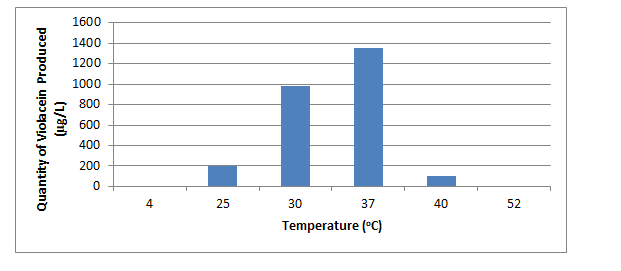
Fiure 1: The effect of temperature (40C – 520C) on violacein production.
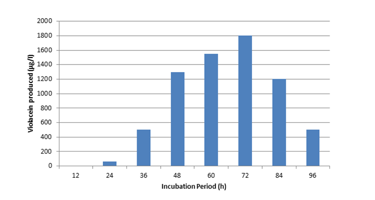
Effect of Incubation Periodon Violacien Produced: Violacein production was determined during different period of incubation (12, 24, 36, 48, 72, 60, 84, 96 hours).
Figure 2 shows that the violacein production was highest at 72hr (1800µg/l) after which the production declined.
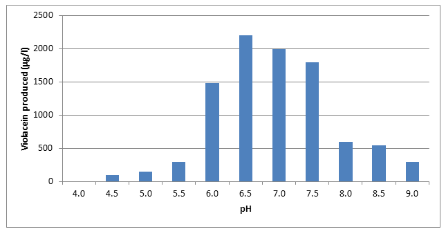
Effect of pH on Violacien Produced: Violacein production at different pH (4.0, 4.5, 5.0, 5.5, 6.0, 6.5, 7.0, 7.5 8.0, 8.5, 9.0) was examined. Figure 3 shows that violacein production was clearly affected by initial pH 4.0. The maximum pH for violacein production was observed at 6.5 (2200µg/l). As the pH increased above 6.5, the production of violacein decreased.
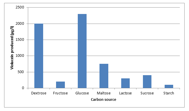
Effect of Carbon Sources on Violacien Produced: Growth of microorganism and the production of metabolites are influenced by the organism’s utilization of carbon sources during fermentation. The growth medium was inoculated with 5% different carbon sources (dextrose sucrose glucose, maltose, lactose fructose and starch). Glucose was the best carbon source for violacein production as it yielded high amount of violacein (2300µg/l) followed by dextrose and maltose. The least was starch (Figure 4).
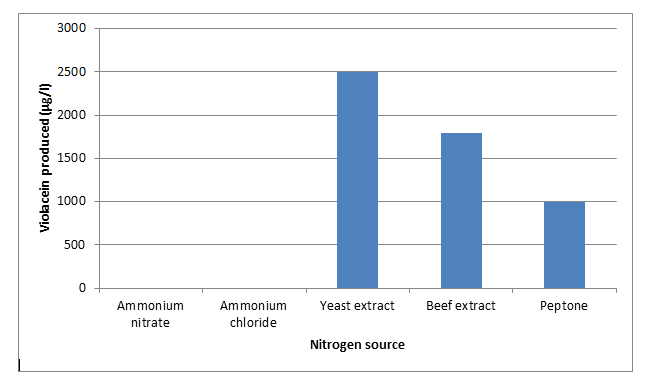
Effect of Nitrogen Source: Various nitrogen sources were examined for their effects on violacein production by the isolate (Chromobacterium violaceum). As shown on Figure 5, organic nitrogen sources were found to yield more violacein than the inorganic. Yeast extract was found to be the most efficient nitrogen source (2500µg/l) followed by beef extract (1800µg/l) and Peptone (1000µg/l).
Antibacterial Susceptibility
The result of antibacterial sensitivity test (Table 4 & 5) shows that Staphylococcus aureus and Escherichia were highly sensitive to violacein at the concentration of 4.4 while Klebsiella aerogenes and Pseudomonas aeruginosa were resistant to violacein.
| 35 | 17.5 | 8.8 | 4.4 | 2.2 | Negative Control (Ethanol) | Positive Control (Ciproflox) | |
|---|---|---|---|---|---|---|---|
| S. aureus | 26±5.66 | 17.50±10.61 | 17.00±4.24 | 15.00±7.07 | 0.00±0.00 | 0.00±0.00 | 22.50±3.54 |
| E. coli | 30±7.07 | 20.00±0.00 | 14.50±0.71 | 17.00±1.41 | 9.00±1.41 | 0.00±0.00 | 26.50±4.95 |
| Klebsiella aerogenes | 14.00±8.49 | 8.00±2.83 | 0.00±0.00 | 0.00±0.00 | 0.00±0.00 | 0.00±0.00 | 11.50±4.95 |
| Pseudomonas aeruginosa | 0.0±0.0 | 0.00±0.00 | 0.00±0.00 | 0.00±0.00 | 0.00±0.00 | 0.00±0.00 | 0.00±0.00 |
| Test Organism | 35 | 17.5 | 8.8 | 4.4 | 2.2 | Negative Control (Ethanol) | Positive Control (Ciproflox) |
| S. aureus | - | - | - | + | - | - | - |
| E. coli | - | - | - | + | - | - | - |
| Klebsiella aerogenes | + | + | + | + | - | + | + |
| Pseudomonas Aeruginosa | + | + | + | + | - | + | + |
Table 4: Antibacterial Susceptibility Test.
Violacein Concentration (mg/ml) and Zone Of Inhibition (mm). Table 4: Antibacterial Susceptibility Test.
Minimum Inhibitory Concentration of Violacein (MIC)
(-) No visible turbidity (No growth) (+) Visible turbidity (growth) The minimum inhibitory concentration (MIC) of violacein was examined and the result showed that the MIC ranged from 35- 4.4mg/ml. Table 5: Minimum Inhibitory Concentration of Violacein (MIC).
Discussion
Chromobacterium violaceum was isolated from Otamiri River in Owerri, Imo State. The isolate is a facultative anaerobe, motile, Gram-negative bacillus. Chromobacterium violaceum has attracted much attention due to its bioactive pigment violacein which is a secondary metabolic [22]. Investigation on the quantification of violacein production by Chromobacterium violaceum isolated from Otamiri River in Owerri showed that 35mg/ml of violaciem was produced at 570nm and this agrees with the work of Gallardo, et al. [23]. Violacein production is an important property of Chromobacterium violaceum so the optimization conditions for violacein production such as growth media, temperature, incubation period, pH, and Carbon and Nitrogen sources are a prerequisite. In the present work, an attempt was taken to analyze the effect of media on violacein production, among the three media used, Nutrient broth resulted in a favourably high violacein production of 1000mg/l, followed by Peptone broth (5000mg/l), Brain Heart infusion showed lower production of violacein (4000mg/l). It corroboration with the work of Ahmed, et al. [19], Vishnu and Palaniswamy, et al. [22]. Nutrient broth gave maximum production of violacein, may be due to the capacity of the isolate to utilize tryptone and yeast extract. In BHI absence of tryptone, pepetone and yeasts extract may lead to lower production. Temperature has got profound effect on the production of violacein. The effect of temperature in the range of 4_°C – 52°C was examined on the violacein production by _C. violacein, there was no growth at 4_°C -52°C, the optimal temperature was found to be at 370c with maximum production of 135mg/ml. In other studies with violacein producing bacteria, the optimal temperature was reported as 28°C for _Chromobacterium violaceum BB-78 [24], 33_°C for _Chromobacterium violaceum CCT 3496 25_°C for Pseudoalteromonas spp X 82144 [25] and 20°C for Dunganelle spp DSM 1953 [18], respectively. Zhang, et al. [26] studied the optimization conditions for violacein production by recombinant _Citrobacter freundi, the maximum violacein production was at 37_°_C, and this result is similar to our present study. Temperature is one of the most important environmental factors affecting the growth of micro-organism.
Incubation time plays a substantial role in the maximum pigment production. The influence of incubation period in violacein production by C. violaceum was studied at interval of 12hr, we observed that the pigment production was increased with respect to high incubation time, from 24hr to 72hr and after, the production declined. The maximum production of violacein (1800µg/l) was observed at 72hr incubation, the lowest production was observed at 24hr of incubation, no production at 12hr incubation. Wang, et al. [18] reported that the maximum production of violacein was at 32hr by a new strain Dunganella spp B2. Sivaranjini [27] reported the maximum production of violacein at the third day of incubation by C violaceum JC1 in solid state fermentation and Vishnu and Palaniswamy, et al. [22] reported the maximum production of violacein to be at 72hr of incubation. In Mendes, et al. [20] the highest crude violacein production was obtained after 36hr which C violaceum CCT3496 and after 40hr for recombinant Citrobacter freundii ATCC05417 in the liquid cultures. Ahmed, et al. [19] reported the maximum production of violacein was at 4th hour after bacterial growth that was at the late exponential phase. Their results suggested that the pigments produced by bacteria are secondary metabolites [28].
The pH of the growth medium plays an important role by inducing morphological changes in microbes. The maximum pH may affect cell membrane function, cell morphology and structure, solubility of salts, the ionic state of substrate, uptake of various nutrient and production biosynthesis. In general, cells can only grow within a certain pH range and metabolite formation is also affected by pH [29]. The pH change observed during the growth of microbes also affects the product stability in the medium [30]. Violacein production at various pH (4.0 – 9.0) was studied and we observed that the maximum pigment production was obtained at pH 6.5.
As the pH increased from 4.5 to 6.5, the pigment production increased. The production was found to decrease beyond pH 7.0. Similar results were obtained in a study of Zhang, et al. [26]. The growth and metabolic production of microorganism are influenced by the organism’s utilization of carbon and nitrogen sources during fermentation. The influence of carbon and nitrogen sources on the pigment production was observed in this study and glucose (2300µg/l) was found to be the optimum carbon source for violacein production followed by dextrose (2000µg/l) and starch was the least (100µg/l). Drew and Demain, et al. [31] reported that the use of carbon sources resulted in a decrease cellular growth and an increase in the secondary metabolite production. Yeast extract was found to be the best nitrogen source for pigment production in our study, whereas ammonium nitrate and ammonium chloride strongly inhibited violacein production. Our results agree with the result of Vishnu and Palaniswamy, et al. [22]. Antibacterial potentials of violacein were investigated in our study and we discovered that staphylococcus aureus and Escherichia coli were highly sensitive to violacein.
Staphylococcus aureus has the highest concentration at 35mg/ml and the lowest at 4.4mg/l with zone of inhibitions of 26 and 15 (mm) respectively. Klebsiella aerogenes has high concentration with zone of inhibition of 14mm in diameter while Pseudomonas aeroginosa was resistant to all concentration. This is in agreement with the work done by Cazoto, et al. [32]. The Minimum Inhibitory Concentration (MIC) of violacein against different organisms was examined and it was discovered that the MIC of violacein was 4.4 against S. aurens and E.coli. Klebsiella aerogenes and Pseudomonas aeruginosa has no inhibitory activity. It corroborates with work done by Subramanian, et al. [10] who discovered MIC for Staphylococcus aureus at 5.7 and resistant to Klebsiella pneumonia and Pseudomonas aeruginosa. The minimum bacteriocidal concentration of Staphylococcus aureus and Escherichia coli was 8.8mg/ml while the zone of inhibition on plate and the MIC observed at 4.4mg/ml for Staphylococcus aureus and Escherichia Coli may be considered as bacteriostatic. The negative controls (Ethanol) did not show any bacteriocidal effect against the test organism on the other hand the positive control (Ciprofloxacin) showed bacteriocidal effect against the aforementioned organisms.
Conclusion
The result of this research has demonstrated the antibacterial potentials of violacein obtained from Chromobacterium violaceum. A significant increase in violacein production was obtained when the organisms was grown at 370c, pH 6.5 for 72 hours in the presence of glucose as carbon source and yeast extract as nitrogen source. The successful inhibition of both Gram-positive organism (Staphylococcus aureus) and Gram-negative organism (Escherichia coli) by violacein depicts it as a potential source for the production of board spectrum antibiotics, which can be used to treat some infections.
References
-
Sneath PHA, Mair NS, Sharpe ME, Holt JG (1986) Bergey’s Manual of Systematic Bacteriology. Williams and Wilkins, Baltimore, MD.
-
Lichstein HC, Sand VFVD (1945) Violacein an antibiotic pigment produced by _Chromobacterium violaceum._ J Infect Dis 76(1): 47-51.
-
Durán N, Antonio RV, Haun M, Pilli RA (1994) Biosynthesis of trypanocide by _Chromobacterium violaceum._ World J Microbiol Biotechnol 10(6): 686-690.
-
Durán N (1990) Violaceina: a descoberta de um antibiotic. Ciencia Hoje 11: 58-80.
-
May G, Brummer B, Ott H (1991) Purification of the antiviral compound violacein from cultures of _Chromobacterium._ Ger Offen DE 3935066.
-
Durán N, Justo GZ, Ferreira CV, Melo PS, Cordi L, et al. (2007) Violacein: properties and biological activities. Biotechnol Appl Biochem 48(pt 3): 127-133.
-
Lopes SCP, Blanco YC, Justo GZ, Nogueira PA, Rodrigues FL, et al. (2009) Violacein extracted from _Chromobacterium_ _violaceum_ inhibits _Plasmodium_ growth _in vitro_ and in vivo. Antimicrob Agents Chemother 53(5): 2149- 2152.
-
Souza AOD, Aily DCG, Sato DN, Duran N (1999) Atividade da violaceina in vitro Mycobacterium turbeculosis H37RA. Rev Inst Adolfo Lutz 58(1): 59-62.
-
Bromberg N, Dreyfuss JL, Regatieri CV, Palladino MV, Durán N, et al. (2010) Growth inhibition and pro- apoptotic activity of violacein in Ehrlich ascites tumor. Chem Biol Interact 186(1): 43-52.
-
Subramaniam S, Ravi V, Sivasubramanian A (2014) Synergistic antimicrobial profiling of violacein with commercial antibiotics against pathogenic microorganisms. Pharm Biol 52(1): 86-90.
-
Leon LL, Miranda CC, Souza AOD, Duran N (2001) Antileishmanial activity of the violacein extracted from Chromobacterium violaceum. J Antimicrob Chemother 48: 449-450.
-
De Carvalho DD, Costa FTM, Duran N, Haun M (2006) Cytotoxic activity of violacein in human colon cancer cells. Toxicol in Vitro 20(8): 1514-1521_._
-
Melo PS, Maria SS, Vidal BC, Haun M, Durán N (2000) Violacein cytotoxicity and induction of apoptosis in V79 cells. In Vitro Cell Dev Biol Anim 36(8): 539-543.
-
Ferreira CV, Bos CL, Versteeg HH, Justo GZ,Duran N, et al. (2004) Molecular mechanism of violacein-mediated human leukemia cell death. Blood 104(5): 1459-1464.
-
Malghani S, Chatterjee N, Yu HX, Luo Z (2009) Isolation and identification of profenofos degrading bacteria. Braz J Microbiol 40(4): 893-900.
-
Lammert JM (2007) Techniques for Microbiology: a student handbook. Pearson Education Inc.
-
Vishnu TS, Palaniswamy M (2016) Isolation andidentification of _Chromobacterium_ sp from different ecosystems. Asian J Phar Clin Res 9(3): 253-257.
-
Wang HS, Jiang PX, Lu Y, Ruan Z, Jiang R, et al. (2009) Optimization of culture conditions for violacein production by a new strain of _Duganella_ sp. B2. Biochemical Engineering Journal 44(2-3): 119-124.
-
Ahmad WA, Yusof NZ, Nordin N, Zakaria ZA, Rezali MF (2012) Production and characterization of violacein by locally isolated Chromobacterium violaceum grown in agricultural wastes. Appl Biochem Biotechnol 167(5): 1220-1234.
-
Mendes AS, Carvalho JED, Duarte MC, Durán N, Bruns R (2001) Factorial design and response surface optimization of crude violacein for _Chromobacterium_ _violaceum_ production. Biotechnology Letters 23(23): 1963-1969.
-
Kirby WM, Bauer AW, Sherris JC, Turck M (1966) Antibiotic susceptibility testing by a standardized single disk method. Am J Clin Pathol 45(4): 493-496.
-
Vishnu TS, Palaniswamy M (2017) Impact of Various Fermentation Conditions on the Production of Violacein by the novel isolate _Chromobacterium vaccinia_ cv5. Int J Pharm BioSci 8(3): 514 -522.
-
Gallardo MJ, Staforelli JP, Meza P, Bordeu I, Torres S (2014) Characterization of _Chromobacterium violaceum_ pigment through a hyperspectral imaging system. AMB Express 4: 4.
-
Riveros R, Haun M, Durán N (1989) Effect of growth conditions on production of violascein by _Chromobacterium violaceum_ BB-78 strain. Braz J Med Biol Res 22(5): 569-577.
-
Yang LH, Xiong H, Lee OO, Qi SH, Qian PY (2007) Effect of agitation on violacein production in _Pseudoalteromonas_ _luteoviolacea_ isolated from a marine sponge. Lett Appl Microbiol 44(6): 625-630.
-
Zhang R, Jiang P, Li C, Xing X (2010) Optimization of fermentation condition for violacein production by recombinant _Citrobacter freundii_. CIESC J 6: 1495-1505.
-
Sivarenjini G (2011) Production of violacein by a newly isolated bacterium _Chromobacterium_ sp JC1. Jawaharlal Nehru Technological University, pp: 154.
-
Lu Y, Wang L, Xue Y, Zhang C, Xing X, et al. (2009) Production of violet pigment by a newly isolated psychrotrophic bacterium from a glacier in Xinjiang, China. Biochemical Engineering Journal 43(2): 135-141.
-
Merlin JN, Christhudas IVSN, Kumar PP, Agastian P (2013) Optimization of growth andbioactive metabolite production: _Fusarium solani_. Asian J Pharm Clin Res 6(3): 98-103.
-
Rani G, Paresh G, Harapriya M, Vinee KG, Bhavna C (2003) Microbial α-amylases: a biotechnological perspective. Process Biochem 38(11): 1599-1616.
-
Drew SW, Demain AL (1977) Effect of primarymetabolites on secondary metabolism. Ann Rev Microbiol 31: 343- 356.
-
Cazoto LL, Martins D, Ribeiro MG, Durán N, Nakazato G (2011) Antibacterial activity of violacein against _Staphylococcus aureus_ isolated from bovine mastitis. J Antibiot 64(5): 395-397.
- Antifungal Activity of New Acetophenone Derivatives
- Interconnected Microbiomes Human Health Within an Environmental Framework
- Silkworm-Based Vaccine Production for H5N1: A One Health Approach to Pandemic Preparedness
- Microbial Diversity and Lipolytic Activity of Bacteria and Fungi from Oil-Contaminated Sites in Makurdi Metroplois
- Antibiotic Resistance Profile of Bacteria Isolated at the Central Laboratory of the National Hospital Center of Nouakchott
- Epidemiology and Sensitivity to Antibiotics of Germs Isolated from Blood Cultures in the Laboratory of the National Hospital Center of Nouakchott-Mauritania