Antifungal Drugs Loaded Nanosponges to Improve Bioavailability of Antifungal Drugs; A Review
Fungal infections have become a major concern in this era for human health leading to morbidity and mortality. A lot of antifungal agents are available for treatment, with poor therapeutic outcomes due to poor bioavailability, toxicity, and resistance challenges. Ordinary dosage forms are unable to meet the clinical needs. Nanosponges provide a promising drug delivery system overcoming major challenges concerned with antifungal agents. Nanosponges improves solubility, permeation, taste masking and stability of antifungal agents. It serves as an important tool for providing controlled release dosage forms. The nanosponges are easier to formulate and convenient for bilk manufacturing. Nanosponges thus provide various benefits to clinician to cope the overcoming demand of fungal infectious disease.
Introduction
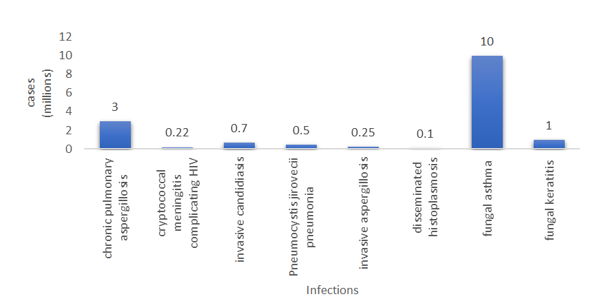
Prevalence of fungal infections has increased in last two decades leading to increased morbidity and mortality rate [1, 2]. Annually about 40 million people not only in underdeveloped as well as in developed countries suffers from fungal infections [3]. About 600 various human infecting fungal species have been identified. Fungai may infect hair, skin, nail, and mucosa, and may also cause allergies [4]. Mostly serious fungal diseases are caused by Candida, Aspergillus, Cryptococcus species, Histoplasma capsulatum, Mucormycetes and Pneumocystis jirovecii fungal pathogens. Candida albicans is mostly responsible for mucosal infections, Aspergillus fumigatus for most of the allergic fungal infections and Trichophyton spp., especially T. rubrum, mostly for skin infections [5]. A recent estimation of globally fungal infection shows 3M chronic pulmonary aspergillosis, 0.22M cryptococcal meningitis complicating HIV/AIDS, 0.7M invasive candidiasis, 0.5M of Pneumocystis jirovecii pneumonia, 0.25M of invasive aspergillosis, 0.1M of disseminated histoplasmosis, 10M of fungal asthma and 1M fungal keratitis cases annually [6, 7, 8].
Infection caused by fungi in human is known as mycosis AA. Fungal infections may be classified as superficial and invasive. Superficial fungal infections also called dermatophytosis are the fungal infections involving skin and skin structures. Superficial fungal infections of nail (onychomycosis) and skin (ring worm, thrush) are the most common, affecting almost 1/4th of the world population [6, 9, 10]. Invasive fungal infections are the infections of the sterile body sites. Invasive fungal infections are less common and are limited to the immunocompromised patients such as cancer patients with chemotherapy, AIDS patients, with higher mortality rate [11, 12, 13].
To treat fungal infections various drugs of different classes are available. These classes include azoles, echinocandins, allylamines, antibiotics, antimetabolites. Azole class is further divided into imidazole and triazoles [14, 15, 16].
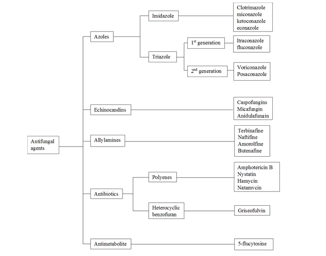
Imidazole (clotrimazoles, miconazole, ketoconazole, econazole) has high side effects, toxicity, and interactions. Azole has better safety profile with border spectrum than imidazole. 1st generation azole drugs (itraconazole, fluconazole) are fungistatic resulting in resistance. 2nd generation azole drugs (voriconazole, Posaconazole) are fungicidal with broader spectrum [17, 18, 19, 20]. Echinocandins (caspofungin, micafungin, anidulafungins) are newer fungicidal agents with better safety profile [21, 22]. Allylamines (terbinafine, naftifine, amorolfine, butenafine) is newer class better than azoles. Antibiotics class is divided further into two subclasses polyenes and heterocyclic benzofuran. Polyene antibiotics (amphotericin B, natamycin, nystatin, hamycin) are fungicidal with border activity and nephrotoxicity limiting only topical use [23, 24, 25]. Antimetabolite includes single agent flucytosine used only in adjunctive therapy [26].
Drug resistance, poor bioavailability, drug interactions and toxicity issues are the challenges making antifungal therapy one of the complicated therapies despite the availability of various drugs. Widespread use of antifungal agents in human and animal health care, agriculture, and timber preservation results in emergence of drug resistance making antifungal therapy challenging [27, 28]. Azoles, echinocandins and polyene classes are facing the drug resistance against Candida species (Candida glabrata, Candida tropicalis, Candida parapsilosis) and Aspergillus species (Aspergillus fumigatus, Aspergillus flavus). Candida auris has raised with alarming multidrug resistances challenge globally [29, 30, 31]. Majority of antifungal agents are lipophilic in nature resulting in low water solubility, poor bioavailability, and formulation challenges. A variety of available dosage forms tablets, capsules, creams, injectables are ineffective to overcome these challenges [32].
Nanosponges are sponge like porous structures of nanometric dimension, having the size of a virus with a diameter of ≤1µm [33]. Nanoporous structures are classified into nanoporous hydrogels, nanoporous membranes and nanoporous particles. Nanosponges belong to nanoporous particles category [34]. Nanosponges have the ability to carry lipophilic as well as hydrophilic drugs efficiently to targeted site. Drug release can be modulated by controlling the polymer crosslinker ratio [35]. It is mainly used for cosmeceutical and pharmaceutical approaches, as it has the advantages of nanosized and microsponges vesicular structure [36]. They improve the bioavailability of poorly water soluble (hydrophobic) drugs by enhancing their wetting and solubility. They provide stability to drug molecules against the harsh physical, chemical and biological environments [37, 38]. They are free flowing powder can be incorporate in different dosage forms (tablets, capsules, emulgels, hydrogels, saline water) facilitating administration through various routes including oral, topical, pulmonary, parenteral [39]. Nanosponges are of different types as classified in Figure 3. Vital constituents used in synthesis of the nanosponges includes polymers, copolymers, crosslinkers and aprotic solvents [40].
Nanosponges are a type of nanoparticles having the ability to incorporate drug into their core. Nanoparticles carry a drug in it by three types:
- Encapsulating nanoparticles: In this type nanoparticles containing hollow cavities like sponges incorporate drug in it. Alginate nanosponges are spongelike nanoparticles having many hollow cavities that incorporate the drug molecules.
- Complexing nanoparticles: Drug molecules are carried due to the electrostatic charges of the nanoparticles and drug molecules.
- Conjugating nanoparticles: Nanoparticles and drug molecules are attached together through covalent bonding [41].
![Figure 3: Vital constituents used in synthesis of the nanosponges includes polymers, copolymers, crosslinkers and aprotic solvents [40].](/fulltextimages/8048/fig_3.png)
This systematic review provides a comprehensive analysis of antifungal drug delivery through nanosponges including fungal infection prevalence, antifungal classes, challenges in antifungal therapy, nanosponges for antifungal drug delivery, their methods of preparation, characterization, and advantages & disadvantages of this drug delivery system.
Methods of Preparations
Nanosponges are the class of nanoparticles that are colloidal having a size range of 1-100 nm, carbon contains polymer, porous in structure. Generally, two methods of preparation of nanoparticles. These are top-down and bottom-up approaches. In the bottom-up approach, the atoms and molecules join to form supramolecular structures e.g., cresol, sol-gel precipitation process. While in top- down approach submicron particles are broken down into nanoparticles by nano mill [42, 43].
| Polymers | Hyper cross linkage polystyrenes, Cyclodextrins (alkyl β-cyclodextrins, alkoxy carbonyl cyclodextrins, hydroxy propyl betadex), and some di block polymers like ethyl cellulose, polyvinyl chloride, etc. |
| Crosslinkers | Diphenyl carbonate, carbonyl di imidazole, pyromellitic di-anhydride, glutaraldehyde, carboxylic acid di-anhydrides, di-isocyanates, and epichloridine [44]. |
| Solvents | Dichloromethane, dimethyl sulfoxide, ethanol, methanol, chloroform, deionized water |
| Copolymers | Ethyl cellulose, polyvinyl alcohol [45]. |
Table 1: Polymers and crosslinkers used in the preparation of nanosponges [44,46-48].
Nanosponges Preparation Methods
Generally, in practical aspects, four methods are used for the preparation of nanosponges.
Solvent Method
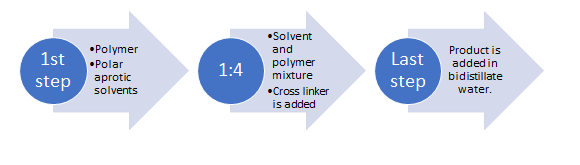
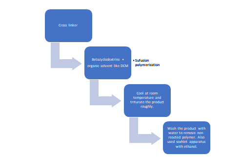
First, is solvent-based synthesis in which dimethylformamide, pyridine, and dimethyl sulfoxide solubilize CDs and requires solvent washing after preparation by Soxhlet apparatus [37]. In this polymer is added in one of these solvents and then cross-linker is added in it in a ratio of 1:4. The previous reaction is operated at 10-12℃ for 1 to 48 hours [49].
Melt Based Synthesis
The second method is fusion melt-based synthesis in which cross-linking agent and cyclodextrins are merged at high temperatures. This method is green because of no use of organic solvents [38].
Ultrasound-Assisted Synthesis
The third method is ultrasound-aided synthesis. The polymer and crosslinker are added to a flask and reacted in an ultrasonic water bath without a solvent. The mixture is heated at 90℃ and that product is sonicated for 5-6 hours. The product is cooled down at room temperature. The excess polymer is removed by water and purification is done by Soxhlet apparatus [43, 50].

Emulsion Solvent Diffusion Method
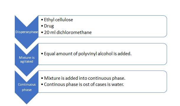
In this method, two phases are used. One is the dispersed phase and the other is the continuous phase. The disperse phase contains ethyl cellulose and drug which is then added in 20 ml dichloromethane. Along with an equal amount of polyvinyl alcohol this mixture is then added to 150 ml of the continuous aqueous phase. This mixture is then magnetically stirred. Then the product is dried [45].
Microwave-assisted synthesis
The microwave-assisted synthesis produces crystalline NS. In this method, microwaves are used for the preparation of nanosponges [43].
From hyper crosslinked β-cyclodextrins
Mostly cyclodextrins are used for the formation of nanosponges as a polymer. They are non-reducing cyclic oligosaccharides having almost 6 or 8 glucose units bounded with ⍺ 1,4- glycosidic linkage having abridged cone structure [34]. Its outer part is hydrophilic and its inner part is lipophilic. Cyclodextrins are prepared by enzymatic action on starch and alpha, beta, and gamma CDs are formed comprise of six, seven, and eight glucopyranose subunits [37]. In nanosponges beta cyclodextrins condensing polymerization reaction takes place. There is regiospecific addition of reactants, balanced reaction conditions, and well removal of excess product in nanosponges preparation. In a solution with small molecules of crosslinking agents that work as small joining hook-ups that connect polymer together, cyclodextrins are heated [34].
Nanosponges are prepared by different methods according to the nature of polymer and crosslinker. Cyclodextrins-based urethane/carbamates Nanosponges were made by reacting B-CD and hexamethylene diisocyanate and toluene diisocyanate in dimethylformamide for 20-24 hours at 70℃ in N2 environment [51].
Cyclodextrins Based Carbonate Nanosponges: Cyclodextrins-based carbonate Nanosponges are frequently used in drug delivery applications. These are prepared by reacting CD with dynamic carbonyl compounds e.g., triphosgene and diphenyl carbonate in the presence of DMF under retort conditions. The use of ultrasound-assisted synthesis results in the fusion of crosslinking agent and combining with CD. After that product is cleaned with water and alcohol mixture to remove unreacted carbonyl derivatives [51].
Cyclodextrins with ether group based Nanosponges: Cyclodextrins with ether group nanosponges are prepared in the presence of a base, CDs with epoxide crosslinkers such as ethylene glycol and epichlorohydrin. This reaction is carried out in aqueous conditions. They are used as tablet disintegrant and other drug delivery systems because of their high chemical resistance and recoverable swelling properties [42].
Cyclodextrins based ester Nanosponges: Cyclodextrin- based ester Nanosponges have high water swelling capacity and form a hydrogel-like network. Swelling is inversely proportional to crosslinking. They are prepared by CDs and dianhydrides or polycarboxylic acids e.g., ethylene diamine tetra acetic acid dianhydride and tetra acetic acid dianhydride and citric acid. Both are soluble in polar organic solvents like DMS, TEA.
Functionalized nanosponges synthesis: Functionalized Nanosponges are prepared either as nanosponges that has been fluorescently labelled or charged side chain nanosponges e.g., Acyclovir nanosponges are prepared by mixing carbonated nanosponges with succinic anhydride in dimethyl sulfoxide [52].
Factors that affect the synthesis of Nanosponges
Different factors that affect the preparation of nanosponges. These are the ratio of hydrophilic and lipophilic components, nature of the drug, natural polymer, and nature of crosslinker. Epichlorohydrin is used as crosslinker to modify the rate of drug release, resulting in hydrophilic nanosponges. While for hydrophobic NS diphenyl carbonate or diisocyanates and carbonyl diimidazole are used to sustain release carriers for peptides and proteins [43].
Examples of Nanosponges Synthesis that are used for Antifungal Drugs
Β-Cyclodextrins Based Itraconazole Nanosponges: Itraconazole nanosponges based on beta cyclodextrins are prepared by Shankar and its coworkers. In these nanosponges, beta cyclodextrins interlink with carbonate bonds. Itraconazole is a BCS class II antifungal drug that has low solubility in water. They used a solid dispersion technique for drug incorporation. In their preparation of nanosponges itraconazole dissolved in DCM and nanosponges added in this mixture with co polyvidone and then pulverized until DCM evaporates. The ratio of drug: nanosponges: polyvidone is 1:1:1. After that nanosponges dried in oven and itraconazole attains improved bioavailability [53].
Hydroxypropyl Β-Cyclodextrin Based Clotrimazole Nanosponges: Clotrimazole BCS II also has poor solubility and a short half-life. Its solubility increased by forming B-cyclodextrin nanosponges that are hyper crosslinked cyclodextrin polymers. In this preparation, dimethyl carbonate is used as a cross-linker, and then it is suitably gelled. Hydroxypropyl-β-CD added in DCM at 90℃ with magnetic stirring and then waits for 5 hr. for the reaction completed. After that allow it to cool and filter it. The solid mass is broken down and the Soxhlet apparatus is used to get rid of excess DCM using ethanol. A high concentration of crosslinker is used; nanosponges formed are stored at 25℃.
Now the introduction of clotrimazole into nanosponges is done by dispersing it into an aqueous suspension of nanosponges in different concentrations. This mixture then agitated magnetically for almost 24 hr. centrifuged at 2000 rpm for 10-15 mins to separate the nonincorporated drug. After that colloidal supernatant are freeze-dried. The resultant nanosponges have an increase in in vitro drug release as well as in vitro bio adhesion. In vivo antifungal activity was also enhanced [54].
β-Cyclodextrins Based Itraconazole Nanosponges: In the preparation of itraconazole nanosponges reaction takes place under sonication at 90℃ for 5 hr. as beta cyclodextrins as polymer, diphenyl carbonate as cross liker [43].
Econazole Nitrate Nano Sponges: Econazole nitrate nano sponges are prepared by the solvent method. In this preparation polyvinyl alcohol is used as a polymer and ethyl cellulose as a cross-linker. Both these increase the topical permeation of econazole nitrate. The bulk phase consists of PVA in 150 ml of water and distributed phase is econazole nitrate (100mg) and ethyl cellulose is dissolved in 20 ml Miconazole Nitrate Nanosponges dichloromethane. The organic component is slightly added into the aqueous phase at 35℃ under magnetic stirring conditions like 1000 rpm [55].
![Figure 8: Preparation of nano sponges by beta cyclodextrins as a polymer and diphenyl carbonate as crosslinker [34].](/fulltextimages/8048/fig_8.png)
One method is reported for the perpetration of miconazole nitrate nanosponges by a solvent evaporation method. They are prepared by diphenyl carbonate as cross- linker and beta cyclodextrins in a 1:1 ratio in almost 100 ml flask. The mixture is heated at 120℃ under magnetic stirring and wait for 5 hours to complete the reaction. Cool the product, break it, and washed with water to clear unreacted diphenyl carbonate and phenol as a by-product. In chloroform miconazole nitrate dissolved separately, nanosponges added to it until solvent evaporated. Clusters should be removed. After that dry them at 50℃ for 12 hours and sieved [56]. Miconazole nitrate nanosuspension and vaginal gels can be prepared from these nanosponges. For miconazole nitrate nanosuspension, dried nanosponges are introduced into a 500 ml flask and add 100 ml methanol in it to remove any powdered drug that is not encapsulated in nanosponges. Filter this solution through 0.22µm and disperse this into the water using ultrasonication. For vaginal gel, previously prepared nanosponges are mixed with methylparaben, propylparaben and this will be incorporated in the dispersion of water with constant stirring [56].
Ethyl Cellulose-Based Fluconazole Nanosponges: Fluconazole nanosponges can be prepared by the emulsion solvent diffusion method in which ethyl cellulose is used as a polymer. Other reagents are polyvinyl alcohol, carbopol-940, and propylene glycol are used to prepare nanosponges-based hydrogel [57]. In the emulsion solvent diffusion method, the researchers prepared 6 batches all of which have different ratios of drug to a polymer. In the first phase, ethyl cellulose and fluconazole are both added in 20-30 ml dichloromethane to form disperse phase with the help of ultra-sonication. In the second phase, 0.5% w/v of polyvinyl alcohol was dissolved in almost 150-200 ml of water to form the aqueous phase by heating on the water bath. After that disperse phase slowly but continuously added into bulk phase by syringe injection method or by magnetically stirred at 1000 rpm for 2 hours [58].
Ethyl Cellulose-Based Voriconazole Nanosponges: In one research article, the emulsion solvent evaporation method for voriconazole nanosponges is reported. These nanosponges are formed by ethyl cellulose as a polymer, polymethacrylate, and Pluronic F-68. Different proportions of polymer and PVA. Disperse phase consists of voriconazole and polymer in 20-25 ml of dichloromethane. This mixture is added into a precise amount of PVA that is dissolved in 100 ml water to form an aqueous continuous phase by magnetic stirring [59].
Ketoconazole Based Nanosponges Gel: Ketoconazole- based nanosponges gel is prepared through hyper cross- linked β-cyclodextrins along with crosslinkers. In their preparation method, dimethyl sulfoxide is taken in a round bottom flask. Anhydrous β-cyclodextrins are added to it and then diphenyl carbonate is added to it and waits for 4 hours. Condensation polymerization takes place and transparent blocks of β-cyclodextrins are formed. This product is then triturated. To remove any excess of dimethyl sulfoxide, the product is washed water. The product can also be washed by Soxhlet apparatus by ethanol. Then this product is dried, ground and fine powder dispersed in water [60]. 6% ketoconazole nano sponge gel can be formulated using carbopol-940.
Cyclodextrins Based Griseofulvin Nanosponges: Cyclodextrins nanosponges of griseofulvin is an oral dosage form for children with improved bioavailability. They also mask the bitter taste of drug. Β-cyclodextrins reacted with diphenyl carbonate as a crosslinker by using the ultra- sonication method. The drug is loaded in nanosponges in the presence or absence of 0.25% polyvinyl pyrrolidone [61].
Terbinafine nanosponges: Terbinafine nanosponges are produced by the solvent emulsion evaporation method. In this, the drug HCL, polyvinyl alcohol as an emulsifier, and ethyl cellulose are used. Phase I, terbinafine HCL, and ethyl cellulose are dissolved in dichloromethane and homogenized. Phase II, polyvinyl alcohol added to water. Phase I preparation slowly added in phase II with constant stirring. Terbinafine nanosponges are formed dried and stored at 25℃ [62].
| Polymers used in the preparation of nanosponges | Cross linkers | Antifungal drugs loaded nanosponges | BCS class | Method used | References | |
|---|---|---|---|---|---|---|
| B-cyclodextrins | Copolyvinodum | Itraconazole (BCS class II) | II | Solid dispersion technique | [53] | |
| Hydroxy propyl B-cyclodextrins | Dimethyl carbonate | Clotrimazole (BCS II and shorter half-life) | II | Solvent evaporation method | [54] | |
| Ethyl cellulose | Polyvinyl alcohol | Econazole nitrate | II | Solvent evaporation method | [55] | |
| B-cyclodextrins | Diphenyl carbonate | Miconazole nitrate | II | Solvent evaporation method | [56] | |
| Ethyl cellulose | Polyvinyl alcohol | Fluconazole nanosponges-based hydrogel | I | Emulsion solvent diffusion method | [57] | |
| Ethyl cellulose | Polymethyl methacrylate | Voriconazole | II | Emulsion solvent evaporation method | [58] | |
| B-cyclodextrins | Diphenyl carbonate | Ketoconazole gel-based nanosponges | II | Condensation polymerization techniques | [60] | |
| B-cyclodextrins | Diphenyl carbonate | Griseofulvin oral liquid nanosponges-based pediatric dosage form | II | Ultrasonication method | [61] | |
| Ethyl cellulose | Polyvinyl alcohol | Terbinafine nanosponges | II | Solvent emulsion evaporation method. | [62] |
Table 2: Methods of preparation of antifungal drugs loaded nanosponges.
Characterizations of Nanosponges
Inclusion complexes or nanosponges are characterized by various following methods [38, 44]:
Loading Efficiency
The loading efficiency of inclusion complexes is determined. This is done by solubilizing the nanosponges in a suitable solvent. Sonicate it and allow it to break. The contents are then analyzed by HPLC or UV spectrophotometer [38].
100 Drug encapsulant
Drug loading capacity
$$ v = \frac {D r u g e n capsulant}{D r u g t o t a l} \times 1 $$
Morphology Evaluation of Nanosponges
Scanning electron microscope and transmission electron microscope are widely used to study different aspects of nanosponges.0.5% W/V suspension of nanosponges is sprayed on copper grids. And this must be air dried before observation. This method indicates the difference of crystallization rate of starting material and product. NIH image software is used [52].
Determination of size, zeta potential and polydispersity
Zeta potential is the measure of surface charge or electrical potential at slipping plane. This can be used by using specific electrode in particle size measurement equipment. For zeta potential determination all nanosponges are diluted with 0.1 mM KCL. Then they enter in electrophoretic cell where electric field is about 15V/cm is applied. Zeta potential of carbonated nanosponges is -25mV which indicated that they are form stable suspensions that don’t sufficiently aggregate [37].
Polydispersity is the variation within particle size distribution. NS sizes and polydispersity parameters are determined by dynamic light scattering. This method uses 90 plus particle sizer with MAS OPTION particle sizing software. 95℃ and 25℃ scattering angles are mostly used. All samples are diluted with deionized water before any measurement [52].
$$ P D I = \frac {\Delta d}{d a v g} $$
“d” is the width of distribution and “d avg” is the average particle size [48].
Fourier Transform Infrared (FTIR) analysis
It is used to determine the possible relationship between the drug and polymer. Carbon blank is used as a reference and sample is scanned from 400-4000 cm-1. The purging of detector is done by helium before analysis. KBr pellets are mostly used [63]. 4000 to 650 cm-1 range spectra of drug, polymer, drug polymer physical mixture, drug loaded nanosponges and blank NS is noted to observe any possible interaction. This method also indicates the hydrophilic and hydrophobic sites in NS [38].
Thin layer chromatography
In TLC evaluation, the Rf value of drug candidate is disappear and thus identify the nanosponges joined with drug. But it is reversible process. So that only drug and nanosponges spots are found on TLC-plate.
In-vitro Drug Release of Nanosponges
Multi-compartment rotating cell is used. It has two compartments one of which is donor that filled with aqueous suspension of nanosponges with drugs and other is receiver compartment filled with suitable PH phosphate buffer. These two compartments are separated by hydrophilic dialysis membrane. The receptor buffer is completely withdrawn at specific time intervals and fresh buffer is added. The sample is than analyzed by HPLC or other tool specified in individual monograph of drug [38].
Infrared Spectroscopy
This technique is used to study the interaction between drug and nanosponges in the solid state. Nanosponges band small changes upon complex formation. IR spectroscopy technique is limited to drugs having carbonyl and sulfonated groups [44].
Phase Solubility Studies
The effect of nanosponges on solubility of drugs is determined by phase solubility studies. In this method, solubility constant is determined by dissolving drug into suitable solvent at its saturated point. This solution is then mixed with different concentration of nanosponges i.e., 1:1, 1:2, 1:3, etc. High concentration of nanosponges interact more drug. A plot is drawn between nanosponges concentration and drug concentration. Solubility constant that obtained gives idea about extent of interaction between nanosponges and drug [43].
Porosity
It gives us estimate about nanocavities formed in the nanosponges. Helium pycnometer is used because it uses helium gas that possesses the ability to penetrate inter and intra molecular channels of NS. The true volume can be calculated by the extent of helium displacement [38].
$$ \% P o r o s i t y = \frac {B u l k v o l u m e - T r u e v o l u m e}{B u l k v o l u m e} \times 1 0 0 $$
Water Uptake and Swelling Studies
For polyamidoamine polymer nanosponges swelling studies can be done by soaking nanosponges in water [43].
Percentage swelling can be calculated by using following formula:
$$ \% o f s w e l l i n g = \frac {S t}{S o} \times 1 0 0 $$
Where “St” means cylinder marking at specified time after soaking and “So” means initial cylinder marking [38].
$$ \% \text{water uptake} = M t / M o \times 1 0 0 $$
Mt is the mass of hydrogel after some time and Mo is the initial mass od dry polymer.
Thermal Analysis
Differential scanning calorimetry (DSC), differential thermal analysis (DTA) and thermogravimetric analysis (TGA) are most important. These studies are used to determine melting point, degree of crystallinity (Xc), crystallization temperature (Tc) and thermostability of nanosponges. In TGA, the sample is heated in N2 environment and loss of mass is noted. Weight loss is another indicator of formation on inclusion complex formation [38]. Then sample is heated in the presence of oxygen and loss of mass and oxidized products are identified. DTA and DSC patterns of nanosponges can be observed for any enlargement, shifting and appearance of new peaks shows the molecular dispersion of drug in polymer [43].
Moisture analysis
Nanosponges are non-hygroscopic. The retention of crystal structure during moisture absorption and desorption can be confirmed by dynamic vapor sorption studies [38].
X-ray diffractometry and single crystal X-ray structure analysis
The diffraction pattern is noted in solid state. In liquid form, mixture not shows any diffraction pattern. The diffraction is completely different of un complexed NS from complexed NS. This difference indicates the complex formation. The diffraction peaks are important to determine the chemical decomposition and complex formation [44]. The single crystal X-ray structure analysis reveals the complete structure and interaction of nanosponges. The precise geometric relationship can be identified [48].
Photodegradation Study
In this study, UV lamp is used. The sample is taken at 10cm from UV lamp. The sample is stirring under dark. The sample is analyzed at different time intervals using HPLC [48].
Benefits Associated with Antifungal Nanosponges
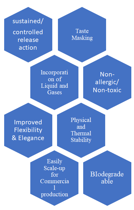
- Nano-sponges offer entrapment and compatibility with wide variety of drugs.
- Side effects are markedly reduced through use of nano- sponges without decreasing the efficacy of drug.
- Formulation flexibility is enhanced, and stability is improved using this system.
- They are non-allergic, non-irritating, non-mutagenic and non-toxic.
- Nano-sponges are free flowing.
- Nano-sponges can incorporate immiscible liquids which improves material processing through this liquid can be converted to powder form [35].
- They are stable over wide pH range and temperature.
- There are self-sterilizing because very low pore size i.e. 0.25 micron, hence bacteria cannot enter into it.
- Controlled / Sustained release action is obtained through this system [64]
- They are cost effective as compared to other formulation.
- They also mask the bitter taste of drug [65].
- They have better thermal and physical stability as compared to other systems.
- Encapsulation of drugs can be done through nano- sponges.
- The superior property of nanosponges have been attributed the ability to control the structure of particles and control the nature and size of aperture. By varying the proportion of cross-linker to polymer, the degree of cross linking can be modulated, which ultimately affects drug loading and release [66].
- They are bio-degradable and protect the drug from degradation.
- Nano-sponges can incorporate gases in it.
- They are easily scaled up for commercial production.
Limitations associated with anti-fungal nanosponges
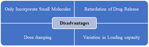
- They can only incorporate small molecule in it. Molecule having more than five condensed rings cannot be incorporated.
- Their loading capacity also varies depending on the degree of crystallization. Para crystalline form has different loading capacity as compared to crystalline form.
- Nanosponges may sometime retard the drug release.
- The phenomena of dose dumping may occur.
Challenges associated with Antifungal Nano-sponges
Solubility Enhancement
Most antifungal drugs i.e itraconazole, fluconazole, miconazole, ketoconazole, clotrimazole, griesofulvin etc, are BCS 2 drugs, having low solubility in the body. Hence nanosponges are made to enhance solubility. The increase in the concentration of nanosponges enhances the solubility of the drug [64]. Further enhancement of solubility can be done through addition of auxiliary components like copolyvidonum [67]. Masking of hydrophobic group by increasing wetting of drug or by decreasing crystallinity of drug nanosponges improves solubility [68].
Increase in Permeability
The permeability of nanosponges is enhanced using nanosponges. The nanosponges permeate deeper into the skin to an extent to which no other formulation can reach. They, hence, improved the efficacy by channelizing the antifungal drugs to their desired targeted site and eradicate fungal infection [69]. The drugs in nanosponges are of small size and are loaded in a precise way, likewise the polymer in nanosponges also contributes to the increase permeability of less permeable antifungals e.g., resveratrol enhances the permeability of nanosponges.
Enhancement of Topical Antifungal Activity
Nano-sponges hydrogels are more effective in inhibiting the fungal growth topically as compared to normal antifungal creams. Usually, creams are of hydrophobic nature due to which they have lower drug uptake causing ineffective treatment of the disease. Skin deposition of terbinafine nanosponges hydrogels is significantly higher than normal hydrogels or creams [70]. Ketoconazole, a drug effective against candiasis and other fungal infection having poor solubility and permeability, can enhance its activity through formulation in to nanosponges [64, 71].
Protection against Degradation
There are some antifungals which are prone to degradation by photolysis e.g., amphotericin B, flucytosine, griesofulvin, voriconazole etc. The antifungal activity of amphotericin decreases within 2 to 4 days of exposure to sunlight. Similarly, clotrimazole photodegradation reaches to 40% when exposed to polychromatic light within 9 hours [72]. Thus, nanosponges may serve as a protective agent to minimize the degradation.
Sustained/Controlled Release of Antifungal drugs
Nanosponges also play an important role in sustained release of drug in the body. Sustained release of a drug maintain the constant drug concentration in the body by releasing the drug programmed rate for long period of time [73]. Sustained release antifungals like miconazole and nystatin helps to inhibit recurrence of denture stomatitis by Candida spp. [74]. Likewise, clotrimazole is effective in vaginal candidiasis by maintaining sustained release in vagina [75]. Hence constant release of antifungals helps to inhibit reinfection. Nanosponges, thus play a promising role in releasing of antifungals over an extended period. The release rate further can be controlled by increasing or decreasing the polymer. Consequently, the sustained release effect in fluconazole and voriconazole is helpful in chronic fungal diseases like Jock itch, Tinea versicolor, Onhycomycosis etc. [60, 62]. miconazole nanosponges also provide sustained released effect in vagina due to its good viscosity and pH [76]. Likewise, euconazole nanosponges serves as sustained release formulation for topical activity in which nanosponges serves as a depot for drug storage [77].
Increase Bioavailability of Antifungals
Bioavailability, the amount of drug which reaches to the systemic circulation, can also be increased by nanosponges [78]. As cyclodextrin based nanosponges may enhance the solubility up to 30 times, dissolution rate also increases to an extent, which consequently increases the bioavailability. Most antifungals like itraconazole, miconazole, ketoconazole, terbinafine, griesofulvin and etc. having poor bioavailability [79, 80].Their bioavailability can be increased by incorporation of these in to the nanosponges. Hence, help them to attain required plasma concentration for the desired therapeutic effects.
Bitter Taste masking
Antifungals especially griseofulvin, terbinafine and amphotericin has very bitter taste render it hard to swallow. Nanosponges may play pivotal role in taste masking of antifungals [64]. The cyclodextrins of nanosponges form inclusion complexes with the drug molecules, thus forming interaction with proteins of taste bud preventing the contact of bitter drugs with the taste buds [81].
Better Stability
They are stable at a very high degree of temperature i.e., up to 130c. it can tolerate and remain stable over broad range of pH i.e. 1 to 11 [82]. It is compatible with various ingredients. It has better chemical, physical and thermal stability [83, 84].
Increase in Patient Compliance
Due to increase in the bioavailability and sustained release action of antifungal nanosponges, the duration of action will be enhanced, and the dosage interval is increased which require less dosing frequency. Consequently, patient compliance is increased.
Improved Oral Drug Delivery
For most antifungals dissolution rate is the rate limiting step in bioavailability of drug when taken orally. Many drugs like miconazole, itracoanzole, ketoconazole, griseofulvin have low bioavailability which make them less efficacious and contributes to decrease antifungal activity. Nanosponges play a vital role in enhancing the dissolution rate, permeability, and solubility of an oral drug B-cyclodextrin based nanosponges will deliver the drug three to five times more to the target site when compared to the injections.
Targeted Drug Delivery
Nanosponges can be tagged with specific linkers for targeted drug delivery to the affected site and diseased cells [34, 85]. Nanosponges are conjugated by the ligands on their surface which make them effective against disease cells thus reducing side effects, dosage frequency and increasing patient compliance [37]. Antifungals have a huge list of side effects; these side effects can be reduced through this technique.
| Antifungal drugs | BCS Class | Polymer used in preparation | Cross Linkers | Drawbacks | Applications | References | |
|---|---|---|---|---|---|---|---|
| 1. Econazole | II | Ethyl cellulose | Polyvinyl alcohol | Decrease penetration in skin. | Increased topical permeation in skin. | [66,77] | |
| 1. Econazole | II | Ethyl cellulose | Polyvinyl alcohol | Low retention time | Sustained release. | [66,77] | |
| 1. Econazole | II | Ethyl cellulose | Polyvinyl alcohol | Poor solubility | Increase in retention time. | [66,77] | |
| 2. Itraconazole | II | B-cyclodextrin | Co-polyvidonum | Decrease bioavailability. | Increased solubility | [67] | |
| 2. Itraconazole | II | B-cyclodextrin | Co-polyvidonum | Low skin permeation | Increased Bioavailability | [67] | |
| 2. Itraconazole | II | B-cyclodextrin | Co-polyvidonum | Low skin permeation | Enhanced permeation | [67] | |
| 3. Fluconazole | I | Ethyl cellulose | Polyvinyl alcohol | Decrease solubility | Improved topical drug delivery. | [60] | |
| 3. Fluconazole | I | Ethyl cellulose | Polyvinyl alcohol | Decrease solubility | Increased solubility | [60] | |
| 3. Fluconazole | I | Ethyl cellulose | Polyvinyl alcohol | Decrease solubility | Enhance bioavailability | [60] | |
| 4. Clotrimazole | II | Hydroxyl propyl B-cyclodextrin | Diphenyl carbonate | Degradation | Increase in half life. | [72] | |
| 4. Clotrimazole | II | Hydroxyl propyl B-cyclodextrin | Diphenyl carbonate | Short half life | Decrease biodegradation. | [72] | |
| 4. Clotrimazole | II | Hydroxyl propyl B-cyclodextrin | Diphenyl carbonate | Short half life | Improved solubility | [72] | |
| 4. Clotrimazole | II | Hydroxyl propyl B-cyclodextrin | Diphenyl carbonate | Short half life | Sustained release | [72] | |
| 5. Miconazole | II | B-cyclodextrin | Diphenyl carbonate | Short half life | Sustained release | [76] | |
| 5. Miconazole | II | B-cyclodextrin | Diphenyl carbonate | Short half life | Increase permeability | [76] | |
| 5. Miconazole | II | B-cyclodextrin | Diphenyl carbonate | Short half life | Improved efficacy | [76] | |
| 5. Miconazole | II | B-cyclodextrin | Diphenyl carbonate | Short half life | Increased duration of action | [76] | |
| 6. Ketoconazole | II | B-cyclodxtrin | Diphenyl carbonate | Less solubility | Increase topical activity | [64,71] | |
| 6. Ketoconazole | II | B-cyclodxtrin | Diphenyl carbonate | Less topical activity | Increase in bioavailability | [64,71] | |
| 6. Ketoconazole | II | B-cyclodxtrin | Diphenyl carbonate | Less topical activity | Increase in solubility | [64,71] | |
| 7. voriconazole | II | Ethyl cellulose | Polymethyl methacrylate | Degradative | Prevent degradation | [62] | |
| 7. voriconazole | II | Ethyl cellulose | Polymethyl methacrylate | Short half lif | Increase half-life. | [62] | |
| 7. voriconazole | II | Ethyl cellulose | Polymethyl methacrylate | Short half lif | Controlled release | [62] | |
| 7. voriconazole | II | Ethyl cellulose | Polymethyl methacrylate | Short half lif | Enhance bioavailability | [62] | |
| 8. Grieseofulvin | II | B-cyclodextrin | Diphenyl carbonate | Bitter taste | Masks bitter taste | [64] | |
| Degradative | Prevent degradation. | ||||||
| Degradative | Enhanced bioavailability | ||||||
| 9. Terbinafine | II | Ethyl cellulose | Polyvinyl alcohol | Bitter taste | Improved topical activity. | [70] | |
| 9. Terbinafine | II | Ethyl cellulose | Polyvinyl alcohol | Less penetration in skin | Bitter taste masking | [70] | |
| 9. Terbinafine | II | Ethyl cellulose | Polyvinyl alcohol | Less penetration in skin | Increase bioavailability | [70] | |
| 10. Amphotericin B | IV | Ethyl cellulose | Polyvinyl alcohol | Bitter taste | Prevent degradation. | [86] | |
| 10. Amphotericin B | IV | Ethyl cellulose | Polyvinyl alcohol | Short half life | Increase in half life. | [86] | |
| 10. Amphotericin B | IV | Ethyl cellulose | Polyvinyl alcohol | Short half life | Bitter taste masking. | [86] | |
| 11. Nystatin | II | Ethyl cellulose | Polyvinyl methacrylate/Polyvinyl alcohol | Less penetration in skin | Sustained release | [87] | |
| 11. Nystatin | II | Ethyl cellulose | Polyvinyl methacrylate/Polyvinyl alcohol | Less penetration in skin | Increased topical activity | [87] |
Table 3: Application of antifungal drug loaded nanosponges and their drawbacks.
Conclusion
The current space of antifungal demands is unlikely to fulfil the clinical needs of patients. Therefore, it requires an extensive drug development in the field of antifungal to meet the needs of the clinician due to its various therapy and formulation problems. These various difficulties demand for an effective drug delivery system i.e., nanosponges.
The nanosponges due to its various properties make it one of the better candidates for the delivery of antifungals. The controlled release properties due to its polymeric nature and the solubility enhancement due to its polymer and cross linkers facilitate the delivery of anti-fungal agents. Likewise, the incorporation of solids and liquids, taste masking, biodegradability, protection against degradation, flexibility and elegance with physical and thermal stability overcomes the needs and hurdles accompanying anti-fungal as a therapeutic agent. Further, the use of antifungal nanosponges in variety of formulations i.e., oral, topical, parenteral etc. give clinical care providers better approach in choice of suitable formulation. The tagging of specific linkers and conjugation of ligands with cyclodextrin or other polymer of nanosponges make them valuable for the targeted drug delivery in different antifungal diseases like onychomycosis etc. Last but not the least, their smaller size even one three thousandth size of RBC’s enables them to hide it thorough immune system which helps them to tackle the destruction cause by immune cells.
Although, a lot of research has been going on and new aspects are being discovered each day, the use of natural exterior and synthetic interior components enhances the complexity. The low solubility and decrease stability are leading cause of structural complications. Thus, nanosponges may open a new era in the field of antifungals therapeutic and treatment system by eliminating old and conventional type of delivery of drugs. But like any other medical field, there are also some risks associated with this system which can be tackle over time.
References
-
Shao PL, Huang LM, Po-Ren H (2007) Recent advances and challenges in the treatment of invasive fungal infections. Int J Antimicrob Agents 30(6): 487-495.
-
Netea MG, Gordon D Brown (2012) Fungal infections: the next challenge. Curr Opin Microbiol 15(4): 403-5.
-
Garg A, Sharma GS, Goyal AK, Ghosh G, Chandra Si S, et al. (2020) Recent advances in topical carriers of anti-fungal agents. Heliyon 6(8): e04663.
-
Brown GD, Denning DW, Levitz SM (2012) Tackling human fungal infections. Science 336(6082): 647.
-
Bongomin F, Gago S, Oladele RO, Denning DW (2017) Global and multi-national prevalence of fungal diseases—estimate precision. J Fungi (Basel) 3(4): 57.
-
Brown GD, Denning DW, Gow NAR, Levitz SM, Netea MG, et al. (2012) Hidden killers: human fungal infections. Sci Transl Med 4(165): 165rv13-165rv13.
-
Denning DJM (2013) Global Fungal Burden: PS1. pp: 1-56.
-
Denning DW (2015) The ambitious ‘95–95 by 2025’roadmap for the diagnosis and management of fungal diseases. Thorax 70(7): 613-4.
-
Santos RS, Loureiro KC, Rezende PS, Andrade LN, Barbosa RM, et al. (2019) Innovative nanocompounds for cutaneous administration of classical antifungal drugs: a systematic review. J Dermatolog Treat 30(6): 617-626.
-
Kelly BP (2012) Superficial fungal infections. Pediatr Rev 33(4): e22-37.
-
Richardson M, Lass-Flörl C (2008) Changing epidemiology of systemic fungal infections. Clin Microbiol Infect 14(4): 5-24.
-
Roemer T, Krysan DJ (2014) Antifungal drug development: challenges, unmet clinical needs, and new approaches. Cold Spring Harb Perspect Med 4(5): a019703.
-
Hof H (2010) IFI= invasive fungal infections. What is that? A misnomer, because a non-invasive fungal infection does not exist!. International Journal of Infectious Diseases 14(6): e458-e459.
-
Mahajan M (2019) Pathogenesis and Practice, Antifungal Agents: Classification. pp: 73.
-
Dixon DM. and T.J.J.M.M.t.e. Walsh, Antifungal agents. 1996.
-
Nett JE, Andes DR (2016) Antifungal agents: spectrum of activity, pharmacology, and clinical indications. Infect Dis Clin North Am 30(1): 51-83.
-
Moriyama B, Henning SA, Leung J, Falade-Nwulia O, Jarosinski P, et al. (2012) Adverse interactions between antifungal azoles and vincristine: review and analysis of cases. Mycoses 55(4): 290-297.
-
Andes DR, Dismukes WE (2011) Essentials of Clinical Mycology. Springer pp: 61-93.
-
Saravolatz LD, Johnson LB, Kauffman CA (2003) Voriconazole: a new triazole antifungal agent. Clinical Infectious Diseases 36(5): 630-637.
-
Chen SC, Sorrell TC (2007) Antifungal agents. Med J Aust 187(7): 404-9.
-
Grover ND (2010) Echinocandins: A ray of hope in antifungal drug therapy. Indian J Pharmacol 42(1): 9-11.
-
Stan CD, Tuchilus C, Stan CI (2014) Echinocandins-new antifungal agents. Rev Med Chir Soc Med Nat Iasi 118(2): 528-536.
-
Shukla PK, Singh P, Yadav RK, Pandey S, Bhunia SS (2016) Past, present, and future of antifungal drug development. Communicable Diseases of the Developing World pp: 125-167.
-
Rotta I, Otuki MF, Sanches ACC, Correr CJ (2012) Efficacy of topical antifungal drugs in different dermatomycoses: a systematic review with meta-analysis. Rev Assoc Med Bras (1992) 58(3): 308-318.
-
Kathiravan MK, Salake AM, Chothe AS, Dudhe PB, Watode RP, et al. (2012) The biology and chemistry of antifungal agents: a review. Bioorg Med Chem 20(19): 5678-5698.
-
Campoy S, Adrio JL (2017) Antifungals. Biochem Pharmacol 133: 86-96.
-
Kontoyiannis DP (2017) Antifungal resistance: an emerging reality and a global challenge. J Infect Dis 216(suppl_3): S431-S435.
-
Fisher MC, Hawkins NJ, Sanglard D, Gurr SJ (2018) Worldwide emergence of resistance to antifungal drugs challenges human health and food security. Science 360(6390): 739-742.
-
Alcazar‐Fuoli L, Mellado E (2014) Current status of antifungal resistance and its impact on clinical practice. Br J Haematol 166(4): 471-484.
-
Perlin DS, Rautemaa-Richardson R, Alastruey-Izquierdo A (2017) The global problem of antifungal resistance: prevalence, mechanisms, and management. Lancet Infect Dis 17(12): e383-e392.
-
Hendrickson JA, Hu C, Aitken SL, Beyda N (2019) Antifungal resistance: a concerning trend for the present and future. Curr Infect Dis Rep 21(12): 47.
-
Soliman GM (2017) Nanoparticles as safe and effective delivery systems of antifungal agents: achievements and challenges. Int J Pharm 523(1): 15-32.
-
Balasaheb M, Patil MP, Jahagirdar AC, Khandekar BD (2015) Nanosponge An emerging drug delivery system. International Journal of Institutional Pharmacy and Life Sciences 5(6): 160-174.
-
Ahmed RZ, Patil G, Zaheer Z (2013) Nanosponges–a completely new nano-horizon: pharmaceutical applications and recent advances. Drug Dev Ind Pharm 39(9): 1263-1272.
-
Tejashri G, Amrita B, Darshana J (2013) Cyclodextrin based nanosponges for pharmaceutical use: A review. Acta Pharm 63(3): 335-358.
-
Ghose A, Nabi B, Rehman S, Md S, Alhakamy NA, et al. (2020) Development and Evaluation of Polymeric Nanosponge Hydrogel for Terbinafine Hydrochloride: Statistical Optimization, In Vitro and In Vivo Studies. Polymers 12(12): 2903.
-
Trotta F, Zanetti M, Cavalli RJ (2012) Cyclodextrin-based nanosponges as drug carriers. Beilstein J Org Chem 8(1): 2091-2099.
-
Sherje AP, Dravyakar BR, Kadam D, Jadhav M (2017) Cyclodextrin-based nanosponges: a critical review. Carbohydrate Polymers 173: 37-49.
-
Panda S, Vijayalakshmi Sv, Pattnaik S, Swain RP (2015) Nanosponges: A novel carrier for targeted drug delivery. International Journal of PharmTech Research 8(7): 213- 24.
-
Pawar S, Shende P, Trotta FJ (2019) Diversity of β-cyclodextrin-based nanosponges for transformation of actives. Int J Pharm 565: 333-350.
-
Trotta F, Vander T, Roberta C, Carlo Maria R, Barbara M, et al. (2009) Cyclodextrin-based nanosponges as a vehicle for antitumoral drugs. 3656: A1.
-
Caldera F, Tannous M, Cavalli R, Zanetti M, Trotta F (2017) Evolution of cyclodextrin nanosponges. International journal of pharmaceutics 531(2): 470-479.
-
Tejashri G, Amrita B, Darshana J (2013) Cyclodextrin based nanosponges for pharmaceutical use: A review. Acta pharmaceutica 63(3): 335-358.
-
Selvamuthukumar S, Anandam S, Krishnamoorthy K, Rajappan M (2012) Nanosponges: A novel class of drug delivery system-review. Journal of Pharmacy & Pharmaceutical Sciences 15(1): 103-111.
-
Krabicová I, Appleton SL, Tannous M, Hoti G, Caldera F, et al. (2020) History of Cyclodextrin Nanosponges. Polymers 12(5): 1122.
-
Shivani S, Poladi KK (2015) Nanosponges-Novel Emerging Drug Delivery System: A Review. International Journal of Pharmaceutical Sciences and Research 6(2): 529-540.
-
Shringirishi M, Prajapati SK, Mahor A, Alok S, Yadav P, et al. (2014) Nanosponges: a potential nanocarrier for novel drug delivery-a review. Asian Pacific journal of Tropical Disease 4(Suppl 2): S519-S526.
-
Silpa GS, Deepa MR, Mathan S, Shaiju SD (2020) Nanosponges: A Potential Nanocarrier: A Review. Journal of Pharmaceutical Sciences and Research 12(10): 1341- 1344.
-
Shende PK, Trotta F, Gaud RS, Deshmukh K, Cavalli R, et al. (2012) Influence of different techniques on formulation and comparative characterization of inclusion complexes of ASA with β-cyclodextrin and inclusion complexes of ASA with PMDA cross-linked β-cyclodextrin nanosponges. Journal of inclusion phenomena and macrocyclic chemistry 74(1-4): 447- 454.
-
Chilajwar SV, Pednekar PP, Jadhav KR, Gupta GJC, Kadam VJ (2014) Cyclodextrin-based nanosponges: a propitious platform for enhancing drug delivery. Expert opinion on drug delivery 11(1): 111-120.
-
Cavalli R, Trotta F, Tumiatti W (2006) Cyclodextrin-based nanosponges for drug delivery. Journal of inclusion phenomena and macrocyclic chemistry 56(1): 209-213.
-
Trotta F (2011) Cyclodextrin nanosponges and their applications. John Wiley & Sons, Inc.: Hoboken, NJ, pp: 323-342.
-
Lembo D, Swaminathan S, Donalisio M, Civra A, Pastero L, et al. (2013) Encapsulation of Acyclovir in new carboxylated cyclodextrin-based nanosponges improves the agent’s antiviral efficacy. International journal of pharmaceutics 443(1-2): 262-272.
-
Swaminathan S, Vavia PR, Trotta F, Torne S (2007) Formulation of betacyclodextrin based nanosponges of itraconazole. Journal of inclusion phenomena and macrocyclic chemistry 57(1): 89-94.
-
Osmani RAM, Kulkarni PK, Shanmuganathan S, Hani U, Srivastav A, et al. (2016) A 32 full factorial design for development and characterization of a nanosponge- based intravaginal in situ gelling system for vulvovaginal candidiasis. RSC advances 6(23): 18737-18750.
-
Sharma R, Pathak K (2011) Polymeric nanosponges as an alternative carrier for improved retention of econazole nitrate onto the skin through topical hydrogel formulation. Pharmaceutical development and technology 16(4): 367-376.
-
Kumar PS, Hematheerthani N, Ratna JV, Saikishore V (2016) Design and characterization of miconazole nitrate loaded nanosponges containing vaginal gels. Int J Pharm Ana Res 5(3): 410-7.
-
Abbas N, Hussain A, Hafiz MA, Kausar P (2017) Formulation and evaluation of fluconazole loaded nanosponges for improved topical drug delivery. British Journal of Pharmacy 2(2): S41-S42.
-
Abbas N, Parveen K, Hussain A, Latif S, Zaman S, et al., (2019) Nanosponge-based hydrogel preparation of fluconazole for improved topical delivery. Tropical Journal of Pharmaceutical Research 18(2): 215-222.
-
Srinivas P, Sreeja K (2013) Formulation and evaluation of voriconazole loaded nanosponges for oral and topical delivery. Int J Drug Dev Res 5(1): 55-69.
-
Jadhao UT, Sayali RP, Gunesh DN, Shital SD, Sneha LS (2021) Formulation and evaluation of nanosponge gel containing ketoconazole. Innov Pharm Pharmacother 9(1): 15-24.
-
Omar SM, Ibrahim F, Ismail A (2020) Formulation and evaluation of cyclodextrin-based nanosponges of griseofulvin as pediatric oral liquid dosage form for enhancing bioavailability and masking bitter taste. Saudi Pharmaceutical Journal 28(3): 349-361.
-
Amer RI, El-Osaily GH, Gad SS (2020) Design and optimization of topical terbinafine hydrochloride nanosponges: Application of full factorial design, in vitro and in vivo evaluation. Journal of advanced pharmaceutical technology & research 11(1): 13.
-
Swaminathan S, Vavia PR, Trotta F, Cavalli R (2013) Nanosponges encapsulating dexamethasone for ocular delivery: formulation design, physicochemical characterization, safety and corneal permeability assessment. Journal of biomedical nanotechnology 9(6): 998-1007.
-
Venkatesh DN, Balamurugan SD, Manisha M, Bhowmik H (2019) Formulation and Characterization of Ketoconazole Loaded Nanosponges in Hydrogel for Treating Topical Fungal Infections. In Conference on Drug Design and Discovery Technologies. Royal Society of Chemistry.
-
Singh V, Xu J, Wu L, Liu B, Guo T, et al. (2017) Ordered and disordered cyclodextrin nanosponges with diverse physicochemical properties. RSC Adv 7(38): 23759- 23764.
-
Swaminathan S, Vavia PR, Trotta F, Torne S (2007) Formulation of betacyclodextrin based nanosponges of itraconazole. Journal of Inclusion Phenomena and Macrocyclic Chemistry 57(1): 89-94.
-
Mognetti B, Barberis A, Marino S, Berta G, Francia SD, et al. (2012) In vitro enhancement of anticancer activity of paclitaxel by a Cremophor free cyclodextrin- based nanosponge formulation. Journal of Inclusion Phenomena and Macrocyclic Chemistry 74(1-4): 201- 210.
-
Ahmed MM, Fatima F, Anwer K, OsmanIbnouf E, Abul Kalam M, et al. (2021) Formulation and in vitro evaluation of topical nanosponge-based gel containing butenafine for the treatment of fungal skin infection. Saudi Pharmaceutical Journal 29(5): 467-477.
-
Amer RI (2020) Design and optimization of topical terbinafine hydrochloride nanosponges: Application of full factorial design, in vitro and in vivo evaluation. 11(1): 13.
-
Murahari M (2019) Conference on Drug Design and Discovery Technologies. Royal Society of Chemistry 355.
-
Sapino S, Carlotti ME, Cavalli R, Ugazio E, Berlier G, et al. (2013) Photochemical and antioxidant properties of gamma-oryzanol in beta-cyclodextrin-based nanosponges. Journal of Inclusion Phenomena and Macrocyclic Chemistry 75(1-2): 69-76.
-
Alasvand N, Urbanska AM, Rahmati M, Saeidifar M, Gungor-Ozkerim PS, et al. (2017) Therapeutic nanoparticles for targeted delivery of anticancer drugs In: Multifunctional Systems for Combined Delivery, Biosensing and Diagnostics pp: 245-259.
-
Neppelenbroek KH (2016) Sustained drug-delivery system: a promising therapy for denture stomatitis?. J Appl Oral Sci 24(5): 420-422.
-
Ning M, Guo Y, Pan H, Chen X, Gu Z (2005) Preparation, in vitro and in vivo evaluation of liposomal/niosomal gel delivery systems for clotrimazole. Drug Dev Ind Pharm 31(4-5): 375-383.
-
Kumar PS, Hematheerthani N, Ratna J, Saikishore V (2015) Formulation and evaluation of miconazole nitrate loaded nanosponges for vaginal drug delivery. IAJPS 2(6): 1028-1037.
-
Sharma R, Walker RB, Pathak K (2011) Evaluation of the kinetics and mechanism of drug release from econazole nitrate nanosponge loaded carbapol hydrogel. Indian Journal of Pharmaceutical Research and Education 45(1): 25-31.
-
Wolverton SE, Wu JJ (2019) Comprehensive Dermatologic Drug Therapy. 4th (Edn.), E-Book.: Elsevier Health Sciences pp: 858.
-
Mady FM, Mohamed Ibrahim SR (2018) Cyclodextrin- based nanosponge for improvement of solubility and oral bioavailability of Ellagic acid. Pak J Pharm Sci 31(5): 2069-2076.
-
Yaşayan G, Sert BS, Tatar E, Küçükgüzel I (2020) Fabrication and characterisation studies of cyclodextrin- based nanosponges for sulfamethoxazole delivery. Journal of Inclusion Phenomena and Macrocyclic Chemistry 97: 175-186.
-
Fulzele S, Rieschl SJ (2015) Taste-masking— Pharmaceutical taste-masking technologies. pp: 15.
-
Pandey SP, Bano N, Ray SK, Shukla T, Upmanyu N, et al. (2019) Multifunctional nanosponges for the treatment of various diseases: A review. Asian Journal of Pharmacy and Pharmacology 5(2): 235-248.
-
Patel G, Patel JJ (2008) Use of a microsponge in drug delivery systems. 158(1).
-
Jain N, Devi V, Dang EB (2013) Microsponges: A novel drug delivery system. 15: 81.
-
Khan KAA, Bhargav E, Rajesh reddy K, Sowmya C (2016) Nanosponges: A New Approach for Drug Targetting. IJPPR 7(3): 381-96.
-
Killedar S (2019) Development and Characterization of Microsponge of Amphotercin B for Topical Drug Delivery. 10(1): 1288-1300.
-
Kanchana C (2016) Formulation, Characterization and Evaluation of Nystatin Nanosponge GEL For the Treatment of Candidiasis. College of Pharmacy Madras Medical College, Chennai.
- Acido Labile or Gastro Irritant Apis and Enteric Release in Galenic Practice: An Overview
- A Study on Knowledge, Attitude and Practice of Hand Hygiene among Healthcare Professionals at a Tertiary Care Hospital, India
- Influence of Inoculum Concentration on In Vivo Incubation Period of Emmia lacerata, Pathogenesis and Management of Wilt in Pepper (Capsicum annuum L.)
- Vanilla’s Chemistry
- Marine Anti-Cancer Compounds and Adverse Effects of Global Warming on Oceans: An Overview
- Serological Investigation of Chikungunya Virus Antibody among Malaria-Suspected Febrile Patients in Some Healthcare Facilities in Rivers State