Development of Analytical Methodology for Determination of Microbeads from Paste in Some Pharmaceuticals and Personal Care Products
Since the study of microplastics has only emerged in the last few years, there is a gap in research in terms of the analysis and quantification of microplastics in cosmetic pastes. Consequently, the main aim of this project was to develop an optimal analytical method for the separation and quantification of microbeads from cosmetic pastes in order to address this emerging global issue. Liquid solid extraction of microplastics from cosmetic paste through filtration under vacuum was implemented. And quantification with standard addition and characterisation via infrared spectroscopy and light microscopy were used. Optimal extraction conditions were established which consists of boiled distilled water and vacuum filtration using Büchner funnel of 125 mm diameter. Recovery from different pastes had 94.64 %, 85.09 % and 92.30 % microbead recovery which indicated that the extraction method proved to be efficient. Repeatability was found to be supportive of findings. The microbeads were analysed under light microscopy where it was established that the microplastics extracted from the cosmetic pastes were smaller than 1 mm in size. An ideal method was developed for the extraction and quantification of microbeads from pastes. From this research project it was also deduced that paste matrix affects the recovery of microbeads from the product. Thus, standard addition approach must be carried out for each paste for quantification with high trueness.
Introduction
Microbeads and their Worldwide Impact
Chemical adsorption to plastic microbeads: Many drugs (such as carbamazepine, diclofenac, paracetamol or ibuprofen) and other products, such as plastics, contain chemicals that persist through STW and consequently are introduced into the aquatic environment [1]. PPCPs and other emerging contaminants (EC) such as plasticisers and perfluorinated compounds have been detected in waters and streams [2, 3]. In spite of the development of knowledge since the 1990s, once these chemical products enter the aquatic environment, their fate remains unresolved [1]. Recently, PPCPs and other ECs have been detected in sewage effluents, surface-and ground-waters and occasionally drinking waters at trace levels [4, 5, 6, 7]. Many studies suggest that PPCPs and ECs may adsorb to plastic microbeads in the environment, hence microbeads become carriers that can accumulate and release contaminants [8, 9, 10, 11, 12, 13].
Ubiquitous in the environment, microbeads, do not biodegrade therefore they persist in the aquatic ecosystem over hundreds of years and may never be completely eliminated.40 consequently; microbeads could become omnipresent in the marine ecosystem allowing PPCPs and EC to adsorb onto the microbeads. Mato, et al. [8] study was one of the first to demonstrate adsorption of chemicals onto microbeads. Chemical compounds such as polychlorinated biphenyl and nonylphenols bound to plastic particles were recovered from the aquatic environment. In addition, Hirai, et al. [10] discovered that hydrophobic contaminants are more likely to adsorb and accumulate onto plastic microbeads. This demonstrates that microbeads serve as vectors in transporting persistent, bio-accumulating and toxic substances such as PPCPs and other ECs in the aquatic environment.
Potential issues to marine organisms caused by microbeads: The Group of Experts on the Scientific Aspects of Marine Environmental Protection (GESAMP), provides scientific advice to organisations and governments supporting the protection and sustainable use of the marine environment [14]. In GESAMP’s 2015 global assessment, it was proposed that microplastic presence is posing a larger threat to marine life than larger plastic fragments [14]. Numerous laboratory experiments have demonstrated that a number of marine invertebrates including ciliates, copepods, amphipods, mussels and fish ingest microbeads [15, 16, 17]. Further studies [9, 18, 19, 20] have detected microplastics in the gut of marine animals, such as fish, whales, seabirds, turtles. Once ingested, microbeads may lead to death of the organisms through the entanglement and blockage of their digestive system or kidneys [21].
Furthermore, microbeads from PCPs transfer adsorbed pollutants to animals that ingest them. Adsorption of toxic substances and metals such as lead onto microbeads allows them to serve as a vector for the transfer of pollutants to organisms. Wardrop, et al. study [22] provides evidence where fish that were exposed to microbeads sorbed with polybrominated diphenyl ethers (PBDE) had significantly higher concentrations than control treatments [22]. Similarly, the uptake of fluorescently labelled micro-sized PS and PE spheres by marine organisms was examined in many studies [23, 24, 25, 26, 27]. It was confirmed that organisms such as the copepod Tigriopus japonicas and the amphipod Allorchestes compressa that were exposed to microbeads had ingested them and therefore could be detected in their gastrointestinal tract (GI) [23, 24, 25, 26, 27].
In addition to being ingested, microbeads may also be taken up by the gill surface via endocytosis, for example in mussels [25, 26]. It was therefore established that the PS microbeads are able to cross the gut epithelium and thus absorb into body tissue of marine organisms. Mussels pre-exposed to PS microspheres were fed to shore crabs, where translocation of microbeads with 0.5 µm diameter was observed from the intestinal tract to the haemolymph, hepatopancreas, ovary and gills [28]. Thus, microbeads transfer across the food chain and trophic levels [26, 28]. The ingestion of contaminated microbeads may cause biological disruption in exposed aquatic organisms [29] and lead to bioaccumulation of adsorbed chemicals in marine organisms that ingest microparticles [13].
Potential issues to humans caused by microbeads: Comprehensive recent reviews such as those conducted by Bouwmeester, et al. [30] and GESAMP [14] have demonstrated that not only do microplastics have a detrimental effect on animals in the marine life, but in return have potential human health effects too. Worryingly, there is no legislation for plastic microbead as contaminants in food and drinking water [31]. Human consumption of fish and shellfish exposed to microplastics has been strongly linked to elevated levels of PBDEs in humans, where Souichi Ohta, et al. [32] study demonstrated that there was a strong positive relationship between dietary intake of fish and shellfish and PBDE concentrations in human milk of nursing women. This raises a concern about PBDE contamination being introduced via microplastics into fish and other foods intended for human consumption where the long term effects and health risks on humans is currently unknown.
Microbeads in personal care products: Exploring public perceptions: Microbead presence in the environment is now an emerging area of research where it has received increasing attention during the last few years [33, 34]. Consumers play a vital role in shaping the demand for PPCPs that contain plastic microbeads and consequently any associated environmental concerns. Therefore, the public perceptions must be understood in order to facilitate the reduction of microbead emissions. Anderson, et al. research [35] explored awareness of plastic microbeads in PCPs in participants that belong to three facets: trainee beauticians, environmental activists, and university students in South West England. During the focus groups, participants were shown microbeads in PCPs and asked several questions to produce qualitative data. Analysis indicated that environmentalists were initially aware of the matter, however, this awareness was lacking in the trainee beauticians and university students. Therefore, public awareness and education regarding plastics in everyday use PCPs and the choices consumers make may have a positive impact in reducing microplastic emissions into the environment. There was a general consensus amongst the groups that microbead use was “unnecessary” and all participants expressed concern about the potential harm microbeads could cause to the ecosystem [35].
Microbeads in current affairs: Plastic microbeads have raised health and environmental concerns for many years and have caused much controversy internationally [36, 37]. Public support has forced policymakers and multinational companies to take action. Although legislations to phase off microbeads (Figure 4) in ‘personal care’ and ‘rinse off-products’ has been proposed, more than 7.3 trillion microbeads will enter the marine ecosystem before the legislation will become effective during summer of 2018 [38].
Recently, a number of companies producing PCPs have announced a phase-out of microbeads in their products.
However, the issue of microplastics in the environment will certainly not be solved by these actions since microbeads are non-biodegradable (Table 1). Thus, such bans may not have a significant effect on the number of microbeads already present in the environment for many hundreds of years. Research should, therefore, focus on determining an efficient method for the determination of microbeads from PCPs in order to assess their presence, identity and potentially quantify their amount which may help us understand their possible environmental implications further. Precisely, this gap of knowledge is going to be addressed in this research project.
| Country | Date of action | Legislation |
|---|---|---|
| United Kingdom | 9-Jan-18 | Ban on the manufacture of products containing microbeads – ban on sale of products containing microbeads will follow later in the year |
| Canada | 1-Jan-18 | Ban on microbeads – the ban effects products containing microbeads ≤ 5 mm in size |
| Ireland | Expected by the end of 2018 | Ban on microbeads |
| New Zealand | 1-Jul-18 | Ban on sale of PCPs containing microbeads |
| Taiwan | Jul-18 | Ban all cosmetic products that contain microbeads |
| United States | Jul-17 | Manufacture of microbeads banned from July 2017 and ban of sale of cosmetic products containing microbeads from January 2018 |
| Gothenborg (Sweden) | Oct-15 | Ban on plastic microbeads |
Table 1: Summary of legislations on the manufacture and sale of products containing microbeads across the world as of early 2018
Separation and Quantification Approaches in Current Literature
Pre-treatment and separation methods: The earliest literature concerning microbeads in PPCPs was published in the 1990s identifying PS and PE particles originating from facial cleaners. These studies formed the basis of the first quantification approaches utilising a sieve or density separation to quantify microbeads in PPCPs [40, 41].
Hidalgo-Ruz, et al. [42] identifies visual sorting, density separation, filtration and sieving as the four main methods for the separation of microbeads. In all reviewed studies [42], separation through visual sorting and examination of the sample containing microplastics remains a mandatory step. This is carried out by direct examination of samples by the naked eye or under a microscope in order to isolate the microbeads from the sample [42, 43, 44]. Visual sorting, however, does not enable microbeads of all sizes to be identified and isolated due to their minute size. Most plastic fragments identified in studies that use visual sorting as a method of separation of microplastics from a sample ranged between 0.25 to 5 mm in size [42, 44]. Consequently, microbeads of a size smaller than that range would not be identified and separated from the sample. Thus, the approach of visual sorting would not be of benefit to this study as it does not meet the criteria of developing an efficient method for the separation and quantification of microbeads of all ranges from cosmetic pastes.
Recently published methods of separation avoid visual sorting [45, 46, 47, 48]. According to Hidalgo- Ruz, et al. review [42] density of plastic microbeads varies considerably depending on the type of polymer and the manufacturing process. Values for density of plastics range from 0.8 to 1.4 g cm-3. Specifically, 0.85 to 0.94 g cm-3 for PP, 0.92 to 0.97 g cm-3 for PE and <0.05 to 1.00 g cm-3 for PS. The difference in density between the microbeads and other sediments was exploited in Hidalgoruz, et al. study [42] to separate the lighter microbeads from the heavier sediment grains via density separation. This was done by adding a saturated solution, such as concentrated saline NaCl (1.2 g cm-3) and shaking the mixture. After mixing, the sediment rapidly settled to the bottom, while the low-density microbeads remained in suspension or floated to the surface of the solution. Similarly, in Imhof, et al. study [47] filtrate was acquired from density separation using ZnCl2 solution. This density- based identification approach implies that separation of microbeads from a sample can be carried out by isolation based on polymer density; however the values do not take into account the effect on density of additives that are added during manufacturing of cosmetic pastes. In addition, the polymer density ranges overlap therefore identification of the polymers based solely on density values may prove to be challenging and inaccurate. In addition, plastic microbead density separation does not take into account the effect of paste matrices during product manufacturing. Consequently, density separation, although proven successful in sediment studies will not determine microbeads from cosmetic pastes due to matrix effect and the incorporation of additives.
Filtration is a method used in most studies [42, 49, 50] to further separate microbeads from the supernatant following pre-treatment steps such as density separation. Plastic microbeads are separated from the supernatant by passing the solution over a filter, usually aided by vacuum. Pore sizes of filter papers used are of a size range of 1 to 2 μm [42, 51]. Although filtration usually aided by vacuum is utilised in these studies, the time needed and the efficiency of this method is not indicated specifically, therefore vacuum filtration may prove to be time consuming. However, this methodology formed the basis of this study where vacuum filtration was trialled and optimum conditions were determined for efficient extraction and quantification of microbeads from cosmetic pastes.
Moreover, another technique discussed in this literature review is sieving [42]. In contrast to filtration, this method involves samples sieved over a 500 μm mesh without the presence of vacuum whereby microplastics can directly be sorted from the sieve. By utilising different mesh sizes, this allows microbeads to be distinguished into different size categories. However, although this methodology is cost- efficient and straightforward, it will be impossible in this study since microbeads are completely integrated into the paste of the cosmetic face and body scrubs.
Furthermore, Fendall, et al. study [48] utilised a plastic syringe with a luer lock fitting attached to a stainless steel microsyringe filter holder containing an 8 μm nitrocellulose membrane filter. The mixture in the syringe was shaken vigorously for 1 minute to get the product into solution with water. Next, the syringe was discharged through the filter and plastic microbeads washed into a small petri dish. This study does not provide percentage microbead recovery nor does it specify time required to carry out the procedure. Therefore, a loss of microbeads could have occurred during the process when the syringe was being discharged through the filter. In addition, it is not clear whether the products used are pastes or not, and thus the products used in Fendall, et al. study could be gel or water based. As a result, this approach for the extraction of microbeads may not be applicable to this research as this study specifically focuses on cosmetic pastes.
Quantification methods: A novel approach for the quantification of microbeads in PCPs was developed by Hintersteiner, et al. [45]. The methodology involves a twofold density separation followed by quantitation with high-temperature gel-permeation chromatography. As a procedure, there are disadvantages to gel-permeation chromatography. The molecular masses of polymer chains may be too close for separation therefore only broad peaks may appear, thus quantification may be inaccurate. Furthermore, there are a limited number of peaks that can be determined within the short time frame of the gel- permeation chromatography run [52].
Due to PCPs manufacturing nature, they consist of diverse and complex product matrices thus microbeads often cannot be completely separated from other matrix compounds. As a consequence, quantitation from weighing microbeads after a physical separation on the basis of polymer density or particle size leads to potential errors. Accordingly, a method is required to disintegrate the paste initially in order to quantify microbeads in PCPs. The sample matrix of the cosmetic face and body scrubs must be taken into consideration, as each interact differently causing a significant effect on the way the separation is conducted and the quality of the results obtained. While face and body gels are by weight mostly liquid, cosmetic pastes are thicker and behave like a solid until sufficient stress is applied. In addition, pastes create a foam and consists a suspension of small particles. Therefore, these characteristics must be taken into consideration when developing a method for the separation of microbeads from cosmetic paste.
Through this critical evaluation of approaches for separation of microbeads, it is evident that a study has yet to determine microbeads from cosmetic pastes specifically. Although there is a wealth of literature regarding identifying microbeads in the environment, there seems to be a gap in literature in terms of the extraction process of microbeads from cosmetic pastes. Therefore, a disintegration method of pastes must be developed in order to progress any further to separate and quantify plastic microbeads in PCPs. Rationale of the investigation: The adsorption of bioactive chemicals from PPCPs and EC to microbeads is a significant topic with threats to marine organisms potentially greater than previously thought. Microbeads are likely to remain of large concern for centuries after legislations take place due to the non-biodegradable nature of microbeads.
Plastic microbeads present an economic importance for the cosmetic industry, where intentionally added microplastics fulfil the purpose of many PCPs. Microplastics are part of the formulation for a variety of everyday use PCPs such as facial cleansers, toothpastes, shampoo, shaving cream, skin creams and make-up foundation [53]. Synthetic polymers achieve a wide range of functions in these PCPs including viscosity regulators, bulking agents, skin conditioning, abrasives and exfoliants [53]. Researchers estimate that in China alone, 209.6 trillion pieces (306,900 kilos) of microplastics enter the surface water from cosmetics and toiletries products every year [37]. This denotes that the cosmetic industry largely depends on the use of microplastics to fulfill the purposes of the PCPs as advertised to consumers. Once the legislative ban on the use of microbeads in cosmetics does take place, companies will be required to reformulate products with alternative raw materials, such as walnut shell or jojoba seeds, to provide similar function. It is estimated that product reformulation could cost up to €100 million which appears unaffordable for many cosmetic companies.
Since the study of microplastics has only emerged in the last few years, there seems to be a gap in research in terms of primary microplastics, where no literature has identified the efficiency of extraction method or whether sample matrix has any effect on the efficiency of the separation methods and hence affect the accuracy of the quantification and knowledge available.
In light of this research gap, microbeads incorporated into cosmetic paste are the main focal point of this research project. Analytical methods should be further developed, in order to assess their presence, identify and potentially quantify their amount in pastes. Thus, the development of an analytical method to determine microbeads from pastes has become crucial in order to address this emerging global issue.
Methods
The developed procedures for the separation of microbeads from pastes are presented in a step-by- step format to allow ease of repetition by researchers in laboratories.
Step 1: Heat 100 mL distilled water in a clean glass beaker on a heating mantle in the fume hood to boiling temperature of 100°C. Step 2: Using weighing scales, squeeze out 1 gram of paste into a clean glass beaker.
Step 3: Once distilled water reaches boiling point, which can be confirmed using a thermometer, pour approximately 40 mL of the boiled distilled water into the glass beaker that contains 1 gram paste. Step 4: Using a clean glass stirring rod, stir the paste and distilled water mixture for approximately 3 minutes until paste completely dissolves and microbeads can be visibly seen floating on the surface. Step 5: Set up vacuum filtration apparatus in the fume hood using a large Büchner funnel of 125 mm diameter and Whattman filter paper, Grade 1. Step 6: Pour the paste-distilled water mixture into the Büchner funnel and carry out vacuum filtration. Rinse the beaker and pour into the funnel to ensure all microbeads are collected and separated. Step 7: Once vacuum filtration is complete, pick up the filter paper using a tweezer and place on a glass plate. Step 8: Place the glass plate into the oven at approximately 60°C for 15 minutes in order to dry the microbeads and get rid of any leftover distilled water used during the extraction process.
Methods of Analysis
IR spectroscopy: Infrared spectroscopy is a type of vibrational spectroscopy commonly used in laboratories that involves infrared radiation interaction with a sample in order to determine different functional groups in a molecule.
Once the infrared frequency is identical to the vibrational frequency of a bond, absorption occurs, and the bond vibrates. Each specific vibration of a bond, which can either be a stretching or bending vibration, produces frequencies which are part of the electromagnetic spectrum. These wavenumbers then correspond to its specific functional group which is reflected in the spectra generated. Different functional groups vibrate at different characteristic energies and this makes possible their identification. The IR used in the work was an ATR from Thermo fisher. By referring to a reference table, the spectra can then be interpreted, and functional groups can be identified. Once the functional groups and bonds present are identified, this can help to form a potential structure of the compound present within the sample.
Micro-sized particles extracted from cosmetic paste may be erroneously characterised as plastic microbeads, a problem that increases significantly with decreasing particle size. Thus, the use of FT-IR spectroscopy is a vital step for micro-sized plastic fragments, because it can determine the chemical composition of microbeads with high reliability. This step is crucial since up to 70 % of particles that visually resemble plastic microbeads are not confirmed as plastics by FT-IR spectroscopy [42].
Light Microscopy: The use of light microscopy is necessary in order to visibly identify microbeads extracted from pastes following the developed sample treatment. It is a type of microscope that utilises visible light and a series of lenses to magnify images of microbeads. A mechanical stage allows accurate movement of microbead specimen and graduated markers allow locating features on the slide. The objective lenses at the bottom of the microscope focus the light transmitted. The light is focused into a narrow beam which passes through the sample and creates an image. A diaphragm controls the diameter of the light beam before it finally passes into the specimen. The image produced can be enhanced or altered via the brightness of the light, zoom on the sample, as well as the resolution which can be controlled by the focus on image. Magnification and resolution are two factors that contribute to clear and high-quality images. Resolution is the ability to differentiate two substances as separate objects, rather than seeing them together and undistinguished. The enlargement of the image when viewed through a microscope is known as the magnification and is dependent upon the degree of light wave bending by the lenses. Magnification usually ranges from 10× to 100×. High magnification and high resolution generate clear images of microbeads that can be analysed. The images produced of microbeads are a useful tool to easily quantify the microbeads and enables the insight of the characteristics and features of microbeads used in pastes.
Results
Microbead Recovery
Below are the results for the protocol test carried out (step-by-step) and standard addition approach on three cosmetic products that contain microbeads: Neutrogena Daily face scrub, Real Shaving Co. face scrub and Senspa Detox body scrub. The protocol test involved the production of a scrub sample resembling a cosmetic wash which was produced in the laboratory using PE microbeads and Clean and Clear cream wash.
Table 2 presents the recovery of microbeads from pastes using the protocol developed. The developed methodology was carried out on pastes produced in the laboratory to resemble an everyday use cosmetic face scrub. Three samples were produced to increase representativeness of results, where microbead recovery obtained was 84 %, 85 % and 74 % with the mean percentage recovery of microbeads of 81 %.
| Parameter | Sample 1 | Sample2 | Sample 3 | Mean | SD |
|---|---|---|---|---|---|
| Paste (g) | 1.0618 | 1.0286 | 1.0362 | 1.0422 | 0.0174 |
| Microbeads added (g) | 0.111 | 0.106 | 0.1055 | 0.1075 | 0.0029 |
| Microbeads recovered (g) | 0.0934 | 0.0896 | 0.0784 | 0.0871 | 0.0079 |
| Recovery (%) | 84.14% | 84.52% | 74.31% | 80.99% | 0.0608 |
Table 2: Tabulated results for the separation of microbeads from a paste produced in the laboratory using 180 µm polyethylene mic
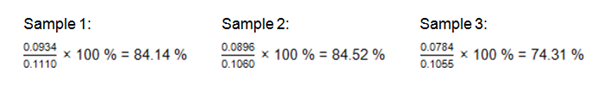
Microbead percentage recovery generated in the experimental procedure was carried out without the standard addition approach. Microbead percentage recovery from paste was calculated by dividing the number of microbeads recovered by the known numbers of microbeads added to the cream after carrying out the developed procedure.
By using microbead recovery equation in part 1of this study (section 1.2.4) and the data generated in Table 2, the following is deduced:
Interpretation of Microbead Infrared Spectroscopy
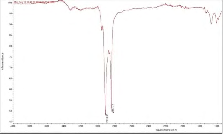
The superficial composition of the beads was characterised with IR as presented in Figures 1-4. The infrared of a dried paste from Clean and Clear face wash was carried out as presented in Figure 1. This was done in order to figure out the possible composition of the paste. The very first sharp peak at 2915.58 cm-1 is for aromatic C–H bonds and the peak at 2847.77 cm-1 is for aliphatic C–H bonds. It is important to note the absence of amines and carbonyls, both of which are polar groups. Therefore, this may explain why NaOH did not disintegrate the paste as there are no carboxylic acids present.
The infrared of the microbeads extracted from the face and body scrubs were also carried out during the study. With reference to the microbeads from the Neutrogena face scrub in Figure 2, the very sharp peak at 2914.72 cm-1 corresponds to sp3 C–H bond, while the 2847.37 cm-1 peak corresponds to sp2 C–H bond. From this infrared spectrum, it can be deduced that these two peaks could infer that the microbeads are synthesised from polyethylene as the FTIR spectrum of PE has absorbance bands located exactly at 2914 cm-1 and 2847 cm-1 [54].
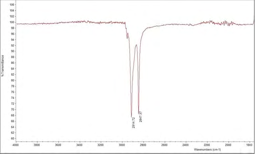
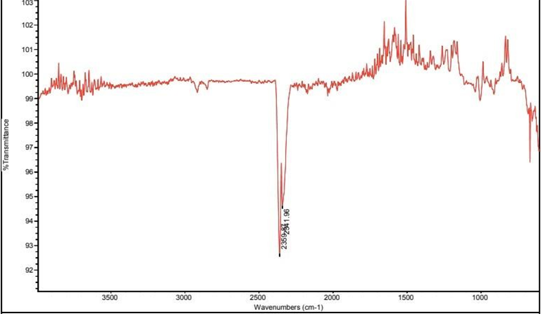
Figure 3 presents the IR of the microbeads extracted from Real Shaving Co. face scrub. There is a strong and sharp peak at 2359.87 cm-1 and a weak band at 2341.96 cm-1. Peaks between 1500 – 2500 cm-1 correspond to C–C aromatic bonds which could indicate the presence of a benzene ring which is a functional group present in polystyrene.
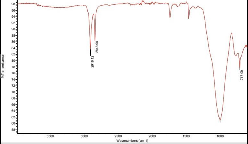
Microbeads analysed from Senspa detox body scrub as shown in Figure 4, have sharp but weak peaks at 2916.12 cm-1 and 2848.90 cm-1 which correspond to sp3– hybridised C–H alkane. In addition, the peak between 985 – 1000 cm-1 could indicate the presence of mono substituted C=C alkene. All microbeads analysed from the face and body scrubs show an absence of any 3200 – 3400 cm-1 O–H alcohol bands or any N–H stretch.
Microbeads Under Light Microscopy
An essential component for the determination of microbeads from cosmetic pastes is to observe the microbeads in a way that enables the analysis both quantitatively and qualitatively. Below (Figure 5), two images were taken per cosmetic paste and it can be observed that Neutrogena face scrub (images A1 and A2) had two different types of microbeads. A 1 & 2 – Neutrogena Daily Scrub B 1 & 2 – Real Shaving Co. face scrub C 1 & 2 – Senspa Detox body scrub
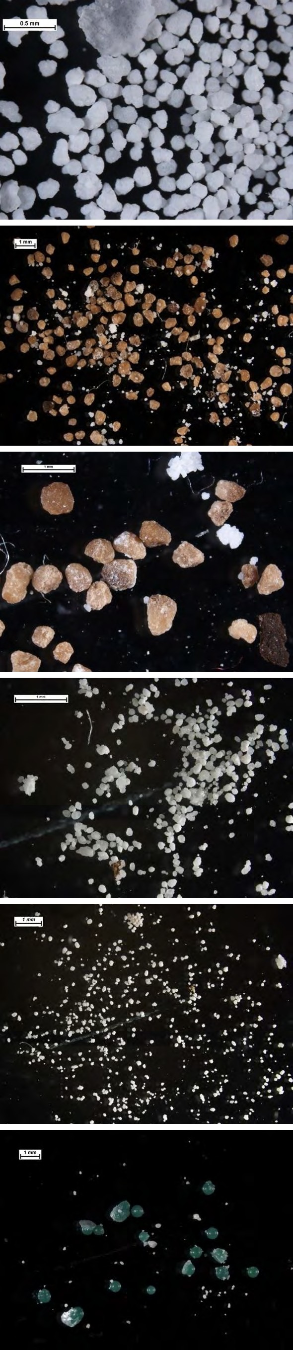
Observation of microbeads under light microscopy is vital as it allows seeing the sample at a very high magnification which means small details that are not visible to the naked eye can now be seen. Below (Table 3) is the qualitative and quantitative data generated from the images taken of the microbeads extracted using light microscopy.
| Brand | Mean (µm) | SD (µm) | Median size (µm) | Size range (µm) | Particle appearance |
|---|---|---|---|---|---|
| Neutrogena face scrub | 157.15 | 55.97 | 142.9 | 71.40 – 285.70 | White, slightly granular, green, rounded |
| RS Co. face scrub | 701.33 | 144.69 | 680 | 440 – 960 | Brown, smooth, slightly rounded |
| Senspa detox body scrub | 116.05 | 38.58 | 111.11 | 74.07 – 185.19 | White, uniform, spherical, rough surface |
Table 3: Characteristics of microbeads in three brands of face and body washes.
(N = 30 microbeads per brand) Table 3: Characteristics of microbeads in three brands of face and body washes.
The microbeads contained in the facial and body cleansers are spherical, granular and vary in terms of texture. The Neutrogena daily face scrub showed a wide range of microbead size with the average microbead being the size of approximately 160 µm with few larger than 200µm. When analyzing the Neutrogena daily face scrub, two types of microbeads can be observed (Figure 5, images A1 and A2). The white coloured microbeads are slightly granular whilst the green microbeads are smoother and rounded. The microbeads extracted from the Real Shaving Co. face scrub were of a larger size (with the mean size being 701.33 µm) than those extracted from Neutrogena face scrub (157.15 µm size). The Real Shaving Co. microbeads were brown in colour and slightly rounded. When quantitatively analyzing the Real Shaving Co. microbeads, they display a larger standard deviation than the microbeads of the other two face and body scrubs, indicating that there is a larger range in terms of microbead size. Microbeads extracted from Senspa detox body scrub have a mean size value of 116.05 µm and a very small standard deviation in comparison to the other two bodies and face wash. It can be deduced, therefore, that the microbeads added into Senspa detox body scrub are of consistent sizes.
Discussion
From this study’s experimental procedure carried out using the developed methodology for the extraction of microbeads form pastes, microbead recovery for each cosmetic paste used can be identified and therefore, the percentage of microbeads was calculated per product. It was found that ranging between 1.98 % to 9.06 % (Table 4). This range corresponds to Gouin T, et al. [55], where the study states that PCPs usually comprise of 0.05 % to 12 % of microbeads.
| Product | Recovery in 1.2 g | Recovery in 1 g | Microbeads in product | (%) of microbeads per product |
|---|---|---|---|---|
| Neutrogena Daily face scrub 150 mL | 0.0816/0.9464= 0.0862 | 0.0718 | 150 mL x 0.0718= 10.77 g | 7.18% |
| RS Co. face scrub 100 mL | 0.0203/0.8509= 0.0238 | 0.0198 | 100 mL x 0.0198= 1.98 g | 1.98% |
| Senspa Detox body scrub 200 mL | 0.1005/0.9230= 0.1088 | 0.0906 | 200 mL x 0.0906= 18.12 g | 9.06% |
Table 4: Calculations of microbeads present in each of the PCPs used in the standard additions approach.
Since 1.2 g of paste was used to enumerate the recovery of microbeads, this value can be used to find recovery in 1 g of paste and consequently calculate microbeads present in the total weight in grams of each PCP used in this study. From the experimental procedure carried out, Table 4 presents the calculations to find the number of microbeads and the percentage of microbeads in each product. The percentage of microbeads per product ranged from 1.98 % to 9.06 %.
Although Hidalgo-Ruz, et al. review [42] proposed methods that could not be directly applied to pastes, identical observations in relation to Hidalgo-Ruz, et al. review were made [42]. In his study, it is mentioned that the difference in density between the microbeads and other sediments was exploited to separate the lighter microbeads from the heavier sediment grains [42]. This was done by adding NaCl solution and shaking the mixture. After mixing, the sediment rapidly settled to the bottom, while the low-density microbeads remained in suspension or floated to the surface of the solution.8 similar observations were made in this research project where once hot distilled water was added to the paste sample and stirred vigorously using a glass stirring rod, the microbeads floated to the surface of the solution. This observation proposes the fact that microbeads released into the aquatic ecosystem will float on the water surface, and therefore become readily available to a wide variety of planktonic organisms feeding in the euphotic zone, as well as fish and seabirds that feed at the water surface.
Plastic microbeads extracted from cosmetic pastes were analysed under IR (Figures 1-4) and found to be hydrophobic. It can be observed that there are many C–H bonds present and no alcohols O–H. This indicates that the hydrophobic microbeads are completely inside the hydrophobic paste and therefore makes it challenging to extract. If the microbeads were hydrophilic there would have been repulsion between the plastic beads and the paste when water is added. Therefore, this research proposes the most difficult case which was to remove hydrophobic beads from hydrophobic matrix using heating.
Conclusion
In this study, it was observed that NaOH dissolves plastic microbeads, and since surface water of rivers tend to be basic (pH= 8), it can be reasoned that although microbeads may be stable, they may dissolve thus release precursors of the polymers. Moreover, through light microscopy, the particle size found in commercial products was less than 1 mm. These microplastics are of a size range that are easily ingested by at least 267 marine species and therefore cause entanglement in their GI tract. Once microplastics are ingested, they are translocated to the circulatory system and persist for more than 48 days. Marine animals consuming microplastics are at particular risk from reduced food consumption, starvation, or intestinal blockage leading to death. Furthermore, they accumulate in species of pelagic fish that are consumed by humans and therefore their persistence in the ecosystem could potentially lead to unknown harm to humans in the long-term.
References
-
Wilkinson J, Hooda P, Barker J, Barton S, Swinden J (2017) Occurrence, fate and transformation of emerging contaminants in water: An overarching review of the field. Environmental Pollution 231: 954-970.
-
Veillette J, Muir DC, Antoniades D, Small JM, Spencer C, et al. (2012) Perfluorinated chemicals in meromictic lakes on the northern coast of Ellesmere Island, High Arctic Canada. Arctic 65(3): 245-256.
-
Filipovic M, Laudon H, McLachlan M, Berger Urs (2015) Mass Balance of Perfluorinated Alkyl Acids in a Pristine Boreal Catchment. Environmental Science & Technology 49(20): 12127-12135.
-
Benotti MJ, Brownawell B (2009) Microbial degradation of pharmaceuticals in estuarine and coastal seawater. Environ. Pollut 157(3): 994-1002.
-
Mahmoud MA, Karrman A, Oono S, Harada KH, Koizumi A (2009) Polyfluorinated telomers in precipitation and surface water in an urban area of Japan. Chemosphere 74(3): 467-472.
-
Silva BFd, Jelic A, Lopez-Serna R, Mozeto AA, Petrovic M, et al. (2011) Occurrence and distribution of pharmaceuticals in surface water, suspended solids and sediments of the Ebro River Basin, Spain. Chemosphere 85(8): 1331-1339.
-
Tijani JO, Fatoba OO, Babajide OO, Petrik LF (2016) Pharmaceuticals, endocrine disruptors, personal care products, nanomaterials and perfluorinated pollutants: a review. Environ. Chem Lett 14(1): 27-49.
-
Mato Y, Isobe T, Takada H, Kanehiro H, Ohtake C (2001) Plastic Resin Pellets as a Transport Medium for Toxic Chemicals in the Marine Environment. Environmental Science & Technology 35(2): 318-324.
-
Teuten E, Saquing J, Knappe D, Barlaz M, Jonsson S, et al. (2009) Transport and release of chemicals from plastics to the environment and to wildlife. Philosophical Transactions of the Royal Society B: Biological Sciences 364(1526): 2027-2045.
-
Hirai H, Takada H, Ogata Y, Yamashita R, Mizukawa K, et al. (2011) Organic micropollutants in marine plastics debris from the open ocean and remote and urban beaches. Marine Pollution Bulletin 62(8): 1683-1692.
-
Wegner A, Besseling E, Foekema E, Kamermans P, Koelmans A (2012) Effects of nanopolystyrene on the feeding behavior of the blue mussel (_Mytilus edulis_ L.). Environmental Toxicology and Chemistry 31(11): 2490- 2497.
-
Rochman C (2016) The Role of Plastic Debris as Another Source of Hazardous Chemicals in Lower-Trophic Level Organisms. The Handbook of Environmental Chemistry.
-
(2018) Can marine debris degrade on its own in the environment?
-
(2015) GESAMP. Sources, Fates and Effects of Microplastics in the Marine Environment: A Global Assessment.
-
Patel MM, Goyal BR, Bhadada V, Bhatt JS, Amin AF (2009) Getting into the brain: approaches to enhance brain drug delivery. CNS Drugs 23(1): 35-58.
-
Zbyszewski M, Corcoran PL, Hockin A (2014) Comparison of the distribution and degradation of plastic debris along shorelines of the Great Lakes, North America. J Great Lakes Res 40(2): 288–299.
-
Setälä O, Fleming-Lehtinen V, Lehtiniemi M (2014) Ingestion and transfer of microplastics in the planktonic food web. Environ Pollut 185: 77-83.
-
Davison P, Asch R (2011) Plastic ingestion by mesopelagic fishes in the North Pacific Subtropical Gyre. Marine Ecology Progress Series 432: 173-180.
-
Murray F, Cowie P (2011) Plastic contamination in the decapod crustacean Nephrops norvegicus (Linnaeus, 1758). Marine Pollution Bulletin 62(6): 1207-1217.
-
Cole M, Lindeque P, Fileman E, Halsband C, Goodhead R, et al. (2013) Microplastic Ingestion by Zooplankton. Environmental Science & Technology 47(12): 6646- 6655.
-
Browne MA, Dissanayake A, Galloway TS, Lowe DM, Thompson RC (2008) Ingested microscopic plastic translocates to the circulatory system of the mussel, Mytilus edulis (L) Environ Sci Technol 42: 5026-5031.
-
Wardrop P, Shimeta J, Nugegoda D, Morrison PD, Miranda A, et al. (2016) Chemical Pollutants Sorbed to Ingested Microbeads from Personal Care Products Accumulate in Fish. Environ Sci Technol 50(7): 4037-4044.
-
Lee K, Shim W, Kwon O, Kang J (2013) Size-Dependent Effects of Micro Polystyrene Particles in the Marine Copepod Tigriopus japonicus. Environmental Science & Technology 47(19): 11278-11283.
-
Chua EM, Shimeta J, Nugegoda D, Morrison PD, Clarke BO (2014) Assimilation of polybrominated diphenyl ethers from microplastics by the marine amphipod, Allorchestes compressa. Environ Sci Technol 48(14): 8127-8134.
-
Von Moos N, Burkhardt-Holm P, Köhler A (2012) Uptake and effects of microplastics on cells and tissue of the blue mussel Mytilus edulis L. after an experimental exposure. Environmental science & technology 46(20): 11327-11335.
-
Rosenkranz P, Chaudhry Q, Stone V, Fernandes TF (2009) A comparison of nanoparticle and fine particle uptake by Daphnia magna. Environ Toxicol Chem 28(10): 2142- 2149.
-
Hämer J, Gutow L, Köhler A, Saborowski R (2014) Fate of microplastics in the marine isopod Idotea emarginata. Environ Sci Technol 48(22): 13451-13458.
-
Farrell P, Nelson K (2013) Trophic level transfer of microplastic: Mytilus edulis (L.) to Carcinus maenas (L.) Environ Pollut 177: 1-3.
-
Derraik J (2002) The pollution of the marine environment by plastic debris: a review. Marine Pollution Bulletin 44(9): 842-852.
-
Bouwmeester H, Hollman P, Peters R (2015) Potential Health Impact of Environmentally Released Micro- and Nanoplastics in the Human Food Production Chain: Experiences from Nanotoxicology. Environmental Science & Technology 49(15): 8932-8947.
-
EFSA Panel on Contaminants in the Food Chain (CONTAM) (2016) Presence of microplastics and nanoplastics in food, with particular focus on seafood. EFSA Journal 14(6): 4501.
-
Ohta S, Ishizuka D, Nishimura H, Nakao T, Aozasa O, et al. (2002) Comparison of polybrominated diphenyl ethers in fish, vegetables, and meats and levels in human milk of nursing women in Japan. Chemosphere 46(5): 689-696.
-
Sutherland W, Clout M, Côté I, Daszak P, Depledge M, et al. (2010) A horizon scan of global conservation issues for 2010. Trends in Ecology & Evolution 25(1): 1-7.
-
Wright S, Thompson R, Galloway T (2013) The physical impacts of microplastics on marine organisms: A review. Environmental Pollution 178: 483-492.
-
Anderson A, Grose J, Pahl S, Thompson R, Wyles K (2016) Microplastics in personal care products: Exploring perceptions of environmentalists, beauticians and students. Marine Pollution Bulletin 113(1-2): 454-460.
-
Browne MA, Crump P, Niven SJ, Teuten E, Tonkin A, Galloway T, et al. (2011) Accumulation of microplastic on shorelines worldwide: Sources and sinks. Environ Sci Technol 45(21): 9175-9179.
-
Cheung P, Fok L (2017) Characterisation of plastic microbeads in facial scrubs and their estimated emissions in Mainland China. Water Research 122: 53- 61.
-
(2018) Microbeads, 5Gyres.org.
-
(2018) The Beat the Microbead campaign is supported by 100 NGOs from 42.
-
Zitko V, Hanlon M (1991) Another source of pollution by plastics: Skin cleaners with plastic scrubbers. Marine Pollution Bulletin 22(1): 41-42.
-
Gregory M (1996) Plastic ‘scrubbers’ in hand cleansers: a further (and minor) source for marine pollution identified. Marine Pollution Bulletin 32(12): 867-871.
-
Hidalgo-Ruz V, Gutow L, Thompson R, Thiel M (2012) Microplastics in the Marine Environment: A Review of the Methods Used for Identification and Quantification. Environmental Science & Technology 46(6): 3060-3075.
-
Rios L, Jones P, Moore C, Narayan U (2010) Quantitation of persistent organic pollutants adsorbed on plastic debris from the Northern Pacific Gyre’s eastern garbage patch. Journal of Environmental Monitoring 12(12): 2226.
-
Doyle M, Watson W, Bowlin N, Sheavly S (2011) Plastic particles in coastal pelagic ecosystems of the Northeast Pacific ocean. Marine Environmental Research 71(1): 41-52.
-
Hintersteiner I, Himmelsbach M, Buchberger WW (2015) Characterization and quantitation of polyolefin microplastics in personal-care products using high- temperature gel-permeation chromatography. Anal Bioanal Chem 407(4): 1253-1259.
-
Wardrop P, Shimeta J, Nugegoda D, Morrison PD, Miranda A, et al. (2016) Chemical Pollutants Sorbed to Ingested Microbeads from Personal Care Products Accumulate in Fish. Environ Sci Technol. 50(7): 4037-4044.
-
Imhof H, Schmid J, Niessner R, Ivleva N, Laforsch C (2012) A novel, highly efficient method for the separation and quantification of plastic particles in sediments of aquatic environments. Limnology and Oceanography: Methods 10(7): 524-537.
-
Fendall L, Sewell M (2009) Contributing to marine pollution by washing your face: Microplastics in facial cleansers. Marine Pollution Bulletin 58(8): 1225-1228.
-
Thompson RC, Olsen Y, Mitchell RP, Davis A, Rowland SJ, et al. (2004) Lost at sea: where is all the plastic?. Science 304(5672): 838.
-
Ng K, Obbard J (2006) Prevalence of microplastics in Singapore’s coastal marine environment. Marine Pollution Bulletin 52(7): 761-767.
-
Sweden KI (2007) Small plastic particles in Coastal Swedish waters. N-Research report, commissioned by KIMO Sweden, pp: 1-11.
-
Skoog DA (2006) Principles of Instrumental Analysis, 6th(edn), Thompson Brooks/Cole: Belmont (Chapter 28), California, pp: 33.
-
HA Leslie (2014) Review of Microplastics in Cosmetics - Scientific background on a potential source of plastic particulate marine litter to support decision-making. IVM Institute for Environmental Studies.
-
Ronald PD’Amelia, Gentile S, Nirode F. W., Huang L (2016) Quantitative Analysis of Copolymers and Blends of Polyvinyl Acetate (PVAc) Using Fourier Transform Infrared Spectroscopy (FTIR) and Elemental Analysis (EA). World Journal of Chemical Education 4(2): 25-31.
-
Gouin T, Avalos J, Brunning I, Brzuska K, de Graaf J, et al. (2015) Use of micro-plastics beads in cosmetics products in Europe and their estimated emissions to the North Sea environment. SOFW J 141: 40-46.
- Hydrogen Peroxide Scavenging by Methanolic Extracts of Coriander: An In Vitro Antioxidant Study
- Aromatherapy in Palliative Care: A Fragrant Quest for Relief
- Empowering Women, Securing Futures: Contraception’s Role in Socioeconomic Progress in India
- Effect of Crospovidone, Croscaramellose Sodium in Combination on the Drug Release of Anti diabetic Medication in Tablet Form
- Knowledge, Attitudes, Anxiety, and Preventive Behaviors Regarding Covid-19 Affliction among Healthcare Workers in Pakistan
- “Competitive Landscape and Brand Equivalents: Implications for ANDA (Abbreviated New Drug Application) Approval”