A Neonate with Amniotic Band Syndrome and Osteomyelitis
Background: Amniotic band syndrome (ABS) comprises of a spectrum of congenital malformations involving the limbs more often, but any other anatomic region of fetus could be involved. Its prevalence has been reported to range from 0.19 to 8.1 per 10000 live births. There are different theories for explaining the etiology of amniotic bands, but it has been considered to have a multifactorial etiology. Fetoscopic release of amniotic bands affecting one or more extremities can prevent limb loss or preserve limb function when distal fellow is still identifiable on preoperative ultrasound scans. ABS occurs in 77% of fetuses with multiple anomalies. Both males and females are equally affected. Osteomyelitis in neonates is relatively uncommon, but burdened with an increased hospital stay and possible long‐term sequela if not diagnosed on time. The long bones are the most frequently affected sites. Case Presentation: Here we are introducing a neonate with left upper extremity necrosis due to ABS which needed amputation and it was associated with severe chronic osteomyelitis. Conclusion: Amniotic band syndrome mostly involves the limbs. It is important to diagnose it as soon as possible, because with the interprofessional teamwork approach, an optimal patient result with fetoscopic or surgery procedures could be achieved.
Introduction
Amniotic band syndrome (ABS) is a set of congenital malformations. Amniotic Band Sequence, Streeter’s dysplasia and ABS are the other names used for this condition. These congenital malformations occur due to fibrous amniotic bands [1]. ABS with multifactorial etiology is a congenital anomaly of the fetus, in which the fetal body parts have deformities due to amniotic band wrapped around them, resulting in fetal structural abnormalities and dysfunctions [2]. Usually the diagnosis is made by deformities that are consistent with amniotic bands (such as limb deformities) in the absence of visible bands [3]. Prenatal diagnosis of ABS mainly depends on fetal ultrasound (US) scans [2]. But it is difficult to identify the amniotic bands with standard prenatal imaging, including ultrasound and MRI [3]. Its prevalence has been reported to range from 0.19 to 8.1 per 10000 live births. There are many theories about the etiology of ABS but none of them support the others, so it might have a multifactorial etiology [4]. As the fetus grow up, amniotic bands wrap the extremities and can cause deformity or amputation of the affected limbs, whereas more complex bands involving the abdominal wall can lead to large abdominal wall defects mimicking gastroschisis or omphalocele [1, 3]. Amniotic band syndrome has shown to be associated with hydranencephaly, porencephaly, craniofacial abnormalities and spinal dysraphism [5].
Most cases are sporadic with no recurrence in siblings of affected patients [6]. Karyotyping is important in counseling the parents about risk of recurrence [4]. Affected neonates need individualized multidisciplinary approach. In fact, amniotic bands isolated to the extremities remain the primary indication for fetoscopic band release. If the bands are interrupting other anatomic regions, they have usually made irreversible defects at the time of diagnosis, and currently there does not appear to be any indication for fetal intervention in those cases [3]. In some cases, post-natal surgical repair is suggested [4]. Osteomyelitis in neonates is usually due to hematogenous spread of bacterial infections or less frequently to direct inoculation as a result of a trauma or surgery [7]. Common symptoms include bone pain, swelling, redness, guarding and failure to move the affected body part (pseudoparalysis). The causative pathogens are different in different countries but the most common pathogen is Staphylococcus aureus, found in 70–90% of culture positive cases [7], Streptococcus mainly group B and Gram-negative enteric bacteria (Escherichia coli and Klebsiella pneumonia and Pseudomonas aeruginosa) are responsible in other cases [8, 9]. Acute haematogenous OM is usually defined acute if the signs or symptoms are present for less than 14 days, and subacute or chronic if signs or symptoms are present for more than 14 days [10]. Imaging (computed tomography (CT) scan, radiography, bone scan, US and/or MRI) is used to identify the site of an infection, the presence of liquid collections for diagnostic aspiration and/ or biopsy, to differentiate a unifocal from multifocal disease and to identify present or impending complications, such as joint or extradural involvement [7, 11].
Case Presentation
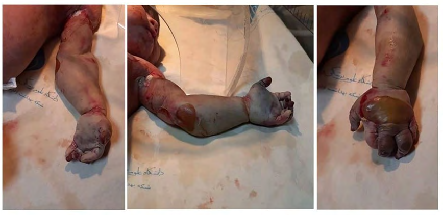
A male infant who was delivered at 35th week and 6 days of pregnancy, by normal vaginal delivery with birth weight of 3250 grams and height of 52 cm. He had normal Apgar score. His mother had gestational diabetes and she was treated by insulin (GDM). He had passed meconium intrauterine but did not need any resuscitation at birth. At birth, his left upper extremity was necrotized probably with amniotic band from proximal humerus to the distal of fingers and it had many wounds on dorsal hand and lateral of forearm. His hand was painful, cold, pulseless and without any movement. Other examinations were normal. His mother`s ultrasonographic imaging in 26th week of pregnancy was reported normal. He had normal vital signs. He was admitted to neonatal ward and after performing a sepsis work-up, antibiotic therapy was started with meropenem and vancomycin. An umbilical catheter was inserted and he was transferred to another hospital for consulting about amputation surgery. After hematologist consultant, Heparin was started and Protein C, Protein S, Factor 5 Leyden, fibrinogen, anti thrombin and D dimer were controlled that all of them were normal. His hand was amputated distal to shoulder at 8th day of life in Hazrat Rasool hospital. After surgery he was slightly tachypneic and he received oxygen with hood which was discontinued after 2 days. The entire lab tests were normal (Tables 1 & 2). At the 10th day of life (two days after surgery), he had weakness and poor feeding and a consultation with pediatric infectious specialist was requested. It was recommended to change the antibiotics to ciprofloxacin, linezolid and metronidazole and an MRI image of amputation site be performed and smear and culture of wound was requested.
| WBC:25200(70% segment,25% lymph) | Platelet: 189000 | Bs:80 |
|---|---|---|
| Bun: 15 mg/dl | ||
| Cr: 1.3 mg/dl | ||
| Hb:14.6 g/L | CRP: 12 rising to 26 | Na: 140 mEq/L |
| K: 5.1 mEq/L | ||
| Hct :45.2% | PT:14.7 s | Ca:8.2 mg/dl |
| PTT:37s INR:1.07 | Mg: 2.1 mg/dl | |
| Blood culture: negative | AST:75 ALT:32 | CPK:1623 |
| Wound Culture: negative | LDH: 1435 |
Table 1: Lab tests.
| WBC:12800(1% band,42% segment, 31% lymph) | Plt:608000 | Na: 138 mEq/L |
|---|---|---|
| Hb:12.2 g/L | CRP less than 6 | K:4.7 mEq/L |
| Hct:35.4% | Bun:9 mg/dl Cr: 0.4 mg/dl |
Table 2: Lab tests after surgery.
The MRI result showed evidence of bone marrow edema in distal of humerus and soft tissue of amputation stump with cortical thickening in distal of humerus in favor of chronic osteomyelitis. Brain and abdominal ultra sonographic scans were normal. Vital signs and general condition of our infant improved after changing the antibiotic therapy, and he was treated for 21 days. The heparin therapy was withdrawn at 14th days after surgery. He was discharged at 24th day of life with good condition and without any antibiotic. After two weeks the neonate was visited again, he was well and there was no problem with the amputation site and the infectious (Figure 1).
Discussion
Amniotic band syndrome (ABS) is a rare birth defect caused by strands of the amniotic sac that separaqte and entangle digits, limbs, or other parts of the fetus. This condition can cause a variety of problems depending on where strands are located and how tightly they are wrapped. It can cause a variety of congenital anomalies which includes disruption, deformation, and malformations of organs which were intended to develop normally [12]. Different theories have attempted to explain the etiology of amniotic band sequence; however, none has individually been able to support each and every defect observed, so it has been considered to be a multifactorial condition [4]. Feto- maternal factors like prematurity, maternal illnesses, low birth weight and maternal drug exposure are predisposing factors for this syndrome [13], although the risk factors are poorly known till now. The approach to ABS is affected by identifying the pre and post natal anomalies. The amniotic bands which are isolated are released by fetoscopy and if it is associated with other anomalies, performing a post-natal surgery repair is recommended. It is also helpful to provide genetic counseling [4]. Clinical presentation can classify into four major categories. Constrictive rings/limb defects/ neural or spine defects/craniofacial defects. The distribution of amniotic bands is also different, for example extremity involvement can account for more than 70% of cases, but any other anatomic region of the fetus can be involved (umbilical cord, abdomen, head, chest, limb-body-wall-complex) [3]. The amniotic bands may be seen confined to the skin or soft tissue and may extend deep into the tissue. If constriction results in amputation in-utero, then amputated part is usually resorbed and not visible after birth. There are no standard guidelines of management of pregnancy complicated with fetal ABS. Follow-up, ultrasound scans and intervention are individually tailored and depend on the severity of ABS complications [14].
The in-utero intervention of amniotic band lysis has the potential to slow down the progression of the effect of constriction and restoring normal flow to the downstream organ. There is a hypothesis that fetal limb recovery is more likely after fetal intervention than postnatal recovery because of the plasticity of tissue healing during fetal life. Although fetoscopic intervention may restore blood flow and save the limb, plastic surgery may still be necessary after birth. However, the efficacy of this intervention is unknown, as there are no set criteria for the selection of candidates and a lack of clinical studies [15]. Prognosis depends on the extension and severity of the defects. Parents should receive counseling from the nursing and clinical interprofessional team. Osteomyelitis (OM) is a severe neonatal infection which usually has a predisposing factor and necrosis of soft tissue and limb surgeries could be a trigger for late infections. The most significant pathogens are those involved in neonatal sepsis with Staphylococcus aureus, found in 70–90% of culture positive cases. Long lasting sequels could be observed if it is not appropriately treated and even with best treatments, shortening of the affected limbs could be observed. Osteomyelitis may become responsible for permanent sequelae in 6–50% like joint disabilities, change in bone growth due to the damage of the cartilaginous growth plate, limb length discrepancies, arthritis, pathologic fractures, and rarely complete destruction of joints. MRI has become the gold standard to evaluate musculoskeletal infection and the positive rate of MRI in detecting osteomyelitis was 100% [16]. It has the capability of assessing the osseous, articular and muscular structures simultaneously and does not require ionizing radiation [7]. Increased marrow intensity with surrounding inflammation are the most suggestive signs of OM. Radiography is usually the first radiological investigation in a neonate with suspected OM, although it is reported that only 20% of the radiographs are abnormal at 10–14 days [16, 17, 18, 19, 20, 21].
There were several differential diagnoses in our case presentation such as congenital malformation, protein c and protein s deficiency, DIC due to sever septicemia, amniotic band syndrome, diabetes of mother. According to the normal range of coagulation proteins, the diagnosis of protein c and s deficiency was omitted. We ruled out the septicemia due to the neonate’s sign and symptoms and the lab data. Based on the mother’s history and the lab tests during pregnancy the diagnosis of GDM was well defined. On the other hand, we surely attributed the diagnosis to the amniotic band syndrome since the amniotic band was whirled toward the humerus meanwhile the delivery.
Conclusion
Amniotic band syndrome mostly involves the limbs. It is important to diagnose it as soon as possible, because with the interprofessional team work approach, an optimal patient result with fetoscopic or surgery procedures could be achieved. Even after surgery, some complications such as osteomyelitis could prolong the recovery process and it might need long term occupational and physiotherapic interventions and limb replacement treatments.
Declaration
Ethic approval and consent to participants: the study received approval form the institutional review board of IUMS
References
-
Kleigman RM, Stanton BF, Geme JWS, Schor NF, Behrman RE (2011) Nelson Text Book of Paediatrics. 19th (Edn.), Saunders, New Delhi, pp: 1-142.
-
Niu Z, Meng H, Zhang X, Ouyang Y, Zhang Y, et al. (2019) Two case reports, Early detection of amniotic band syndrome by adhesion between hand and umbilical cord at 11 to 14 weeks’ gestation. Medicine (Baltimore) 98(50): e18302.
-
Richard JM, Avroy AF, Michele CW (2020) Fanaroff and Martin`s Neonatal-perinatal medicine. 11th(Edn.), Elsevier, Philadelphia.
-
Muñoz EL, Solano LEB (2018) An update on amniotic bands sequence. Arch Argent Pediatr 116(3): e409-e420.
-
Hata T, Tanaka H, Noguchi J (2011) 3D/4D sonographic evaluation of amniotic band syndrome in early pregnancy: a supplement to 2D ultrasound. J Obstet Gynaecol Res 37(6): 656-660.
-
Allington NJ, Kumar SJ, Guille JT (1995) Clubfeet associated with congenital constriction bands of the ipsilateral lower extremity. J Pediatr Orthop 15(5): 599- 603.
-
Decembrino L, Decembrino N, Stronati M (2017) Neonatal Osteomyelitis. In: Mauricio Barría R (Ed.), Selected Topics in Neonatal Care. IntechOpen, pp: 97- 107.
-
Qadir M, Ali SR, Lakhani M, Hashmi P, Amirali A (2010) Klebsiella. osteomyelitis of the right. humerus involving the right shoulder joint in an infant. J Pak Med Assoc 60(9): 769-771.
-
Korakaki E, Aligizakis A, Manoura A, Hatzidaki E, Saitakis E, et al. (2007) Methicillin-resistant Staphylococcus aureus. osteomyelitis and septic arthritis in neonates: Diagnosis and management. Jpn J Infect Dis 60(2-3): 129-131.
-
Berberian G, Firpo V, Soto A, Lopez MJ, Torroija C, et al. (2010) Osteoarthritis in the neonate: Risk factors and outcome. Braz J Infect Dis 14(4): 413-418.
-
Montgomery CO, Siegel E, Blasier RD, Suva LJ (2013) Concurrent septic arthritis and osteomyelitis in children. J Pediatr Orthop 33(4): 464-467.
-
Seeds JW, Cefalo RC, Herbert WN (1982) Amniotic band syndrome. American J obstetric Gynecol 144(3): 243- 248.
-
Foulkes GD, Reinker K (1994) Congenital Constriction Band Syndrome: A seventy-year Experience. J Pediatr Orthop 14(2): 242-248.
-
Gueneuc A, Chalouhi GE, Borali D, Mediouni I, Stirnemann J, et al. (2019) Fetoscopic Release of Amniotic Bands Causing Limb Constriction: Case Series and Review of the Literature. Fetal Diagn Ther 46(4): 246-256.
-
Javadian P, Shamshirsaz AA, Haeri S, Ruano R, Ramin SM, et al. (2013) Perinatal outcome after fetoscopic release of amniotic bands: a single-center experience and review of the literature. Ultrasound Obstet Gynecol 42(4): 449- 455.
-
Zhan C, Zhou B, Du J, Chen L (2019) Clinical analysis of 17 cases of neonatal osteomyelitis, A retrospective study. Medicine (Baltimore) 98(2): e14129.
-
Hind A, Chaara H, Attar I, Jayi S, Alaoui FZF, et al. (2019) Amniotic band syndrome: prenatal diagnosis and management challenges (about 2 cases of lethal malformations). Pan Afr Med J 32: 116.
-
Derderian SC, Iqbal CW, Goldstein R, Lee H, Hirose S (2014) Fetoscopic approach to amniotic band syndrome. J Pediatr Surg 49(2): 359-362.
-
Nyberg DA, Mahony BS, Pretorius DH (1991) Diagnostic ultrasound of fetal anomalies. The New England Journal of Medicine, Boston, 324(13): 930-930.
-
Ronald PR, Jean LB, Joseph LJ (2009) Dermatology, 2nd Edition, 2-Volume Set. Dermatology Online Journal 15(1): 17.
-
Ajay PS, Sudheer RG (2022) Amniotic band syndrome. StatPearls Publishing, Treasure island.
- Understanding Pediatric Multiple Sclerosis: Clinical Presentation, Diagnostic Criteria, Therapeutic Advances, and Supportive Care Approaches
- Hemophilia in Children
- Xia-Gibbs Syndrome- A Case Report
- A Study to Assess Effectiveness of Play Therapy in Reducing Post-Operative Pain among Children Age 2 To 5 Year who have Undergone General Surgeries in Selected Pediatric Hospitals of Vadodara
- Preterm Birth: Scope of the Problem, Cost of Care, Potential Complications and Current Guidelines for Management
- Noradrenaline: Can we Use it to Manage Hemodynamic Instability among Neonatal Septic Shock at the NICU?