Study of Karyotyping and the Genotoxic Effects of Taxol Drug on the Somatic Bone Marrow Cells of Albino Wister Rats
In this study, the chromosome 2N was calculated for the somatic cells of the male albino (Albino Wister Rats) obtained from the Animal House at the Faculty of Science, Misurata University. The study also identified some chromosomal stages, such as the metaphase. The results indicated that the chromosome number 2N=42, in addition to measuring the total and relative length of the chromosomes, and then arrange the chromosomes descending on their lengths. The chromosomal abnormalities resulting from colchicine exposure (such as the scattered metaphase, the ring chromosome, the chromatid separation and the chromosomal break) were also shown, while the chromosomal abnormalities caused by the taxol treatment were: agglomerated colchicine metaphase, associated chromosomal endings, chromosomal viscosity, multiple chromosomal group and lack of chromosomal group).
Introduction
Cellular genetics studies provide more important and more accurate information than phenotype studies. Increasing knowledge about chromosomes can provide reliable information on the evolution of breeds, understanding and interpreting the best relationships between species, as well as their importance in classification, evolution and inheritance [1]. Chromosomes are threaded objects that appear when dark color is scattered in the nuclear sap. The chromosomes are derived from the ancient Greek language and composed of two sections, Chroma means color and Soma means body, and was used for the first time by researcher Waldyer in (1880) to refer to the structures of the thread in the nucleus. In 1903 Sutton pointed out that genes are based on these structures. In Study of Karyotyping and the Genotoxic Effects of Taxol Drug on the Somatic Bone Marrow Cells of Albino Wister Rats his experiments on the drosophila, Morgan has shown the presence of genes on chromosomes, which characterize these structures as self-replicating and maintain functional and morphological characteristics during cell division [2].
Analysis of the chromosome (the Karyotype or chromosome pattern) is important to describe numerical changes and structural changes in chromosomes, which can vary even between very close relatives. Each organism is characterized by its own chromosomal pattern. To determine the chromosome pattern, it is necessary to study the cells at the split stage. This study can be completed on bone marrow cells; the cells have sufficient numbers of cells in the dividing stage [3, 4, 5].
Virol Immunol J
When studying the karyotype, the chromosome number, which is fixed for one species of animal organism, must be defined. This number depends on the type, genetic and taxonomic relationships [6]. Several preliminary studies have indicated a significant variation in the chromosome double number, ranging from 38=2n to 42n=2 in different rat species of rattus Rattus, from different regions of the world [7, 8, 9]. Genotoxicity, a group of negative symptoms, and changes in the organism due to toxicity, cytogenetic cytogenetic symptoms, may, of course, be reflected in the activities and functions of the different organism [10]. Cytogenetic analysis is therefore necessary to detect cytogenetic analysis For various genetic diseases caused by major disorders or chromosomal abnormalities [11]. Several studies of chromosomal aberrations have indicated that all types of abnormal variables appear on chromosomes, which can be seen under the light microscope, such as numerical variables in chromosomes, which are the imbalances that lead to an increase or decrease in the normal number of chromosomes in cells, In aberrations-Numerical, while the change in shape of chromosome is known as structural-aberration [12]. The percentage of chromosomal abnormalities varies depending on the species of the mutagen, the amount of the exposed dose, the type of cells exposed, and the organism [13, 14]. Some chemical drugs used in treatments directly affect cell DNA, while others inhibit the cancer cell's use of the nutrients needed for division and reproduction, thereby disrupting the cell's life cycle in one of its phases, thus hindering its survival.
Colchicine is a drug used to treat gout, has effects on bone marrow cells, is also a cause of chromosomal abnormalities, and some damage to DNA, according to the manufacturer of the drug Wockhardt Company 1982. Colchicine is a chemical that stops the spindle thread and stabilizes cell division at the metaphase stage. The chromosomes are clear and large, and the cells are then treated with a hypotonic saline solution to increase cell capacity and separate the chromosomes [12]. Taxol is an anticancer drug used for the treatment of cancer, as it is derived from the bark of the trees Tksos Pacific [15], and the World Cancer Center referred to the most important cancer drug [16]; for a response in approximately 60% of people treated from Breast cancer, and ovarian cancer [17].
Cytogenetic Analyzes extensively used an indicator to detect the physical and chemical susceptibility of genetic mutations in the living cell. The changes that these factors make in the genetic material are an indication of the seriousness of these factors on the health of living organisms. Genetic analyzes, such as Chromosomal Aberration Assay, a cellular cytological analysis used to detect the ability of physical and chemical effects to induce structural chromosomal changes, that can be observed when the cell in the metaphase of the division, that is include mainly a chromosomal break, ring chromosome, and numerical changes [18]. The present study aimed to describe the chromosome pattern and calculate the 2N chromosome diploid number in the somatic cells of the bone marrow in the male albino rats. As well as the study of cellular genetic effects and structural and numerical chromosomal abnormalities resulting from one type of chemotherapy, known as Txol (mg / kg 1.7).
Materials and Methods
Experiment Animals
8 males of Albino Wister Rat were used in this study. Their weights ranged from 150 to 200 g. They were selected from the Animal House of the Department of Zoology at the Faculty of Science Misurata University (Libya). , and were randomly divided into two groups. Each group included four rats, were raised in plastic cages and in appropriate conditions, where 12 hours in light and 12 hours in darkness, at 25°C.
Materials
Solution (1) Potassium Chloride Solution (Hypotonic
KCL Solution) 0.075 %: Dissolve 0.56 g of potassium
chloride salt in 50 ml distilled water, then complete the
volume to 100 ml of distilled water and keep in the
incubator at 37°C, where this solution increases cell size.
Solution (2) Colchicin Solution: Was prepared with a
0.5 mL weight in 200 ml distilled water and was used
immediately after preparation. Each animal injected 2 mL
of this solution into the peritoneal membrane.
Solution (3) Taxol drug: Injected each animal with a
dose of 0.3 ml of this drug for 18 hours in the peritoneal
membrane, and the dose was calculated 1.7 mg / kg of the
following calculation equation, according to (2017):
$$ \text{Animal} \quad \text{equivalent} \quad \text{dose} = \text{human} \quad \text{dose} $$
concentration×5x conversion factor: The conversion
factor was calculated based on surface area and body
weight, which differ from one animal to another. The
conversion factor for a 200 g rat is 0.018, which was
adopted from Dhanarade and Pandya.
Solution (4) Fixative Solution: Immediately prepare the
solution when preparing the chromosome preparations
by mixing 3 volumes of methyl alcohol with one volume of acetic acid, and refrigerate at 4°C. Chloroform Solution was used to anesthetize experimental animals to ensure that the somatic cells were not damaged.
Methods
The animals were divided into two groups. Four animals were assigned to each group and injected through peritoneal membrane. In this group, animals were injected with 2 ml of colchicine solution. After 3 hours of injection, the animals of this group were anesthetized. The group was treated with Taxol. The animals were injected with Taxol (0.3 ml). The dose was 1.7 mg / kg for 18 hours and 3 hours after injection with 2 ml of colchicine was anesthetized in preparation for the subsequent step. Bone marrow cells were prepared according to the method described in Tresukosol D and Preston RJ [18, 19]. The rats were killed and explained to obtain femur from each rat by cutting these two bones in a cavity by acetabulum cavity, And Tibia on the other. The other muscles and tissues surrounding these bones were completely removed and cleaned thoroughly with distilled water. Inside a clean sterile petri dish , cut off the epiphyses of the bone with fine, sharp scissors to facilitate the entry and exit of the solution used in their cavity. Pump the KCL solution (0.075%) and receive the leachate in a 12 mL centrifuge tube using a 2 mL medical syringe. Repeat the bone rinse 2-3 times to ensure that all bone marrow cells are extracted in the tube, Incubator (100- 100 Beschickung loading model) at 37°C for 20 minutes. Transfer the leachate to the Centrifuge 5810 R at 1000 rpm for 5 minutes. Pull the solution out of the centrifuge tubes by pipette and with extreme caution, and keep the precipitate at the bottom of those tubes. 5 ml of the newly installed stabilizer, consisting of absolute ethyl alcohol and 3: 1 glacial acetic acid, was added to each tube by adding drops of the drip stabilizer on the pipe wall gradually, so that the tube is tilted with continuous stirring. Apply the solution well with a pipette and leave it for 20:30 min at room temperature before centrifuging it again at 1000 rpm for 5 minutes. Remove the top solution, retain the deposit and repeat the fixation 3 times. Added 0.5: 2 ml of the newly prepared fixation (depending on the amount of cellular precipitation) added 1: 2 drop of cellular suspension was dropped onto clean, dry glass microscopic slides and then left to dry.
The slides were stained for 15- minute Geimsa stain and rinsed in water to remove the stain. The slides were covered with appropriate glass covers, using D. P. X, and then left to dry in preparation for the subsequent step.
The slides were examined using a light microscope (Nicon-C-Sw), where the cells were scanned using the objective lens (X10), and the metaphases were then sorted using the oil lens (100x) to detect possible chromosomal abnormalities. The length of the chromosomes was calculated by Nuc Type program (version 1.5), the relative length of the chromosome by the following equation: Total length of chromosome / total lengths. Image Processing Software (Version 9.5 Pics art)
Results
Karyotype Analysis
The slides prepared for the somatic cells of the bone marrow were examined for the males of untreated albino rats and treated with taxol, chromosomes were seen in their normal state, as were the taxol-treated chromosomes (Figures 1 & 2).
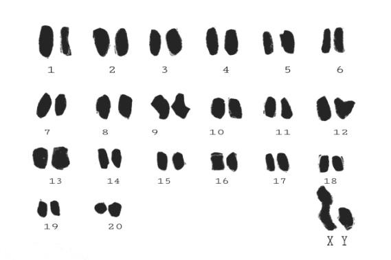
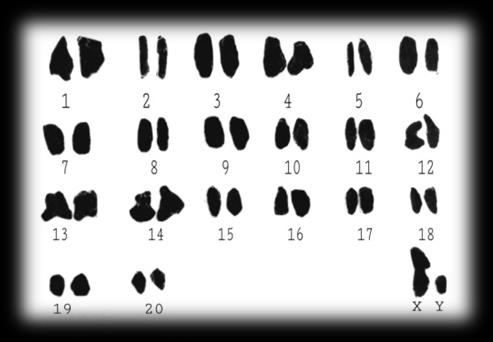
The current study showed that the double number of chromosomes was 42 = 2n. It is clear from Table 1 that the taxol-treated group chromosomes are 100% applicable Control group by chromosome number.
| Chromosome | Compatibility | ||||||
|---|---|---|---|---|---|---|---|
| Numerical | Appearance | ||||||
| Somatic | 100% | Length | Size | ||||
| 65% | 25% | ||||||
| Sexy | - | 0% | 0% |
Table 1: Degree of numerical and phenotypic compatibility of chromosomes in the somatic cells of experimental animals.
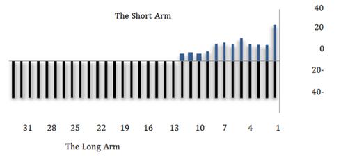
The total and relative length of chromosome 1 in the control group is 77.95 and 0.0325 μm respectively, while the same measurements for the corresponding chromosome in the treatment group were 105.10 and 0.0341 μm, respectively. Comparison of chromosome 40 in the control group and treatment with Taxol showed a difference between the total and relative length, with 39.23 and 0.0163 μm respectively in the control group, 51.17 and 0.0166 μm respectively in the treatment group (Table 2).
| Chromosome Couples Order | Control Group (Colchicine) | Treatment Group (Taxol) | ||||||||||
|---|---|---|---|---|---|---|---|---|---|---|---|---|
| Total Length | Relative Length | Total Length | Relative Length | |||||||||
| 1 | 77.95 | 0.0325 | 105.1 | 0.0341 | ||||||||
| 2 | 77.62 | 0.0323 | 105.08 | 0.0341 | ||||||||
| 3 | 73.84 | 0.0307 | 90.6 | 0.0294 | ||||||||
| 4 | 73.36 | 0.0305 | 90.02 | 0.0292 | ||||||||
| 5 | 68.65 | 0.0286 | 85.72 | 0.0278 | ||||||||
| 6 | 68.06 | 0.0283 | 85.21 | 0.0276 | ||||||||
| 7 | 67.78 | 0.0282 | 84.5 | 0.0274 | ||||||||
| 8 | 67.51 | 0.0281 | 84.08 | 0.0272 | ||||||||
| 9 | 65.87 | 0.0273 | 84.04 | 0.0272 | ||||||||
| 10 | 65.01 | 0.0271 | 84 | 0.0272 | ||||||||
| 11 | 62.81 | 0.0261 | 82.94 | 0.0269 | ||||||||
| 12 | 62.06 | 0.0259 | 82.45 | 0.0267 | ||||||||
| 13 | 58.96 | 0.0245 | 80.75 | 0.0262 | ||||||||
| 14 | 58.9 | 0.0245 | 80.62 | 0.0261 | ||||||||
| 15 | 58.76 | 0.0245 | 76.7 | 0.0248 | ||||||||
| 16 | 58.76 | 0.0245 | 76.62 | 0.0248 | ||||||||
| 17 | 58.26 | 0.0242 | 75.84 | 0.0246 | ||||||||
| 18 | 58.14 | 0.0242 | 75.53 | 0.0245 | ||||||||
| 19 | 55.69 | 0.0232 | 73.7 | 0.0239 | ||||||||
| 20 | 55.33 | 0.023 | 73.24 | 0.0238 | ||||||||
| 21 | 54.82 | 0.0228 | 68.4 | 0.0238 | ||||||||
| 22 | 54.8 | 0.0228 | 68.09 | 0.0222 | ||||||||
| 23 | 54.67 | 0.0227 | 66.28 | 0.0221 | ||||||||
| 24 | 54.45 | 0.0227 | 66.09 | 0.0215 | ||||||||
| 25 | 52.26 | 0.0217 | 64.34 | 0.0214 | ||||||||
| 26 | 52.03 | 0.0216 | 64.13 | 0.0209 | ||||||||
| 27 | 51.38 | 0.0214 | 61.25 | 0.0208 | ||||||||
| 28 | 51.26 | 0.0213 | 61.02 | 0.0199 | ||||||||
| 29 | 50.75 | 0.0211 | 60.2 | 0.0198 | ||||||||
| 30 | 50.38 | 0.021 | 60.07 | 0.0195 | ||||||||
| 31 | 50.31 | 0.0209 | 57.46 | 0.0194 | ||||||||
| 32 | 50.06 | 0.0208 | 57.21 | 0.0186 | ||||||||
| 33 | 46.51 | 0.0193 | 55.86 | 0.0186 | ||||||||
| 34 | 46.4 | 0.0193 | 55.04 | 0.0181 | ||||||||
| 35 | 46.26 | 0.0192 | 53.44 | 0.0179 |
Table 2: The number of chromosomal pairs of the group shows the combined taxol in length with the control group.
| 36 | 46.09 | 0.0192 | 53.44 | 0.0173 |
|---|---|---|---|---|
| 37 | 42.95 | 0.0179 | 52.32 | 0.0173 |
| 38 | 42.63 | 0.0177 | 52.23 | 0.0169 |
| 39 | 39.23 | 0.0163 | 51.26 | 0.0169 |
| 40 | 39.23 | 0.0163 | 51.17 | 0.0166 |
| X | 93.52 | 0.0389 | 131.73 | 0.0427 |
| Y | 35.02 | 0.0146 | 93.34 | 0.0302 |
Table 3: The number of chromosomal pairs of the group shows the combined taxol in length with the control group.
The chromosome phenotype study of the treatment group with taxol was compared with the control group in colchicine (Table 1). Somatic chromosomes were found to be 65% in length and 25% in size. The heterozygous chromosomes of the taxol group did not appear in the phenotype with the control group (treated Colchicine) (Table 3 & Figure 3).
| Length | Control Group | Treatment Group Taxol | ||||||
|---|---|---|---|---|---|---|---|---|
| 2±39 | 1 | 0 | ||||||
| 2±42 | 1 | 0 | ||||||
| 2±45 | 2 | 0 | ||||||
| 2±48 | 2 | 0 | ||||||
| 2±51 | 2 | 3 | ||||||
| 2±54 | 3 | 1 | ||||||
| 2±57 | 3 | 1 | ||||||
| 2±60 | 1 | 2 | ||||||
| 2±63 | 1 | 1 | ||||||
| 2±66 | 2 | 2 | ||||||
| 2±72 | 1 | 1 | ||||||
| 2±75 | 1 | 2 | ||||||
| Total | 20 | 13 |
Table 4: The number of chromosomal pairs of the group shows the combined taxol in length with the control group.
Chromosomal Aberration Assay
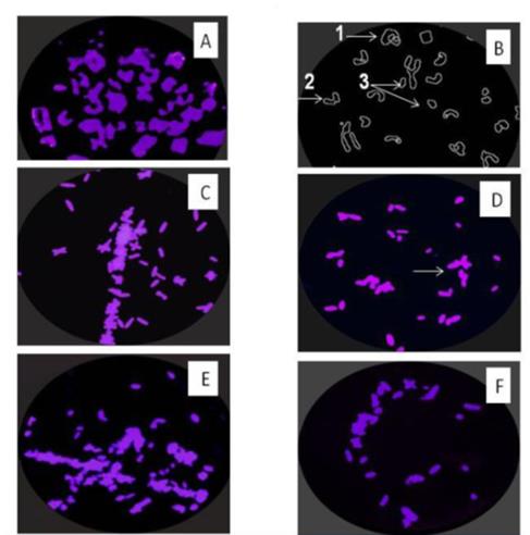
The results showed the chromosomal and structural abnormalities of chromosomes, as colchicine caused some chromosomal abnormalities, such as scattered metaphase, Chromosomal ring, chromatide separation and chromosome break were observed in addition to the appearance of chromosome break Figures 4 A & 4B. Figures 4C & 4D shows the chromosomes of the treated group with Taxol (1.7 mg/kg), where we observe structural and numerical abnormalities of chromosomes. The agglomerated colchicine metaphase due to treatment with Taxol, stickiness chromosome and end-end association is a structural abnormality observed during this study. Numerical chromosomal abnormalities were observed, including multiple of chromosomal group or polyploidy Figure 4E, and lack of chromosomal group Figure 4F.
Discussion
The results of the current study of karyotype results of bone marrow cells for male albino rats indicate that the diploid number of chromosomes for these rats is 42 = 2n. The results of the present study were agreed with a study, which was carried out on the Norwegian albino rat, where the chromosome pattern consisted of 21 pairs of chromosomes (42 chromosomes), as well as both males and females of albino rats, where 2n = chromosome 42, Current study agreed with a study done on the male albino rats [20, 21, 22]. The current study differed with a study carried out where chromosome number 38 = 2n. This variation in chromosomal numbers explains the difference in species belonging to the same sex in addition to losses during the preparation of chromosomal preparations, or additions from adjacent cells, or excessive treatment with a hypotonic solution [23, 24].
The results of the current study of the Karyotype of control group showed some structural chromosomal abnormalities. The present study agreed with the study that when the cells were treated with colchicine, they inhibited spindle and metaphase Colchicine scatter. The appearance of the previous phase as a type of chromosomal abnormalities, due to the assembly of chromosomes at the center of the cell, as these chromosomes do not occur on the central plate as in the natural metaphase, and this is due to the weakness of spindle threads in the work. The chromosomal break and the ring chromosome observed in this present study agreed with what he observed [1, 4, 20]. Several studies have suggested that the cause of breaks of chromosomes is the entry of external factors, such as chemicals, while the ring appearance of some chromosomes is likely to be removed at times from one end of the chromosome so that there are two viscous ends, leading to the adhesion of the peripheral arms to each other. The chromosome into a loop body [12]. In his study suggests that chromosomal abnormalities and chromosomal break are natural, because the chromosome may be exposed to many forms of continuous damage, due to different conditions inside and outside the cell, such as temperature and UV radiation, and that most of this damage is repaired by the cell a reform mechanism for DNA systems [24].
The results obtained in this study of the karyotype in the Taxol group included structural chromosomal abnormalities such as associated chromosomal endings, agglomerated colchicines metaphase and numerical abnormalities, were observed in a study with the use of Taxol [25]. The viscosity (which resulted in the appearance of the degenerate phase) was explained by the increased activity of DNA polymerase responsible for DNA where DNA polymerization inhibited the sequence following the split [26, 27, 28]. The association of chromosomal endings due to the intensification of DNA strands, and the cohesion of chromatin fibers between chromosomes lead to secondary correlations between chromosomes [10]. The total numerical abnormalities were a combination of two cells contains two nucleolus without cytokinesis [2].
In the present study, some chromosomes in the treatment group with Taxol were observed, according to a study [12]. Chromosomal dysfunction caused by chromosomal viscosity and chromosomal abnormalities, as well as numerical anomalies when using taxol alone or in combination with other anticancer agents, has an effect on cellular genetics and leads to chromosomal damage. Imbalance of chromosomes [25].
Conclusion
We conclude from the present study; the results of the chromosomal analysis of the somatic cells of the bone marrow of male albino rats proved that they contain 42=2n. furthermore, the study of the Karyotype of cellular genetic studies important in detecting changes in the genetic material such as structural and numerical changes that occur on the chromosomes, and the emergence of chromosomal and synthetic chromosomal abnormalities in the use of colchicine and Taxol properties. These abnormalities vary according to dose and duration of exposure. They can be repaired by DNA repair mechanisms if the exposure period is not continuous. Taxol, a chemotherapeutic drug, has high toxic effects on chromosomes, which increases the activity of the DNA polymerase enzyme, which causes the appearance of chromosome viscosity, as well as other types of chromosomal abnormalities.
Acknowledgement
The researchers extend their thanks to the Center for Research and Bio-Consultancy and to the master Wissam Al-Turgoman for his efforts in helping to take care of the study samples.
References
-
Al Khayat ASI, Heba HRS (2016) Study of the Chromosomal Structure in Mystus pelusius (Solander in Russell, 1749) Fish. Ibn al Haytham Magazine 29(2): 292-303.
-
Alrabaie Abbas (2014) Entrance to the genetics. Almanhajia Publishing & distribution, Amman-Jordan.
-
Syrio, Ali (2010) Foundations of Medical Genetics. Shuaa Publishing & Science, Syria-halab.
-
Salem, Salem M (1999) The cell is constructed and physiologically. National Center for Scientific Research and Consultancy, National Book House, Benghazi-Libya.
-
Abdi Ali, Abbas (2013) chromosomal number and Karyotype studies of Study of Lycium barbarum L. (Solanaceae) in some region of southe of Iraq. Missan Journal of Academic Studies 12(23): 162-171.
-
Amaka J (2014) Studies on the Karyotype of the black rat (Rattus rattus), collected at the university of Nigeria a zoological Garden. IOSR Journal of Pharmacy and Biological Sciences 9(5): 64-69.
-
Makino S (1953) The Karyotype of the Japanese population of the Rat, Rattus rattus, Journal of the faculty of Science, Hokkaido University 9: 19-23.
-
Oguma K (1935) Karyological studies of Black Rat, Rattus rattus. Journal of the Faculty of Science, Hokkaido University 4: 35-39.
-
Paget GE, Barnes JM (1964) Evaluation of drug activities. Pharmacometrics pp: 474.
-
Yoshida TH, Nakamura A, Fukaya T (1965) The chromosome number of the Black Rat, Rattus rattus, Chromosoma 16: 70-73.
-
Fadhil GM (2013) Detection of cellular and molecular toxicity of aloe vera gel Crude Extract on the Roots of the Allium cepa. Journal of Applied Science 29(1): 204-319.
-
Ali MA (2011) Study of Genetic Aberration of Sex Chromosomes of Turner Syndrome in the Peripheral Blood of Iraqi Patients. Iraqi Center for Cancer Research and Medical Genetics 4(1): 83-88.
-
Al sharif MMZ (2012) Studies on the Genotoxic effects of anticancer drug paclitaxel (Taxol) in Mice. Semantic Scholar 16(7): 989-997.
-
Piesova E, Sivikova K (2003) The induction of micronuclei in bovine lymphocytes by exposure to benzene and S9 mix. Ann Agric Environ 10(2): 261- 263.
-
Siddik ZH (2002) Mechanisms of action of cancer Chemotherapeutic Agents: DNA-Interactive Alkylating Agents and Antimour Platinum-Based Drugs. The University of Taxas MD Anderson Cancer Center, Houston, TX, USA, pp: 1-16.
-
Spratlin J, Sawyer MB (2007) pharmacogenetics of paclitaxel metabolism. Crit Rev Oncol Hematol 61(3): 222-229.
-
Salmon ES, Sartorelli AC (2001) Cancer chemotherapy. In: Bertram G, (Eds.), Basic and Clincal pharmacology Katzung, 8th (Edn.), Lange Medical Books/Mcgra Hill, London.
-
Tresukosol D, Kudelka AP, Edwards CL, Charsangavej CN, Narboni, et al. (1995) Recurrent ovarian granulose Cell tumor: Acase report of adramatic response to Taxol. Int J Gynecol Cancer 5(2): 156-159.
-
Preston RJ, Dean BJ, Galloway S, Holden H, Mefee AF, et al. (1987) Mammalian in vivo cytogenetic assay: Analysis of chromosome aberrations in bone marrow cells. Mut Res 189(2): 157-165.
-
Al Hawary BA (1988) Efeects of some medical drugs on the chromosomes of bone marrow cells of mice. Thesis, King Saud University, pp: 93.
-
Ahmad SI (2014) Genotoxic effects of mercuric chloride in the albino rats, rattus norvegicus. Research Journal of Animal, Veterinary and Fishery Sciences 2(7): 6-9.
-
Abousalem M, Elgerwi A, El mashad A (2003) Genotoxic and histotoxic effects of air pollutants at a benzene station on albino rats. IJBCP 3(1): 144 -150.
-
Aljanga AA, Fatima S, Warda MA (2016) Evaluation of Genotoxicity of Ethanol Extract of Citrulluscolocynthis plant Seeds on Mitosis Chromosomes of Bone Marrow Cells of Albino Rats. Journal of the University of Sabha 5(2): 15-27.
-
Patil AJ (2013) Karyotype analysis of male rat (Rattus rattus) from Amalner, Maharashtra, India 2(2): 103- 105.
-
Amnate AA (2008) Chromosomal structure and mitotic Index in blood of Awassi Ewes. Academic Scientific Journals 1(1): 93-100.
-
Burgaz S, Ozadmar YN, Karakaya AE (1988) Asignal assay for the detection of genotoxic compounds; Application on the urine of cancer patients on chemotherapy and of nurses handling cytotoxic drugs. Hum Toxicol 7(6): 557-560.
-
Elgubbi HS, Teka IA (2014) Effect of colchicines drug and water extract of Peganum harmala seeds on the induction of chromosome abnormality and mutation in peas plant. E-marefa 17(2): 1-15.
-
Elgubbi H, Abdri Al, Al Enezi AK, Sulaiman M (2015) Effect of hydrocortisone concentration of 0.05% on phenotypic Characters, pp: 139-145.
- hMPV: Is It Another Covid-19 Like Situation?
- Streptomyces: Sources of Novel Discoveries in Antibiotic Research to Combat Antimicrobial Resistance
- A Review of Mosquitoes (Diptera: Culicidae) and Their Biodiversity, Medical and Veterinary Importance
- Past and Current Immunotherapy in Cancer
- Hematological Cancer and Viral Infection
- The Growing Threat of Antimicrobial Resistance in India: Challenges and Solutions