Intraabdominal IUD Removal Perlaparascopy, A Case Report From Dr. Zainoel Abidin Hospital in Banda Aceh
Background: Intra-Uterine Device (IUDs) is the most effective long-term contraceptive methods, easy to get and easy to use for trained health workers. IUD users are estimated more than 100 million in the world. In Indonesia, the percentage of IUD users is 6.97%. Serious complications from an IUD both during insertion and using are rare; dislocation is one of the complications. The initial detection of dislocation is by touching the thread at the cervix, examining the sondage and pelvic photographs. Management of IUD dislocation is by laparotomy or laparoscopy. A case of a 25-year-old woman, Para1Abortus 0, complaining about cannot palpable IUD thread and not seen IUD for 1 years before operation and now she wants to remove the IUD. Abdominal x-ray appears IUD outside the uterine cavity and from ultrasound cannot find the IUD intra uterine. We did laparoscopic exploration for IUD removal, showed a perforation in the posterior wall of the uterus that had been covered by the omentum, and IUD was removed, both the adnexa and uterus were in normal limit and after making sure there was no abdominal cavity bleeding : was complete. Conclusion: This is a rare case and the risk of complications should be minimized if it is done at the right time and by a well-trained staff, early detection of simple IUD dislocation with IUD thread control. In the case of dislocation, laparoscopic exploration is safe and the main choice if the facility and expert resources were available.
Introduction
Intrauterine device (IUD) is contraceptive startegy that prevents pregnancy effectively, safely, and reversibly [1]. It is estimated that more than 100 million women around the world use IUD. In 2015, data form 71,963 participants indicated that the percentage of women in Indonesia who used an IUD was 6.97% with 11.9% using an IUD postpartum and 88.1% using an IUD at intervals [2, 3]. The ideal time for inserting IUD is within the first 7 days of the menstrual cycle or 6-8 weeks after giving birth [4, 5, 6]. IUD side effects are include abnormal uterine bleeding, dysmenorrhea, expulsion, uterine perforation, pelvic infection, ectopic pregnancy, anemia, dyspareunia, leucorrhea, menstrual spotting, pain and cramps [7].
Perforation of the uterus and abnormality location (dislocation or translocation) of IUD is a rare complication but has serious effect; occur in up to 1/1,000 cases. Although not many risk factors are documented, abnormalities of the uterus and the experience of the clinician contribute to an increase in the likelihood of perforation and if they occur in the abdominal cavity there is risk of peritonitis, abdominal adhesions, abscess and organ erosion, small intestine laceration, and cystic pelvic cavity [8, 9]. Here we reported an IUD dislocation from a patient and provided the success management.
Case Illustration
A 25-year-old Acehnese woman, parity 1 abortion
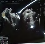
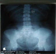
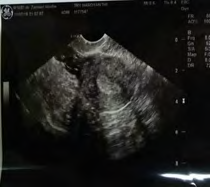
Figure 1: Imaging of the patients. (A) Detection of the intrauterine device (IUD) by plain abdomintal x-ray radiography, the IUDs is seen outside the uterine cavity. (B) Transvaginal ultrasound indicated the IUD was not seen in the uterine cavity. (C) Uansabdominal ultrasound examination revealed an anteflection uterus size 7x3x6 cm, right ovary size 2x2 cm, left ovary size 2x3cm, the IUD was not seen in the uterine cavity.
Laboratory tests obtained Hb / Ht / E / L / Tr: 11.8 / 35 / 4.0 / 8,400 / 285,000, HBsAg; Negative, SGOT / SGPT: 23/26, GDP: 106, Ca 125: 14.07, the patient’s lab results are still within normal limits.
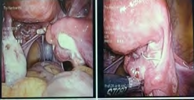
The surgery was performed using laparoscopic exploration procedure, it found an IUD outside the uterus
0, came with the chief complaint unable to palpable IUD thread on cervix. IUD was inserted 3 years ago at midwife and did not do a routine control of IUD position. From vital sign, generalized examination and gynecologic examination were within normal limit. Abdominal x-ray (Figure 1A) and ultrasound examination (Figures 1B&1C) was performed.
covered by omentum and fibrotic tissue at posterior part of uterus (Figures 2A&2B). IUD removal was performed and continued with evaluation of both of tubes by chromotubation (Figure 2C); the bilateral tubes are patent. Abdominal cavity was cleaned by normal saline and ensured there was no bleeding.
Discussion
IUD is the first choice of contraception for women; it is suitable for women who experience difficulty come to hospital to do recontrol [10]. Absolute contraindications to use of an IUD are pregnancy and active pelvic inflammation. Suspected cervical carcinoma, suspected uterine corpus carcinoma are also included in absolute contraindications in some literature. Relative contraindications include ovarian tumors, uterine abnormalities (such as fibroids and polyps), gonorrhea, cervicitis, menstrual abnormalities, dysmenorrhea, cervical canal stenosis and uterine length less than 6.5 cm [11].
Surgical technique using laparoscopy is a safe, minimally invasive technique and is a cosmetically method for removing an IUD [12]. We have performed IUD removal by laparoscopic procedure to a woman with indication of intraabdominal IUD translocation. The IUD has been used for 3 years before the laparoscopic procedure. Laparoscopic exploration procedure was conducted because, based on authors experience, it gives the operator an opportunity to well explore the abdominal cavity, provides a panoramic picture directly from the pelvic reproductive anatomy when compared with laparotomy, and the facilities and expert are available.
Intraoprative procedure, both ovaries and both tubes were within normal limit. We could not see the IUD intraabdominal clearly. On exploration, IUD was seen at posterior corpus of uterus and covered by omentum and fibrotic tissue. If we saw from the position of IUD when we did removal at the lower right posterior uterus, it was impressed that the midwife who performed an IUD insertion did not conduct uterine sondage properly so that it could not identify the position of the uterus and insert an insertor that penetrated the posterior uterus. Post insertion evaluation should be required 1 month later, or immediately if the patient complaining of severe pain in the pelvic area [8].
From our case report one of risk for dislocation IUD is the midwife who performed an IUD insertion did not conduct uterine sondage properly. In addition, most of the patients with the uterine perforation had received an IUD at a lower level of health care centers. Many health providers in lower of health had insufficient training on handling of IUD insertion, which may directly cause perforation at the insertion time. As it was reported before, IUD migrations at the time of insertion may directly contribute to uterine perforation [13, 14].
In China, among 29 cases with uterine perforation, the common part of IUD appeared at myometrium on the top of the scattered pattern of organ injury, and greater omentum, followed by the sigmoid colon, left sacrouterine ligament, and other parts.14 In our case, the IUD located at posterior corpus of uterus and covered by omentum and fibrotic tissues. Pelvic adhesions as a consequence of uterine perforation also frequently occurred in the patients, but the mechanism is still unknown; it may due to different actions of the devices [15].
Conclusion
Dislocation of IUD is a rare case. It should not be happen if the midwife performed the insertion carefully especially when the did sondage of the uterus.To avoid complications of IUD use, it is recommended to control to healthcare professionals if there are any complaints such as severe pain soon after post insertion. If the diagnosis of dislocation is established, IUD removal per laparoscopic is preferred when the facility and expert are available since it is a minimally invasive procedure.
References
-
Syaifuddin AB, Djajadilaga, Affandi B, Bimo (1996) National Reference Book for Family Planning Services. The Foundation of The Bina Pustaka Sarwono Prawirohardjo. Jakarta 9: 1-9.
-
(2015) BKKBN/National Family Planning Coordination Board. Contraceptive Services Feedback Report. Jakarta, BKKBN.
-
Suratun S (2008) Family Planning and Contraceptive Services, 3rd (Edn.), Jakarta, Trans Info Media.
-
Shukla M, Qureshi S, Chandrawati (2012) Post-Placental Intrauterine Device Insertion - A Five Year Experience At A Tertiary Care Centre In North India. Indian J Med Res 136(3): 432-435.
-
(2015) Public Health England. Your Guide to the IUD. The Sexual Health Line FPA, England.
-
Hartanto H (2011) KB Family Plnning and Contraception. CV Mulia Sari, Jakarta.
-
Cunningham (2013) Obstetri Williams. 23rd (Edn.), EGC, Jakarta.
-
Boortz HE, Margolis DJ, Ragavendra N, Patel MK, Kadell BM (2012) Migration of Intrauterine Device: Radiologic Finding and Implications for Patient Care. Radiographics 32(2): 335-352.
-
(1992) Prescibing information Tcu 380A. FEI Products, inc. N. Tonawanda, New York.
-
Glaiser A (2006) Family Planning and Prosperous Family. EGC, Jakarta..
-
Prawiroharjo S (2005) The Foundation of The Bina Pustaka Sarwono Prawiroharjo, Midwifery. Jakarta.
-
Zi D, Duan K, Fu K, Mengyue Y, Hanlin Y, et al. (2019) Single-Incision Laparoscopic Surgery for Removal of Ectopic Iud with Bladder Repair. J Minim Invasive Gynecol 26(7): S57.
-
Van Houdenhoven K, van Kaam KJ, van Grootheest AC, Salemans TH, Dunselman GA (2006) Uterine perforation in women using a levonorgestrel- releasing intrauterine system. Contraception 73(3): 257-260.
-
Sun Xin, Xue Min, Deng X, Lin Y, Tan Y, et al. (2018) Clinical Characteristic and Intraoperative Finding of Uterine Perforation Patients in Using of Intrauterine Devices (IUDs). Gynecol Surg 15(1): 3.
-
Kaislasuo J, Suhonen S, Gissler M, Lahteenmaki P, Heikinheimo O (2013) Uterine perforation caused by intrauterine devices: clinical course and treatment. Hum Reprod 28(6): 1546-1551.
- The Need for Partner Education and Mental Health Support During Pregnancy and the Postpartum Period
- Application of Combined PGT-A and PGT-M for Reproductive Management in a Couple Carrying GCDH Mutations with Prior Affected Offspring: A Rare Case Report
- The Effect of Using a New Technique Karman Injector (Elif Technique) on the Healing Process of Wound Infection-Case Series
- GSM: Counseling Points to Discuss with Women Fearful of Vaginal Estrogen
- Antenatal Diagnosis of Meckel Syndrome: A Case Report
- Discrimination and Workplace Harassment (Mobbing) against Women in the Post-Pandemic Era