Evaluating Strength Gains Using Blood Flow Restriction and Myofascial Release
High intensity strength training leads to increased muscle strength. Research shows that blood flow restriction (BFR) combined with low intensity strength training exercises produce similar results. Flexibility is important for efficient movement. Myofascial release (MFR) uses a gentle sustained pressure to stretch the Myofascial tissue and increase flexibility which promotes efficient movement. The purpose of this study is to assess strength gains of the pectoralis major muscle in the nondominant upper extremity (UE) after implementing BFR and MFR. For four weeks, 27 subjects participated in a randomized control study where they either completed the BFR protocol on the chest-fly machine (control group) or received MFR for three minutes followed by completing the BFR protocol on the chest-fly machine (experimental group). Pre- and post-intervention measurements were taken to determine strength gains and muscle length changes. A significant difference was noted in nondominant pectoralis major muscle strength for the control group. A significant difference was noted in pectoralis major muscle length for the control group in pre- and post-training for the non-dominant pectoralis major. A significant difference was noted in one repetition maximum (1 RM) for both the control and experimental groups for the non-dominant pectoralis major. This study conveys the effectiveness of BFR on strengthening the pectoralis major muscle. Potential implications for clinical practice include using BFR to improve pectoralis major muscle strength in populations who have muscle tightness and are unable to exercise at high-intensity levels.
Introduction
High intensity strength training leads to increased muscle strength. High intensity, strength training leads to muscle strain and injury as well [1]. Research shows high intensity exercise coupled with decreased flexibility lead to muscle strain and injury in the pectoralis major muscle [1]. Blood flow restriction (BFR) combined with low intensity exercises has been shown to decrease the likelihood of muscle strain and injury and improve muscle strength and hypertrophy in healthy individuals, post-surgical patients, and recovering patients after an injury [2, 3]. In a study by Green, et al. completing BFR at 20% of one repetition maximum (1RM) produces the same strength gains in the pectoralis major muscle as high resistance exercise [4]. Based on this information, strength gains may occur with the implementation of BFR training with low intensity exercise and flexibility training, and muscle strain and injury may also be prevented.
Blood flow restriction works by occluding the arterial blood flow by either 50% in the upper extremity (UE) or 80% in the lower extremity with a pressurized tourniquet and using low-intensity resistance exercises with 20-30% of a 1RM [5]. The occlusion reduces the amount of oxygen that reaches the tissues and, in turn, via anaerobic metabolism releases lactate, which is responsible for energy production and results in hypertrophy of the muscle along with cell swelling [5, 6].
Flexibility affects muscle strength and is important for efficient movement [7, 8]. Myofascial release (MFR) uses a gentle sustained pressure to stretch restricted fascial tissue, increase flexibility, and promote efficient movement [9, 10]. Direct and indirect applications of MFR also reduce muscle tightness, decrease pain, and increase range of motion [11, 12]. For example, in a study by Rodriguez-Huguet, et al., neck pain decreased, and pressure pain thresholds improved using MFR [13]. The cross-hand MFR technique is a direct technique administered for at least three minutes to improve posture, restore optimal tissue length, decrease pain, and improve function [10].
Protein supplements are frequently consumed with resistance training and have been shown to have benefits. Leucine is an essential amino acid contained in whey protein. Leucine has been shown to be necessary for starting the rebuilding process post-exercise and promoting muscle protein synthesis [14]. Ingestion of protein before and during exercise has less influence on muscle protein synthesis [15]; however, consumption of whey protein immediately after exercise results in greater muscle fiber hypertrophy [16]. The consumption of whey protein following a workout effectively stimulates postprandial muscle protein accretion, [17] reduces muscle soreness, and improves muscle function and performance in the next workout [18]. The consumption of whey protein supplementation enhances whole body anabolism, and resistance trained individuals could benefit from protein supplementations after exercise [19].
Many studies have examined BFR and the consumption of protein. In a study conducted by Centner, et al. [20]. subjects in an experimental group were provided 15 grams of collagen hydrolysate. Results of the study indicated no significant difference was seen between the experimental and control group, who received a placebo regarding overall strength gains [20]. In a study by Sieljacks, et al. 20 grams of whey protein isolate were provided to subjects in the BFR group and high load resistance group [21]. Results of their study indicated both groups showed increased protein turnover and increased muscle strength [21]. To this date, no studies have evaluated the effects of BFR, MFR, and the consumption of whey protein to determine strength gains for the pectoralis major muscle.
The purpose of this study is to assess the strength gains of the pectoralis major muscle in the non-dominant UE after implementing BFR and MFR with supplementation of whey protein. We hypothesized strength gains would occur on the non-dominant pectoralis major muscle. This research may assist in understanding the effects of BFR and MFR on healthy subjects so these techniques can be more effectively applied to rehabilitation populations limited in their muscle length and ability to perform high resistance exercise.
Methods
Subjects
This study was approved by the Institutional Review Board at the research institution. Informed consent was obtained from each subject, and the rights of the subjects were protected. Subjects were acquired on a voluntary basis via flyers, informational sessions, and advertisements. In total, the study garnered 27 subjects (15 male, 12 female). The mean age for the experimental group was 22 and 23.2 for the control group. Each participant was provided a consent form that instructed them of any potential risk(s) as well as benefits involved in the study. After consenting to participating in the study, all subjects were educated on BFR and MFR prior to initial baseline measurements and the 4-week intervention.
Participant eligibility included the following exclusion criteria: a diagnosis of diabetes; a diagnosis of hypertension or the use of any medication related to hypertension or other cardiac issues; sickle cell anemia; circulatory or cardiopulmonary conditions; significant UE injury or operation in the last six months; pregnant; history of deep vein thrombosis; and active infection. Undergraduate and graduate students between the ages of 18 and 30 who did not present with any of the listed exclusion criteria participated in the study.
Subjects were randomly assigned to either the control or experimental group. The control group only received BFR during the study while the experimental group received both BFR and MFR. Both groups were provided one scoop of a whey protein powder after each session. Before the start of the study and the participant’s first session, additional information was gathered. Examples of additional information include prior experience with BFR, exercise regimens, 24-hour dietary recall, a nutritional self- assessment, dietary status, and subject’s perception of both exercise regimen status and dietary status.
All subjects reported they exercise regularly. Except for one subject in the experimental group, all subjects reported they complete a warm-up prior to exercise. Whereas six out of 12 subjects in the experimental group reported they do not cool-down post exercise, five out of 15 of the subjects in the control group reported they also do not cool-down. Subjects in each group reported they complete strength training for at least 30 minutes. Four out of 12 subjects in the experimental group and four out of 15 of the subjects in the control group reported they complete strength, endurance, and flexibility training. Seven out of 12 subjects in the experimental group and eight out of 15 of the subjects in the control group reported they do not consume protein after they exercise.
Nutritional Self-Assessment Prior to Intervention
On a scale of 0 to 10, with 0 indicating poor, 5 indicating fair, and 10 indicating good, 77.7% of subjects reported their nutritional status between 5 and 10 or fair and good. All subjects ranked the importance of nutritional status. All reported their nutritional status as important to extremely important based on a scale ranging from 0 to 10 with 0 indicating nutritional status as not important and 10 indicating nutritional status as extremely important.
A five-star scale was used to indicate variety or balance of diet with 1 indicating no variety or balance of diet, 3 indicating fair variety or balance of diet, and 5 indicating excellent variety or balance of diet. Subjects ranked their variety or balance of diet as fair or 40.7%. The variety intake of the subjects was based on intake of fat, carbohydrate, protein, water, and hydration status. Most subjects ranked their fat intake at 63% or fair, and 66.7% ranked their carbohydrate intake as good. Most subjects ranked their protein intake at 40.7% or fair. Water intake (44.4%) and hydration intake (40.7%) were ranked as excellent. The weight and height of each subject were also obtained. The mean body mass index (BMI) was 22.75 and 25.8 for the experimental and control groups respectively.
Subjects also ranked their perception of UE strength. They ranked their self-perceived UE strength on a scale from 0 to 10 with 0 indicating poor, 5 indicating fair, and 10 indicating good. A total of 88.9% of subjects ranked their UE strength as fair to good or between 5 and 10.
Experimental Approach to the Problem
Prior to the study, student researchers were trained on the MFR cross-hand pectoral release application and on properly using the Delfi Personalized Tourniquet system (Delfi Medical Innovations, Inc., Vancouver, BC Canada) BFR unit. Two student researchers were taught how to assess pectoralis major muscle length and strength. Inter-rater reliability testing was performed to ensure testing was reliable.
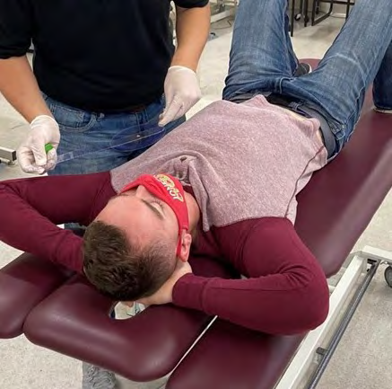
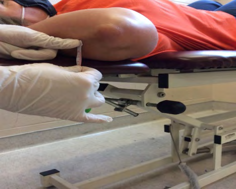
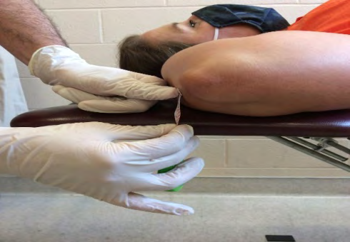
Baseline measurements were taken for each participant using a JTECH dynamometer (JTECH Medical Industries, Inc., Midvale, UT) to measure the strength of the pectoralis major muscle for their non-dominant UE. Three trials were conducted to achieve an average of the participant’s overall strength and to decrease the likelihood of bias. To ensure compensations were not made due to pectoralis major tightness, the subject laid supine with their knees flexed and backs flat against the plinth. The subject’s shoulder was abducted to 135 degrees as measured with a goniometer. Pectoralis major muscle length was then assessed by using a tape measure to determine the distance from the posterior aspect of the subject’s olecranon process to the top of the plinth. Lastly, the subject used a chest-fly machine to determine their 1RM. These baseline measurements were performed 2-weeks prior to the beginning of the 4-week intervention and again the week following the conclusion of the study. Additional baseline measurements obtained from the subjects included reporting average amounts of daily protein consumption and their physical activity level, regime, and frequency, if any.
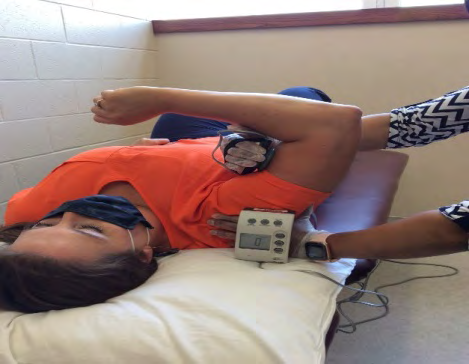
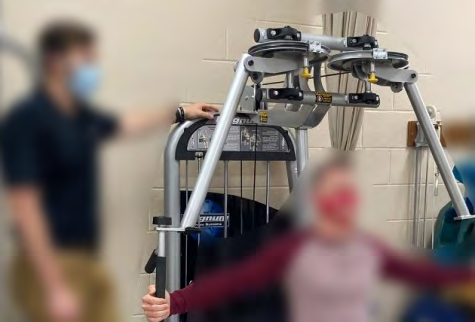
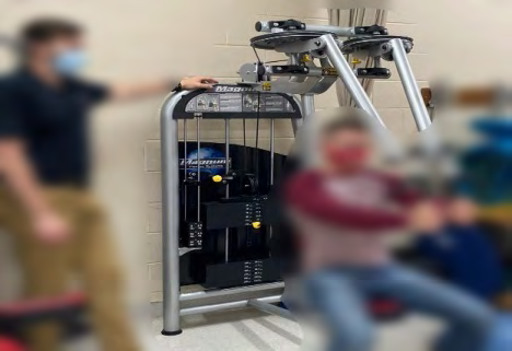
At the beginning of each session, the researcher assessed the subject’s blood pressure. The subjects in the experimental group received three minutes of cross-hand MFR on their pectoralis major muscles prior to beginning BFR. To complete the BFR portion of the intervention, subjects laid supine on a table. Using Delfi’s Personalized Tourniquet system, occlusion pressure was obtained with a cuff applied to their non-dominant UE and 50% of their blood flow was occluded. Once the participant’s personalized tourniquet press was determined, subjects performed one set of 30 repetitions, followed by three additional sets of 15 repetitions each with an approximate 30-second rest period between each set. Repetitions were completed at 20% of the subject’s 1RM and total duration of the BFR intervention was seven minutes. Prior to beginning BFR and at the beginning of their final set of BFR, the subjects rated their level of exertion on a scale from six (no exertion) to 20 (maximal exertion) using Borg’s Rating of Perceived Exertion chart. The intervention for both groups was conducted on two non-consecutive days with one rest day in-between.
Upon completion of the intervention, each subject’s blood pressure was taken again by the researcher. Subjects were also provided one scoop of a whey protein powder, which provided 30 grams of protein, and water for mixing the protein drink. Subjects consumed this protein drink within 20 minutes of their intervention. Following the subject’s final session, they completed a 24-hour dietary recall form for analysis. The 24-hour dietary recall form was compared to the original form to determine if any dietary patterns had changed over the course of the study and to determine if any nutrient deficiencies impacted the overall purpose of determining if MFR aided in the development of increased strength in the pectoralis major muscle. Refer to Figures 1-7.

Results
An intake of 2000 calories based on MyPlate was used to assess the diet quality of each subject. According to the MyPlate recommendations, grains, vegetables, fruits, dairy, and meat and beans were recorded in the 24-hour recall. Each subject was provided a link to an electronic recall form the day before and after the study was completed. A total of 40 recalls were returned, giving the researchers a 74% response rate. Overall, no subject consumed the recommended intake of all food groups. Many reported not consuming breakfast or just drinking coffee. Participants who did consume breakfast largely consisted of protein and grains.
More vegetables were consumed at lunch and dinner, but this consumption only appeared in 12.5% of responses. Reported snacking throughout the day was poor and consisted primarily of candies and processed convenience foods. Upon reviewing the coded dietary recalls, the overall nutritional intake for subjects in both groups was poor. The pre- and post-study recalls were very comparable indicating there was not a significant difference with intake for the two groups; therefore, dietary factors did not impact the results of this study. Other measurements taken included blood pressure and perceived exertion. Blood pressure measurements taken pre- and post-intervention were within normal range for all subjects. On Borg’s Rating of Perceived Exertion scale, most subjects rated their physical activity level as six (no exertion) prior to BFR. Perceived exertion prior to the last set of the BFR protocol was also assessed. Results revealed subjects in the experimental group had a mean perceived exertion level of 10.66 (light) out of 20 with a standard deviation of 2.03. In contrast, subjects in the control group had a mean perceived exertion level of 9.65 (very light) out of 20 with a standard deviation of 2.19.
All statistical analyses were performed with SPSS software, Version 27. To determine the pre-test and post-test effects of pectoralis major muscle length, strength, and 1RM within the control group and MFR group, a paired t-test was performed. Means improved for both groups for dominant and non-dominant strength and dominant and non-dominant pectoralis major muscle length. However, only dominant (p = .009) and non-dominant (p = .004) strength, and dominant (p = .017) and non-dominant (p = .045) pectoralis major muscle length were statistically significant in the control group. The mean for 1RM improved in both groups, and there was a significant difference noted for 1RM in the control group (p = .001) and experimental group (p = .024).
To compare the pectoralis major muscle length, strength, and 1RM between groups, independent t-tests were performed. Statistical analysis was set a priori at p <0.05. There was not a significant difference in strength for pre- or post-training in the dominant or non-dominant UE. There was not a significant difference in pre- or post-training for pectoralis muscle length in the dominant or non-dominant UE. Furthermore, there was not a significant difference in pre- or post-training for 1RM. Refer to Tables 1-3.
| Experimental | Control | |||
|---|---|---|---|---|
| Pre (Mean+/-SD) | Post (Mean +/-SD) | Pre (Mean +/-SD) | Post (Mean +/-SD) | |
| Pectoralis Major | 22.84+/-5.93 | 24.99+/-7.44 | *22.49+/-5.16 | *24.78+/-5.36 |
Table 1: Dynamometer Results for the Nondominant Pectoralis Major for the BFR and Control Groups.
*Significant difference p < 0.05 Table 1: Dynamometer Results for the Nondominant Pectoralis Major for the BFR and Control Groups.
| Experimental | Control | |||
|---|---|---|---|---|
| Pre (Mean+/-SD) | Post (Mean +/-SD) | Pre (Mean +/-SD) | Post (Mean +/-SD) | |
| Pectoralis Major | *37.75+/-17.59 | *40.45+/-19.49 | *41.04+/-19.84 | *44.72+/-21.58 |
Table 2: 1RM Results for the Nondominant Pectoralis Major for the BFR and Control Groups.
*Significant difference p < 0.05 Table 2: 1RM Results for the Nondominant Pectoralis Major for the BFR and Control Groups.
| Experimental | Control | |||
|---|---|---|---|---|
| Pre (Mean+/-SD) | Post (Mean +/-SD) | Pre (Mean +/-SD) | Post (Mean +/-SD) | |
| Pectoralis Major | 0.67+/-1.17 | 0.54+/-1.23 | *0.57+/-.92 | *0.23+/-0.42 |
Table 3: Pectoralis Major Length Results for the Nondominant Pectoralis Major for the BFR and Control Groups.
*Significant difference p < 0.05 Table 3: Pectoralis Major Length Results for the Nondominant Pectoralis Major for the BFR and Control Groups.
Discussion
The purpose of this study was to determine whether combining BFR and MFR with the consumption of whey protein would increase strength in the pectoralis major muscle in the non-dominant UE in the healthy, young adult population. To our knowledge, this is the first study to examine the effects of BFR and MFR on pectoralis muscle strength. We hypothesized those who performed a low intensity lifting protocol concurrent with BFR and MFR would experience greater gains in pectoralis major muscle strength compared to those in the control group. Based on the results, the hypothesis could not fully be accepted.
Results of the study indicate the following. The experimental group rated their physical activity at a higher rate (light) than the control group (very light) prior to completing the last set of the BFR protocol. There were no significant differences between the two groups for age, height, weight, and BMI. Not only did BFR significantly improve strength in the dominant and non-dominant pectoralis major muscles in the control group, but significant improvements were also noted in pectoralis major muscle length in the dominant and non-dominant UE in the control group (p<0.05). In a study by Yasuda, et al., 1RM significantly increased with the incorporation of BFR [22]. similarly, 1RM significantly increased in both groups in this study. A significant difference was not noted between groups for pectoralis major muscle strength, pectoralis major muscle length, or 1RM. The results of this study suggest BFR showed improvements in pectoralis major muscle strength and length and 1RM without the incorporation of MFR.
The following limitations were noted with this study. The power of the results from this study would be greater with a larger sample size. The results of males versus females were not compared. The only limitation on physical activity was no BFR exercise for the UE outside of the intervention; therefore, subjects could have altered their exercise regimen to include more UE flexibility and/or strengthening activities. Because research shows muscles begin to hypertrophy at six to eight weeks, the time frame of this study poses a limitation [20]. This study was only conducted within a 4-week period due to time constraints. Discriminative validity could also be considered a limitation for this study since this study was not a double-blind or single-blind study. Subjects and researchers were aware of group placement for the subjects. Furthermore, not all subjects in the experimental group received MFR pectoral release from the same student researcher. For this reason, different pressures could have been applied to the muscle.
Conclusion
This study explored whether combining BFR and MFR with the consumption of a whey protein would increase strength in the pectoralis major muscle in the non-dominant UE in the healthy, young adult population. The study compared the strength, length, and 1RM gains in the pectoralis major muscle within groups and between two groups.
References
-
West DWD, Sawan SA, Mazzula M, Williamson E, Moore DR (2017) Whey protein supplementation enhances whole body protein metabolism and performance recorded after resistance exercise: A double-blind crossover study. Nutrients 9(7): 735.
-
Laimi K, Makila A, Barlund E, Katajapuu N, Oksanen A, et al. (2018) Effectiveness of myofascial release in treatment of chronic musculoskeletal pain: A systematic review. Clinical Rehabilitation. 32(4): 440-450.
-
Thomas DT, Erdman KA, Burke LM (2016) Position of the academy of nutrition and dietetics, dietitians of Canada, and the American College of Sports Medicine: Nutrition and athletic performance. Journal of the Academy of Nutrition and Dietetics 116(3): 501-528.
-
Yasuda T, Loenneke JP, Thiebaud RS, Abe T (2012) Effects of Blood Flow Restricted Low-Intensity Concentric or Eccentric Training on Muscle Size and Strength. PLOS ONE 7(12): 1-16.
-
Barnes J (1990) Myofascial release the search for excellence: A comprehensive evaluatory and treatment approach. Paoli, PA: National Library of Medicine.
-
Yasuda T, Fujita S, Ogasawara R, Sato Y, Abe T (2010) Effects of low-intensity bench press training with restricted arm muscle blood flow on chest muscle hypertrophy: a pilot study. Clinical Physiology and Functional Imaging 30(5): 338-343.
-
Umehara J, Sato Y, Ikezoe T, Yagi M, Nojiri S, et al. (2021) Regional differential stretching of the pectoralis major muscle: An ultrasound elastography study. Journal of Biomechanics 121(24):1-8.
-
Pennings B, Boirie Y, Senden JM, Gijsen AP, Kuipers H, et al. (2011) Whey protein stimulates postprandial muscle protein accretion more effectively than do casein and casein hydrolysate in older men. The American Journal of Clinical Nutrition 93(5): 997-1005.
-
Pearson SJ, Hussain SR (2015) A review on the mechanisms of blood-flow restriction resistance training-induced muscle hypertrophy. Sports Medicine. 45: 187-200.
-
Serrano B, Serrano J (2019) the efficacy and validity of blood flow restriction training in clinical and post-surgical populations. International Journal of Physiotherapy 6(5): 155-159.
-
Ada L, Canning C, Dwyer T (2000) Effect of muscle length on strength and dexterity after stroke. Clinical Rehabilitation 14 (1): 55-61.
-
Suzuki Y, Iijima H, Tashiro Y, Kajiwara Y, Zeidan H, et al. (2019) Home exercise therapy to improve muscle strength and joint flexibility effectively treats pre- radiographic knee OA in community-dwelling elderly: a randomized controlled trial. Clinical Rheumatology 38: 133-141.
-
Pasiakos SM, McClung HL, McClung JP, Margolis LM, Andersen NE, et al. (2011) Leucine-enriched essential amino acid supplementation during moderate steady state exercise enhances post exercise muscle protein synthesis. The American Journal of Clinical Nutrition 94(3): 809-818.
-
Kuruma H, Takei H, Nitta O, Furukawa Y, Shida N, et al. (2013) Effects of myofascial release and stretching technique on range of motion and reaction time. Journal of Physical Therapy Science. 25(2): 169-171.
-
Pasiakos SM, Lieberman HR, McLellan TM (2014) Effects of protein supplements on muscle damage, soreness and recovery of muscle function and physical performance: A systematic review. Sports Medicine 44: 655-670.
-
Manini TM, Clark BC (2009) Blood Flow Restricted Exercises and Skeletal Muscle Health. Exercise and Sport Science Reviews 37(2): 78-85.
-
Rodriguez Huguet M, Gil Salu JL, Rodriguez Huguet P, Cabrera Afonso JR, Lomas Vega R (2018) Effects of myofascial release on pressure pain thresholds in patients with neck pain. American Journal of Physical Medicine & Rehabilitation 97(1): 16-22.
-
Barnes MF (1997) The basic science of myofascial release: morphologic change in connective tissue. Journal of Body and Movement Therapies 1(4): 231-238.
-
Centner C, Zdzieblik D, Roberts L, Gollhofer A, Konig D (2019) Effects of blood flow restriction training with protein supplementation on muscle mass and strength in older men. Journal of Sports Science and Medicine 18(3): 471-478.
-
Green LL, Cupp JL, Cole E, Craig D, Crawford K, et al. (2020) Investing strength effects at the shoulder using blood flow restriction. Annals of Physiotherapy & Occupational Therapy 3(4): 1-6.
-
Esmarck B, Andersen JL, Olsen S, Richter EA, Mizuno M, et al. (2001) Timing of postexercise protein intake is important for muscle hypertrophy with resistance training in elderly humans. Journal of Physiology 535(1): 301-311.
-
Sieljacks P, Wang J, Groenne Baek T, Rindom E, Jakobsgaard JE, et al. (2019) Six weeks of low-load blood flow restricted and high-load resistance exercise training produce similar increases in cumulative myofibrillar protein synthesis and ribosomal biogenesis in healthy males. Frontiers in Physiology 10: 1-16.
- Electrolyte Considerations for Athletes
- Comprehensive Rehabilitation in Adults with Diabetic Peripheral Neuropathy: A Literature Review on Frequency, Intensity, and Duration Parameters
- Exercise Duration and Its Association with ADHD Symptom Severity in Children and Adolescents: A Parent-Reported Survey Study
- Adaptation of the Adult Neurophysiology of Pain Questionnaire for Use in Pediatrics
- A Non-Pharmacological Multidisciplinary Pain Program within a Hospital Wellness Program: A Mixed Methods Study
- The Effect of Frenkel's Exercise with PNF on Functional Reach in Stroke Survivors: A Randomized Control Trial