In vivo Study of Proliferative Effect and Growth Promoting Effect of Ultra Diluted Phytolacca decandra in Chick Embryo by Using Cam Assay
Homoeopathy and complementary integrative medicine treatments aimed at decreasing the side effects of anti-cancer therapy and cancer symptoms, as well as providing nutritional and lifestyle advice to help patients with breast cancer and enhance their quality of life. Angiogenesis can be used to investigate phase-specific processes in both physiological and pathological situations. Phytolacca decandra has a specific effect on breast cancer ulceration and scar tissue. The current study deals with the angiogenesis activity by using CAM Assay. Phytolacca decandra was taken in account and it increases angiogenesis in a CAM assay, indicating that it is not hazardous to normal cells. It also improves blood supply, which aids in proliferation, and has no side effects on normal cells.
Introduction
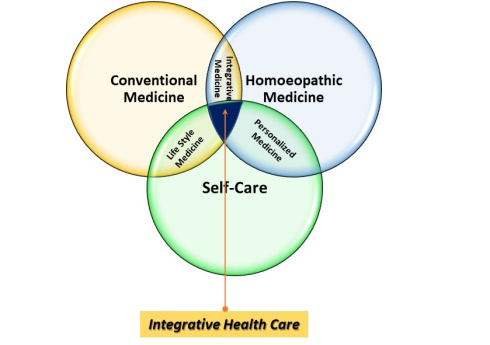
Integrative health is the practise of combining traditional medicine, complementary medicine that includes Homoeopathy and lifestyle medicine. It is the result of a multidisciplinary team’s coordination of patient-centred, science- and evidence-based health practises. Its goal is to enable the return or maintenance of health in a state of optimal well-being. Its uniqueness stems from the inclusion of patients and their own representations of care in overall health management (Figure 1). Integrative health care allows caregivers to link treatment goals with good and new practises for and with the patient by treating the whole person, understanding the needs and values of each patient, and focusing on quality of life as much as curing [1].
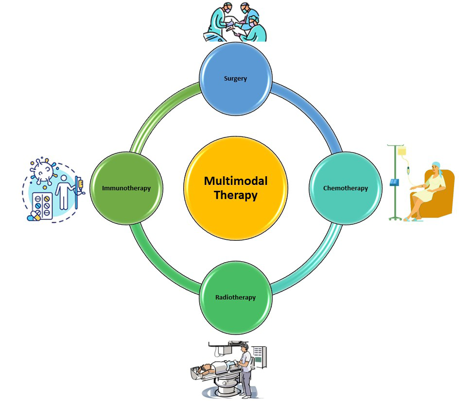
Homeopathy and conventional medicine complement each other well. In cancer patients, additive homoeopathy improves quality of life. In cancer patients, additive homoeopathy improvessurvival [2]. Treatment with homoeopathy and complementary integrative medicine aimed at reducing the side effects of anti-cancer therapy and cancer symptoms, as well as providing dietary and lifestyle advice to improve the quality of life of breast cancer patients. The incorporation of evidence-based complementary treatments into conventional cancer care enables medical doctors and healthcare professionals to respond more effectively to cancer patients’ requests to reduce many side effects of anti-cancer therapies while also improving their quality of life by providing both safety and equal access in public healthcare systems. The exchange of knowledge and experiences is critical in this integration process. As a result, physicians (particularly oncologists) and other cancer-related healthcare professionals must be adequately informed about the potential benefits of complementary and integrative medicines [3]. A recent study investigating the effects of radiotherapy alone or in combination with chemotherapy as post-surgical treatment reveals that a multimodal approach (Figure 2) involving radiotherapy improves survival rates, particularly in Stage II patients [4].
Sufficient blood supply plays one of the most important roles in the entire body for the maintenance of body homeostasis. To guarantee nutrition and metabolism for tissue and organ function, a vascular system with an appropriate circulation is required. Especially in the wound healing of the skin, the restoration of the blood supply over the entire period is an indispensable process. This can be seen impressively in both the acute and chronic forms of wound healing [5].
Whereas in acute wound healing, example after a cut, the vascular system already initiates wound healing from the very beginning by vasoconstriction and activation of the coagulation cascade, one recognizes the importance of the vascular system in the absence or limited function in wound healing disorders (Figure 3). The processes of neovascularization and angiogenesis in skin repair are highly complex and must be subdivided, because each phase of wound healing requires contradictory processes. To investigate wound healing, models are needed that can reflect the physiological or pathological conditions in humans [5].
In recent years, many new in vitro and in vivo models have been developed, which consider the pathogenesis of wound healing and the identification of new drugs or biomarkers (because of the goal to reduce animal experiments). Since a wide variety of wound healing models have been described, this article focuses on the processes involved in vascularization and microcirculation in the skin and presents new results, models and methods for their analysis. It is the aim of this review to summarize established and new in vivo and in vitro models for wound healing, to illustrate their advantages and disadvantages and to describe the key points of implementation of the respective models [5].
In vivo Models
In vivo models still represent the gold standard in the investigation of physiological and pathological processes. They ensure the analysis of relevant questions in a whole organism and thus all possible influencing factors and their effects on the expected outcome [1].
Animal models are widely used in the preclinical phase of the development of new pharmaceutical agents for both risk assessment and pharmacokinetic studies. In this context, however, the transferability of results from animals to humans must be questioned critically, and thus the use of animals as experimental subjects in general. In the case of in vivo models, various models have been described for large and small animals and for humans, but over the last 20 years’ mouse, human, rat and pig models have prevailed. Other species, such as rabbits, goats, hamsters, frogs, or zebra fish are scarcely used as wound healing models. The transferability of data from animals to humans always plays an important role in this context [5].
Chorioallantoic Membrane Assay
The increasing importance of distress reduction in animal experiments and the persisting limitations of in vitro systems in angiogenesis research demand complementary models. The chorioallantoic membrane (CAM) of the chicken embryo has therefore been established as a standard angiogenesis model over recent decade. The CAM is an extra embryonic structure with a dense capillary network mediating gas and nutrient exchange until hatching on day 21. The capillary bed connects arterioles (10-85µm) and venules (10-115µm). During embryonic development, the CAM grows from 6cm2 at day 6 to 65cm2 at day. Capillary angiogenesis is completed until day 11. Therefore, two phases have to be considered: day 1 to 11 enables experiments on rapidly developing vessels and day 12 to 20 enables research on mature vessels. The extracellular matrix between the vessels comprises fibronectin, laminin, collagen type IV and glycosaminoglycans. Single-layer epithelium covers the CAM [5].
The chick embryo CAM was used as an in vivo wound healing model. The validity of this experimental model appears to be confirmed by the fact that we were able to reproduce all the critical events controlling the wound healing process, such as re-epithelisation, angiogenesis, formation of an inflammatory infiltrate and deposition of one of the main constituents of the extracellular matrix, such as fibronectin1.The CAM was used as an in vivo wound healing model. A full excision of a 1 mm2 CAM area was filled by a granulation tissue after 96-120 h, which eventually formed a scar in 75% of the cases. In the remaining 25%, a solution of continuity was left which, however, was smaller in size than the one observed immediately after the excision. The validity of this experimental model appears to be confirmed by the fact that we were able to reproduce all the critical events controlling the wound healing process, such as re-epithelisation, angiogenesis, and formation of an inflammatory infiltrate and deposition of one of the main constituents of the extracellular matrix, such as fibronectin [6].
Phytolacca decandra acts on the whole glandular system, but especially on the breasts and the glands of women, which are prone to become cancerous in those of cancer constitution, Pains are Burning in character in the ulcerations of the Cancer. Phytolacca decandra functions on the entire glandular system, particularly on the breasts and glands of women, which are prone to becoming malignant in individuals with cancer constitution. Cancer ulcerations have a burning sensation [7].
Biomaterials for angiogenesis and vasculogenesis covers the application of materials designed to encourage new blood vessel formation. Angiogenesis and vasculogenesis play an important role in tissue engineering and regenerative medicine research by promoting vascular networks inside engineered tissues and thereby increasing tissue healing and regeneration. However, researchers are faced with the challenge of finding suitable materials for improving angiogenesis and vascular formation in assays. Broad range of biomaterials for the promotion of blood vessel genesis, from polymers and bioactive glass, to nanomaterial scaffolds and 3D antigenic constructs. Variety of applications for biomaterials in tissue repair and regeneration, including cardiovascular regeneration, liver tissue engineering and much more. Biomaterials for angiogenesis and vasculogenesis serve as a detailed reference for researchers in academia and industry, working in the fields of biomedical science and engineering, materials science, regenerative medicine and translational medicine [8].
The glands harden and become irritated. It causes painful throats and inflammation of the neck glands, especially the sub maxillary and parotid glands. Bellis pernnis, Conium, or Phytolacca decandra are tiny groups of drugs that correspond to trauma-related disorders Phytolacca decandra holds a place between Bryonia and Rhus tox.; cures when these fail, despite being adequately indicated.; effects on scar tissue Syphilitic bone aches; chronic rheumatism Sore throat, quinsy, and diphtheria Tetanus and opisthotonus weight loss [9].
The wound healing process involves several distinct phases in which the formation of new blood vessels plays an essential role. This report describes the angiogenic activity of a freeze-dried aqueous extract of the flowers of Calendula officinalis L. utilizing the CAM assay. The CAM assay is a standard and well-established method for assessing the antigenic activity in impure and pure preparations and is suitable for studies requiring examination of large numbers of sample test materials. The antigenic activity of calendula was measured by examination of CAMs using stereomicroscopy [10].
Materials and Methods
Preparations of Phytolacca decandra 6CH
Phytolacca decandra 6CH were purchased from Schwabe India, New Delhi. GMP certified company.
Experimental Protocol
Fertilized eggs were obtained from poultry products. Selected eggs of similar size and weight were used. Surface of the eggs were disinfected with ethanol. They were divided into two groups each containing one egg. The groups were
Treatment Groups
The treatment groups are divided into two groups. They are
- Viable eggs with developing embryo Distilled water
- Viable eggs with developing embryo+Phytolacca decandra 6CH The angiogenesis activity on the eggs was checked using the CAM assay, and viable eggs with growing embryos were chosen and incubated. The eggs were candled and tested for blood vessels on the sixth day of incubation. The eggs were washed with 70% ethanol and a window was cut in the shell with a sterile needle and forceps on the broad end of the egg. The eggs were implanted with Whatman’s sterile filter paper discs holding the test compounds, and the window was covered with a porous plaster. On the eighth day, after 48 hours of incubation, these windows were reopened and angiogenesis was studied.
Results
CAM Assay - Angiogenesis Activity
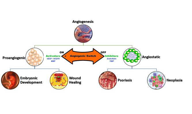
Angiogenesis is a very important event during healthy and diseased conditions. This process normally involves vasodilatation and an increase in vascular permeability. The ability to inhibit angiogenesis by Phytolacca decandra (6CH) extract was evaluated using a modified CAM assay technique. Chorio- allantoic membrane of chick embryo is a highly vascularized extra embryonic membrane, which carries out several functions. The extent of angiogenesis in the untreated control group was found to be decreased compared to the groups treated with the Homeopathic medicines, indicating its growth promoting effect in normal cells.
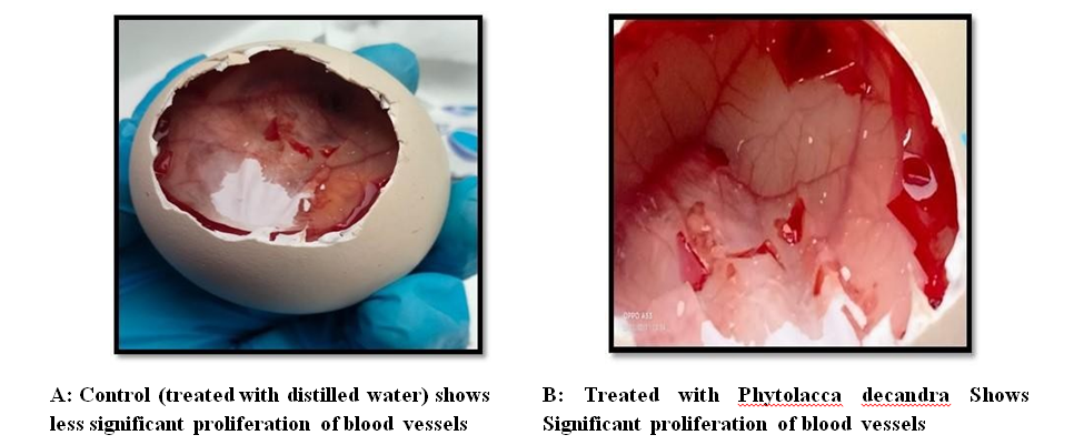
Ability to influence the process of angiogenesis was also tested using chick embryo. Phytolacca decandra increase the process of angiogenesis which shows they are not toxic to normal cells and improved blood supply with help in proliferation and will not affect normal cells unlike chemotherapeutic agents which will kill all the cells including normal cells. It may also aid in wound healing which can be further exploited.
Discussion
Other than demonstrating patterns of cellular toxicity that are comparable to in vitro models, the CAM model is known as an intermediary model that can bridge the gap between cell-based and animal-based studies [11]. Since nature is an endless source of bioactive compounds, extensive research has also been done on the appropriate study of wound healing through the use of medicinal plants. Many plants have been identified by traditional folk medicine as being helpful in the treatment of ischemic heart conditions and in the healing of wounds. These plants have recently been examined for their effect on the development of blood vessels in vitro and in vivo, with a focus on the impact on Vascular Endothelial Growth Factor (VEGF) expression. The following species of plants with substantial proangiogenic activity have been identified from studies on plants used in traditional medicine: Aloe vera, Patrinia villosa Juss., Hippophae rhamnoides L., Angelica sinensis, Cinnamomum cassia, Astragalus membranaceus, Stewartia koreana, Uncaria rhynchophylla, Salvia miltiorrhiza, and four ginsengs: P. schinsen, or Panax ginseng [12]. The normalisation of the tumor’s microenvironment and vasculature may be aided by the enhanced vascularization of CAM following the combination of etoposide and leaf extract treatment, which would increase the effectiveness of the drug administration and reduce hypoxia [13]. There are numerous Phytolacca- based medications on the market, as well as different kinds of Phytolacca extracts, all of which exhibit a range of pharmacological actions, including anti-inflammatory, anti- parasitic, antifungal, anticancer, and insecticidal effects. The significant anti-inflammatory properties of phytolacca extracts promote its use in the treatment of cancer as well as conditions like arthritis, nephritis, and rheumatism. Numerous glycosylated saponins, such as esculentosides and phytolaccosides, as well as a few flavones (cochliophilin A) and phytosterols (-spin sterol), which support Phytolacca’s anti-inflammatory and/or anticancer activities, have been found from the plant [14]. The present study shows the ability to impact the angiogenesis process was also examined using chick embryos. Contrary to chemotherapy, which kills all cells, including normal ones, Phytolacca decandra increases angiogenesis, demonstrating that it is not hazardous to healthy cells. It also improves blood supply and aids in cell proliferation. It might help speed up wound healing, which is something else that can be used.
Conclusion
CAM assay demonstrates the pattern of angiogenesis observed on treatment with Phytolacca decandra 6CH, which shows the significant proliferation of blood vessels. Homoeopathy medicines can be incorporated in the multimodal treatment. Side effects by Chemotherapy drugs can be reduced by Phytolacca decandra. Ethanolic extract of Phytolacca decandra (PD), used in homeopathy for the treatment of various ailments like chronic rheumatism, regular conjunctivitis, psoriasis [15]. The ultimate goal of Homeopathic medicine Phytolacca decandra eradicate disease without side effects. The preliminary results warrants further in vivo and in vitro to evaluate the potential and biological action of Phytolacca decandra. Further prospects to find out action of Phytolacca decandra in wound healing by using scratch assay. The Phytolacca decandra have another therapeutic effect towards chronic rheumatism, regular conjunctivitis, psoriasis, and in some skin diseases [16]. Nanoparticles paves many recompenses as drug carrier, there are still many restrictions to be solved such as unfortunate oral bioavailability, instability in circulation, inadequate tissue distribution, and toxicity [17].
References
-
Bagot JL, Theunissen I, Mouysset JL, Wagner JP, Magné N, et al. (2021) Integrative Health Care: Definition and examples of implementation in Oncology in France. La Revue d’Homéopathie 12(4): e61-e66.
-
Frass M, Lechleitner P, Gründling C, Pirker C, Grasmuk Siegl E, et al. (2020) Homeopathic Treatment as an Add‐ On Therapy May Improve Quality of Life and Prolong Survival in Patients with Non‐Small Cell Lung Cancer: A Prospective, Randomized, Placebo‐Controlled, Double‐ Blind, Three‐Arm, Multicentre Study. The oncologist 25(12): e1930-e1955.
-
Rossi E, Noberasco C, Picchi MP, Di Stefano M, Nurra L, et al. (2018) Homeopathy and complementary integrative medicine, dietary and lifestyle advices to reduce adverse- effects of anti-cancer therapy: a cohort study with breast cancer patients. OBM Integrative and Complementary Medicine 3(3): 17.
-
Molinari L (2022) Mesothelioma multimodal therapy: Types, side effects & amp. Mesothelioma.com.
-
Grambow E, Sorg H, Sorg CG, Strüder D (2021) Experimental Models to Study Skin Wound Healing with a Focus on Angiogenesis. Medical Sciences 9(3):55.
-
Ribatti D, Vacca A, Ranieri G, Sorino S, Roncali L (1996) The chick embryo chorioallantoic membrane as an in vivo wound healing model. Pathology-Research and Practice 192(10): 1068-1076.
-
Murphy R (2003) Lotus materia medica. B Jain Publishers.
-
Kargozar S, Mozafari M (2022) Biomaterials for vasculogenesis and angiogenesis. 1st (Edn.), Woodhead Publishing.
-
Kent JT. Lectures on Homoeopathic materia medica.
-
Patrick KF, Kumar S, Edwardson PA, Hutchinson JJ (1996) Induction of vascularisation by an aqueous extracts of the flowers of Calendula officinalis L. the European marigold. Phytomedicine 3(1): 11-18.
-
Kue CS, Tan KY, Lam ML, Lee HB (2015) Chick embryo chorioallantoic membrane (CAM): an alternative predictive model in acute toxicological studies for anti- cancer drugs. Experimental animals 64(2): 129-138.
-
Hoseinkhani Z, Norooznezhad F, Rastegari Pouyani M, Mansouri K (2020) Medicinal plants extracts with antiangiogenic activity: where is the link?. Advanced Pharmaceutical Bulletin 10(3): 370-378.
-
Yamuna ST, Sowndharyalatha B, Poornima A, Sumathi S (2020) An In-vtro Study on the Anti-Angiogenic Effect of Majorana Hortensis Leaves. International Journal of Pharmaceutical Sciences and Research 12(5): 2939- 2949.
-
Bailly C (2021) Medicinal properties and anti- inflammatory components of Phytolacca (Shanglu). Digital Chinese Medicine 4(3): 159-169.
-
Ghosh S, Bishayee K, Paul A, Mukherjee A, Sikdar S, et al. (2013) Homeopathic mother tincture of Phytolacca decandra induces apoptosis in skin melanoma cells by activating caspase-mediated signaling via reactive oxygen species elevation. Journal of Integrative Medicine 11(2): 116-1124.
-
Kathirvelraja MU, Krishnakumari Amma CR, Winston Vargheese V and Gopukumar ST (2022) In vitro Anti- Cancer, Anti Metabolic and Anti Proliferative Activity of Phytolacca decandra 6CH and 200CH in Breast Cancer CellLines. Nanomedicine and Nanotechnology Open Access 7(2).
-
Akhila R, Praseetha PK, Ariharan VN, Gopukumar ST (2019) In-vitro chick chorioallantoic membrane study of Chitosan capped 5-fluorouracil conjugated gold nanoparticles. J Pharm Sci & Res 11(5): 2090-2094.
- The Muculent Bleb-Mucinous Cystic Neoplasm-Hepatobiliary Region
- Insulin Sensitizers as Anti-Aging Agents: Unveiling Synergies with Albumin, GLP-1RA, Klotho Protein, and Metformin in the Quest to Combat Aging
- Reprogramming of GLP-1 Response at Prediabetes for the Prevention of Type 2 Diabetes: The Role of Albumin and GLP-1 Receptor Agonists
- The Mingled Allies-Combined Hepatocellular Carcinoma and Cholangiocarcinoma
- Compilation and Embodiment-Leydig Cell Tumour Testis
- Glucolipotoxicity: A Novel Different Perspective on the Causes of Cancer