Epidemiological and Clinical Studies of the Chronic Leuts of the Legs at the Friendship Hospital of Bangui, Central African Republic
<p>Introduction: The purpose of this work was to describe the epidemiological and clinical aspects of leg ulcers in Bangui, Central African Republic Material and methods: This was a descriptive cross-sectional study from October 2014 to June 2016 including patients of both sexes, over 18 years old, treated for chronic ulcers in the Surgery Department of the Hospital of Friendship of Bangui.</p> <p>Results: Of a total of 1089 patients in the department, 41 were leg ulcers, with a hospital frequency of 3.8%. The average age was 50.2 years. The most affected age group was over 40 years old and over (78.0%). The male sex (58.5%) was more represented with a sex ratio equal to 1.4. Patients exercising an activity requiring prolonged standing were more likely to have ulcer in 53.7%. The circumstances of ulceration were infection (41.5%), trauma (34.1%), pruritus (12.2%), insect sting (7.3%) or burn (4.9%). The duration of clinical signs was less than 12 months (63.4%), 1-5 years (34.1%) and 6-10 years (2.5%). Among the favored conditions, HIV infection (17.1%), followed by diabetes (14.6), hypertension + diabetes (9.8%), under nutrition (7.3%), HIV + diabetes (2.4%), hypertension (2.4%). Leg ulcers were more localized on the right leg (36.6%). They were unique (73.2%) and multiple (26.8%). The diameter was 10 to 19 cm (36.5%); less than 10 cm (24.4%); greater than 30 cm (24.4%) and 20 to 30 cm (14.6%). The signs associated with the ulcer were edema (46.3%), pain (24.4%), absence of pedal pulse (7.3%), and ankylosis of the ankle (7.3%). Etiologies were established in 68.29% of cases, including infectious (28.57%), necrotic angiodermia (21.42%), arterial (21.42%), venous (17.85%) and pyoderma gangrenosum (10.4%).</p> <p>Conclusion: Ulcers are more common from age 40 years old and men are the most affected. Infectious and therefore avoidable causes dominate. It is essential to inform the patients, to sensitize the population and to train the nursing staff in order to reduce the prevalence and to improve care.</p>
Introduction
Chronic wounds are by definition wounds whose healing process is impaired [1]. When the wound sits on the lower limbs, we talk about ulcers of the leg. Leg ulcers occur around the age of 70 with female predominance [2]. In 80% of cases, necrotic angiodermitis affects women over 60 years of age. Arteriolar involvement is secondary to high blood pressure in 90% of cases and diabetes in one third of cases. Other factors are found associated with this type of wound, 50% of patients have arterial disease of the lower limbs, 33% suffer from venous insufficiency [2, 3, 4, 5]. Data is non-existent in the Central African Republic. The aim of this work was to describe the epidemiological and clinical aspects of leg ulcers in Bangui, Central African Republic.
Material and Methods
It was a descriptive cross-sectional study that took place from October 2014 to June 2016, ie 21 months. The study population was represented by all patients of both sexes treated at the General Surgery Department of the Friendship Hospital. The sample consisted of patients of both sexes with leg ulcers. We included all patients 18 years of age and older, both sexes, who had a loss of skin substance with no tendency to spontaneous scarring, and who had been in surgery for 4 to 6 weeks sitting in the leg or ankle General. Patients with recent loss of substance sitting in the leg and other parts of the body were not included. The recruitment of patients was done on individual cards open on arrival of each patient. The data was collected using a pre-established questionnaire and was captured and analyzed using Excel and epi info 7. The chi-square test was performed to compare proportions with a significance level of 5%.
Results
Of a total of 1089 patients in the department, 41 were leg ulcers, representing a hospital frequency of 3.8%. Table I shows the distribution of patients by sex and age.
The average age was 50.2 years with extreme ages of 18 and 88 years. The most affected age group was over 40 (78.0%). The male sex (58.5%) was more represented with a sex ratio of 1.4 and predominated in all age groups with no statistically significant difference.
Male Female Total
Age / Sex
Number % Number % Number %
˂ 20 years 1 4% 0 0% 1 2,6%
20-39 years 6 25% 2 12% 8 19,5%
40years and + 17 71% 15 88% 32 78%
Total 24 58,5% 17 41,5% 41 100
X2= 25,54; ddl = 26; p= 0,488
Table I: Distribution of leg ulcer cases by age group and sex. The majority of patients (78.1%) came from Bangui. Patients in occupations imposing prolonged standing (grower, law enforcement officers, orderly, nurse, teacher) were more affected with ulcerin 53.7% than seated officials and traders with 24.4% and 19.4% respectively. The circumstances of ulceration were infection (41.5%), trauma (34.1%), pruritus (12.2%), insect sting (7.3%) or burn (4.9%). The duration of clinical signs was less than 12 months (63.4%), 1-5 years (34.1%) and 6-10 years (2.5%). The distribution of cases by identified risk factors is presented in Table 2.
| Frequency | Percentage | |
|---|---|---|
| Alcohol | 16 | 39,0 |
| Alcohol+Tobacco | 8 | 19,5 |
| Alcohol + tobacco Contact with chemical products | 3 | 7,3 |
| Obesity | 2 | 4,9 |
| Orthopedic treatment | 2 | 4,9 |
| Nothing | 10 | 24,4 |
| Total | 41 | 100 |
Table 1: Distribution of leg ulcer cases by identified risk
Advancing ground (antecedent) was HIV infection (17.1%), followed by diabetes (14.6), hypertension + diabetes (9.8%), under nutrition (7.3%), HIV + diabetes (2.4%), HTA (2.4) and no identified ground (19.5). Of the female subjects, 64.7% were at menopause. Table III shows the distribution of leg ulcer cases by seat.
| Seats | Number | Percentage |
|---|---|---|
| Right leg | 15 | 36,6 |
| Left leg | 9 | 22 |
| Right Malloleole | 7 | 17 |
| Left Malloleole | 5 | 12,2 |
| Back side right foot | 1 | 2,4 |
| Back side left foot | 2 | 4,9 |
| Right Achilles' tendon | 2 | 4,9 |
| Total | 41 | 100 |
Table 2: Distribution of leg ulcer cases by seat.
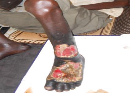
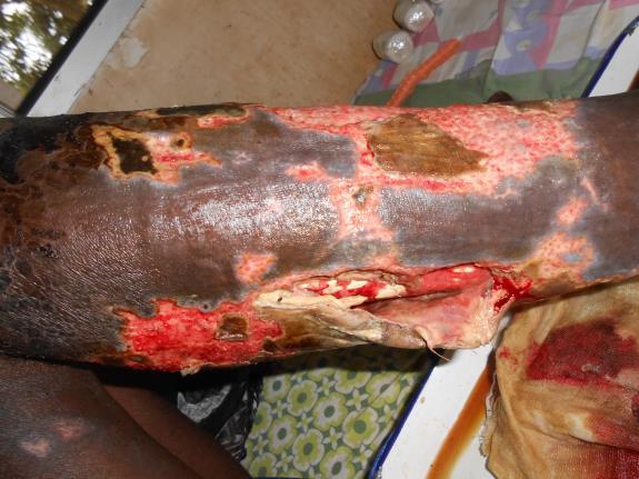
Table 3: Distribution of leg ulcer cases by seat. Leg ulcers were unique in 73.2% and multiple in 26.8% (Figure 1). Most leg ulcer lesions had a diameter of between 10 and 19 cm (36.5%), followed by less than 10 cm (24.4%), greater than 30 cm (24.4%), and 20 to 30 cm (14.6%). The signs associated with the ulcer were edema (46.3%), pain (24.4%), absence of pedal pulse (7.3%), ankylosis of the ankle (7.3%) and no sign (14.6%) of the cases. The etiologies were established in 28 out of 41 cases, or 68.29%, of which infectious causes accounted for 28.57%, ulcer by necrotic angiodermia (21.42%), arterialulcer (21.42%), venousulcer (17.85%) and pyoderma gangrenosum (10.71%) (Figure 2).
Discussion
This work was intended to contribute to the study of the leg ulcers. However, this is a clinical study based on clinical criteria for diagnosis, in an adult surgery department. The low purchasing powers of patients, the less equipped technical platform are all difficulties that have hindered the smooth progress of the study. However, this study was conducted rigorously and may be replicated with a higher sample. In terms of epidemiology, leg ulcer represented 3.8% of the pathologies treated at the General Surgery Department of the Friendish Hospital during this period of study. This frequency is lower than that obtained by So Niang, et al. [6] in Senegal who reported 4.4% of ulcer frequency in the dermatology department. Our frequency was well below the 24% noted by Zaraa I, et al. [7] in a dermatology department in Tunisia. This could be attributed to the expectation of life? Moreover, in our context, leg ulcer is a pathology whose severity is often unknown and often outpatient treatment at the peripheral level. In our study, we noted a young average age of onset of leg ulcer at around 50, 21 years of age. This corroborates data from African studies where the average age is generally between 33 and 50 years old [5, 8, 9]. Leg ulcer predominated in male patients in 58.5% with a sex ratio of 1.4 in our study. This male predominance is observed by African authors [10, 9, 6]. By contrast, in the West, 3 women are affected for 2 men [11]. This female predominance is explained by the high longevity and female predominance of varicoseveins. Our patients came from the city of Bangui and its suburbs (78%). These data are higher than those of So Niang, et al. [6] in Senegal where 59% of patients came from Dakar and its suburbs and 41% from other regions of Senegal.
This would be related to the state of the roads, the insecurity in the country decreasing the accessibility to care of patients from provinces. In addition, the role of traditional healers who often delay these types of pathologyis not to be neglected. Analysis of the patients' profession reveals that 53.6% had activities that required prolonged standing. This result is similar to that of Bakkal [10] in Morocco, which reports the presence of orthostatism in 51% of cases, but it remains higher than that of So Niang, et al. [6] in Senegal which found 28% of the patients exercising an activity involving a prolonged standing position predisposing to venous pathologies such as varicoseveins, the main causes of leg ulcers. In our study, the occurrence of legulcer was a skin infection in 41.5%, followed by trauma in 34.1%. In similar studies, no analysis of this situation has been made. Clinically, the majority of our patients had to self-medicatein 48.8% and others were among traditional healers in 29.2% before their hospitalization. No analysis of this situation has been made in other similar series. Among the pre disposing factors, those favoring the alteration of the immune functions (HIV, Diabetes, Sickle Cell Disease, Undernutrition) and those favoring the alteration of the vascular walls (alcohol, tobacco, HTA) were identified during our study. These factors were also identified by Bakkal [10] in Morocco and by Ndiaye, et al. [12] in Senegal. Most of our patients had consulted within less than 12 months. However, in 34.1% of cases, this period was between 1 to 5 years and 2.4% between 6 to 10 years. This result corroborates that of So. Niang, et al. [6] where 56% of patients usually consulted within less than 1 year. Regarding the number, the ulcerative lesion was unique in the majority of cases. However, it was multiple in a little more than a quarter of cases. Indeed single ulcers are the most common and would be venous cause compared to multiple ulcers that would be arterial cause. Ndiaye [12] in Senegal reports that single ulcers accounted for 76% and multiple ulcers 24%. These results remain superior to those of Bakkal [10] in Morocco and Souissi [9], who reported respectively 57.9% and 53% of single ulcer. In our series, the whole leg was affected in 24.4% of cases with local consequence of a lack of lymphatic circulation resulting in edema of the foot. When the muscle tissues tendons are affected, they lead to ankylosis, which we observed in 7.3% of cases. Disabling lesions, such as underlying bone lesions, were rarely observed in our study (24.4%). In Bakkal's study [10], 82.5% of ulcers were superficial and large in diameter beyond 5 cm in 51% of cases. Very few paraclinical examinations were performed by patients during our study. The search for infectious stigma and HIV immunodepression are the main examinations. Vascular explorations such as Doppler ultrasound and arteriography were not available at the facility level. In the Souissi [9] and Zaraa [7] studies in Tunisia, Doppler ultrasound was performed in 52.2% and 68% of cases, respectively, allowing a etiological diagnosis of certainty. Vascular ulcer accounted for 41.46% of etiologies in our series. This diagnosis was reported respectively in 64.8%, 48% and 61% of cases in the studies of Souissi, Zaraa and Bakkal [9, 7, 10]. Vascular ulcer is prevalent in African countries. The infectious origin of the leg ulcer remains difficult to explain in our region where super infection is frequent. This pathology accounted for 28% of the cases in our study. Bakkal and Zaraa [10, 7] reported the infectious etiology in 13.6% and 5%, respectively.
Conclusion
Leg ulcers are increasingly a problem in our hospitals. Our study showed the profile of ulcerative leg disease in Bangui. They affect men more than women with middle- aged youth. Menopause is the first favoring ground. The main revealing signs are edema. The most common ulcers are infectious and therefore avoidable ulcers. It is essential that strategies based on the information of the patients, the sensitization of the population and the training of the medical and paramedical staff are developed in order to reduce the long duration of evolution of the disease and therefore of management.
References
-
Levy E, Levy P (2001) Management of venous legular by french physicians, diversity and related costs : a prospective medico-economic observational study. J Mal Vasc 26(1): 39-44.
-
ElBakhal A (2011) Les ulcères de jambes : étude prospective à propos de 57 cas. Mémoire DS Med Rabat, pp: 55.
-
Teot L, Meaume S, Dereure O (2001) Wounds and scarring. Masson (Ed), pp: 351.
-
Chaby G (2015) Ulcères de jambe d’origine veineuse ou mixte à prédominance veineuse. EMC – Dermatol pp: 98-570-A-10.
-
Bureau J, Debure C (2006) Leg Ulcers. EMC- Angéiologie, pp: 19-0540.
-
Niang S, Dieng M, Kane A, Cissé M, Diallo M, et al. (2007) Ulcères de jambe à Dakar: 75 cas. Ann Dermatol Venereol 134(11): 868-870.
-
Zaraa I, M’na Ben A, Zribi H, Trojjet S, Euch D El, et al. (2012) Leg ulcer in the young Tunisian. Annales de Dermatologie 139(12S): B206.
-
Abdallah M, Fenniche S, Mahfoudh A, Mokhtar I (2009) Ulcère de jambe : particularités épidémiologique de l’ulcère de jambe en Tunisie. Ann Dermatol Venereol 136(HS1): A150-A151.
-
Souissi A, Tekaya Ben N, Youssef M, Cherif F, Mokni M, et al. (2005) Leg ulcers: clinical and epidemiological study of patients hospitalized in Tunis. Ann Dermatol Venereol 132: 1010-1012.
-
Beguaud B (2005) Epidémiologie des ulcères de jambes. Ann Dermatol Venereol 129: 1225-1226.
-
Haute Autorité de Santé ‘HAS (2006) Prise en charge de l’ulcère de jambe à prédominance veineuse hors pansement : argumentaire, pp: 23.
-
Ndiaye M, Niang S, Diop A, Diallo M, Diaz K et al. (2016) Leg ulcers during sickle cell disease: a retrospective study of 40 cases. Ann Dermatol Venereol 143(2): 103-107.
- Epithelioid Granuloma; 3cases with Different Clinical Features
- Advancing Representation in Dermatology Clinical Trials: Ethical, Scientific, and Regulatory Imperatives for Inclusion Across all Fitzpatrick Skin Types
- A Case of Atopic Dermatitis with Concurrent Psoriasis Vulgaris: Successful Treatment with Upadacitinib
- Innovation Lifting Eyeshadow: A Synthesis of Makeup and Optical Illusion
- Distinguishing Superficial Actinic Porokeratosis from Actinic Keratosis with UVF Dermoscopy: A Case Report
- High Mobility Group Box 1 (HMGB1) in Cutaneous Inflammation: An Immune Modulator Bridging Cellular Stress, Ferroptosis and Danger Signaling