Large Infantile Hemangioma on the Face: Let’s ‘PHACE’ It
Background: Infantile hemangiomas (IH) are the most common benign (vascular) tumors in infancy but due to their natural self-limiting course do not necessarily need treatment; in case of ulceration, bleeding or potential deformity, treatment is necessary. Large segmental facial hemangiomas may be associated with PHACE syndrome. Case presentation: We present a case of a boy with a large segmental facial IH. Ulceration, size and location were indications for starting propranolol up to a dose of 3 mg/kg/day in three doses, eleven days after birth. Because of the location and distribution, PHACE syndrome had to be xcluded: the patient was checked for possible brain, cardiac and vascular malformations and visual impairment. The IH improved. At the age of nine months, there were no adverse effects or IH progression. Discussion: Propranolol is a beta-blocker effective in treating IH. Its working mechanism has not been completely clarified. We provide a thorough description of our approach to the early propranolol treatment of this large segmental facial IH. Conclusion: Complicated IH like the one described here can be treated safely and effectively with propranolol; very early treatment initiation is recommended to prevent the development of anatomic deformities. Our patient was treated with propranolol 3 mg/kg/day in three doses in order to prevent future disfigurement when it is located in a high-risk area like the face. Further research is needed to create a uniform approach and universal guideline for the treatment of IH with propranolol.
Introduction
Infantile hemangiomas (IH) are the most common benign tumors in infancy. This vascular tumor occurs in approximately 5-10% of children, though most of them do not have a lesion at birth but develop one within the first days to months [1, 2]. Precursor lesions of IH are found in up to 65% of children with IH [3]. Suspected predisposing factors for developing IH are low birth weight, prematurity, female sex, twin birth, and advanced maternal age [4].
The natural course of an IH is a four month period of rapid growth followed by a slow growth phase until twelve months. 80% of all IHs reach their final size within three to four months of age. After completion of the growth phase the IH merges into regression, or involution, around 9-12 months. Complete involution will in 90% be reached after four years [5]. Though complete involution does not necessarily mean ‘normal’ skin appearance; half of the patients has residual signs of scarring, atrophy, telangiectasias or fibro-fatty tissue [2]. Most IH are solitary, and can be divided into subtypes: superficial, deep subcutaneous, or a combination of these. A superficial IH, or strawberry-like patch, is a bright red slightly raised plaque, patch or nodule, whereas a deep IH is a skin colored or bluish nodule, sometimes with central telangiectasias. The combined variant has characteristics of both subtypes [2]. Predilection sites are the head and neck area (60%), trunk (25%), and extremities (15%) [1].
In most cases a clinical diagnosis is sufficient, but if an underlying syndrome is suspected, especially in larger and/ or segmental IH in the facial or diaper area, further evaluation is required. An important, and not uncommon, spectrum of anomalies is PHACE syndrome, a neuro- cutaneous disorder. PHACE is an acronym for: posterior fossa and other brain malformations; hemangioma; arterial anomalies of cervical and cerebral vessels; cardiac defects (such as aortic coarctation); and eye anomalies. In some cases an S is added which stands for sternal defects or supra-umbilical raphe. Patients with large IH of the face or scalp should undergo imaging of the head, neck and chest, and ophthalmologic evaluation to rule out PHACE syndrome [6, 7, 8].
Rapidly proliferating lesions are more likely to become ulcerative. In a large cohort study ulceration was observed in 16% of the patients [9]. This serious complication can lead to pain, bleeding, scarring, secondary infection and permanent functional impairment. Other complications are related to localization of the IH in the orbital, auricular or nasal area, which can consequently cause visual problems like amblyopia, ptosis and strabismus, and serious facial anatomy disfigurement. Possible visual or hearing problems are often objectified at the children's health clinic [10]. Segmental IH, most importantly of the face, is associated with worse outcome and a higher complication rate [11].
Most IH are uncomplicated and do not necessarily need treatment due to their self-limiting nature [1, 2]. Treatment is required for IH that will potentially cause disfigurement and functional impairment in the absence of therapy. Other indications for treatment of cutaneous IH are rapid growth, ulceration or facial location [1, 2].
The current gold standard treatment of IH with (potential) complications is systemic treatment with the beta-blocker propranolol.
Case Presentation
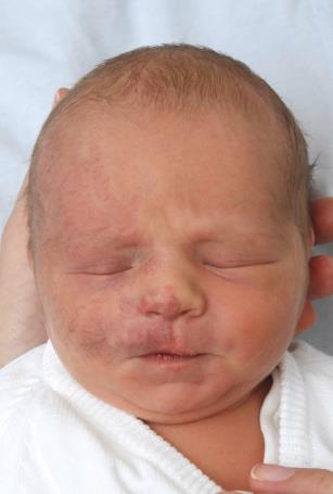
A healthy full-term newborn boy with a birth weight of 2915 gram presented at birth with a light purple patch with diffuse telangiectasias on the right side of his face and an asymmetric upper lip Figure 1. The discoloration intensified with crying. There were no complications during pregnancy or birth. The mother gave birth at the age of 36.
The patch evolved, became darker and more extensive, and we clinically diagnosed him with a segmental IH. Because of its extensiveness we sought advice from the Dutch expertise center in Nijmegen (HECOVAN). The patient went there for a single consultation; they recommended to rule out PHACE syndrome on account of the size and facial involvement and start treatment early. The pediatrician in our center found no clinical signs of syndromal involvement, but additional evaluation was scheduled.
In the meantime, at one week old, the upper lip became ulcerative and the IH thickened in the peri-orbital region.
To prevent further progression and disfigurement, treatment with propranolol was started in collaboration with the pediatrician. At eleven days old, figure 2, the boy was admitted to the children’s ward and after examination of cardiac and pulmonary function and glucose status propranolol was started at a dose of 2.25 mg a day (0.75 mg/kg/day) divided in three doses with feedings. He was also prescribed local Vaseline zinc oxide cream and systemic amoxicillin/clavulanic acid to prevent secondary infection of the ulceration. In five days the dose was slowly increased 3 mg/kg/day in three doses without adverse effects after which he was discharged from the hospital.
Meanwhile, the results of the additional testing for PHACE syndrome came back; we found no signs of PHACE syndrome. MRI/MRA in consultation with the pediatric neurologist showed normal anatomic variation of the brain and its vasculature, echocardiography showed no coarctation of the aorta or other deformities, and ophthalmologic evaluation was without anomalies. Inspection of the mouth and nose mucosa by an otolaryngologist showed no abnormalities and later, the children's health clinic’s evaluation of growth, motor development and social, cognitive and communicative development were within the normal range.
Ulceration healed within one week. After four months of monthly evaluation at the outpatient clinic, the IH had faded. The treatment was very effective and the parents had not noticed any adverse effects. However, after five months, a darker area of proliferating IH on the right cheek developed. His general condition was still good, and he weighed 6 kg. The dose of propranolol was increased to 15 mg a day (2.5mg/kg/day) divided in three doses in accordance with his weight gain. Now at eleven months, only teleangiectasias and scarring are visable.
Discussion
Propranolol was very effective for this large congenital segmental ulcerative and potentially disfiguring IH of the face. Standard dosage of the beta-blocker seemed insufficient to reach optimal effect. Under close evaluation, the dosage was increased without causing any adverse effects and with a positive effect on the IH.
Since the serendipitous discovery of the effectiveness of propranolol in 2008, treatment of IH with this non- selective beta-blocker has been investigated thoroughly [10, 12, 13]. In 2014 propranolol was FDA approved for the treatment of complicated IH and has been the first line treatment for IH worldwide ever since [2]. Before 2008, common treatments were intralesional or systemic corticosteroids, imiquimod, chemotherapeutic agents like vincristine and α-interferon, laser therapy, surgical treatment or a combination of the former [1].
Propranolol has an effect on both β1 and β2-receptors, but its mechanism of action is not fully understood. Theories are that it is a combination of 1) vasoconstriction 2) decreased expression of angiogenic factors like basic Fibroblast Growth Factor (bFGF) and Vascular Endothelial Growth Factor (VEGF), 3) apoptosis of endothelial cells caused by hypoxia and suppression of Glucose Transporter Type 1 (GLUT1), which is an important marker for IH, and 4) inhibition of the renin angiotensin aldosteron system (RAAS) which prevents further proliferation [1, 2, 10].
Multiple studies show that the adverse effects like hypotension, bradycardia, hyperkalemia, bronchospasms, and hypoglycemia are infrequently reported. Restless sleep, gastrointestinal tract problems, and cold extremities are more common [1, 2, 13]. In a trial with 250 cases of IH none of the occurring adverse effects were life- threatening [13]. Initiation of propranolol treatment was, because of these possible serious side-effects, initially exclusively carried out in expertise centers and always combined with admittance to the hospital. Recent developments show a trend of propranolol treatment initiation in an outpatient setting [2, 14].
Our approach is to at least admit the children younger than one month, ex-prematures younger than two months, children with threatened airway, a higher risk of cardiac adverse effects or hypoglycemia (simultaneous prednisone treatment), with a complicated medical history and/or relative contraindications for propranolol treatment.
There is no universal international or even national guideline; every institution seems to have its own protocol [14.15].
Our approach has recently been recorded in a hospital-wide used guideline. Prior to treatment we evaluate possible pulmonic and cardiovascular disease, and ECG is performed. Because infants and pre-terms are more prone to hypoglycemia, because of higher glucose employment and lower stores, checking serum glucose pre-treatment is included in our guideline, it is however not agreed upon world-wide [13, 15]. If all is well, we recommend admitting patients like ours (with complicated IH and age under one month old) for at least three days under supervision of a collaborating dermatologist and pediatrician. The propranolol starting dose should be 0.75-1.0 mg per kg daily in three divided doses with regular intervals of approximately 5-6 hours. One could for instance administer doses at 7 AM, 1 PM and 7 PM with feedings.
For example, if the child weighs 3 kg and starting dose is 0.75 mg/kg/day this results in 0.75 mg three times daily, 2.25 mg/day. The second day it is increased to 1.5 mg/kg/day, 4.5 mg/day in three doses, and the third day to 2.25 mg/kg/day, 6.75 mg/day in three doses. In some cases it is necessary to deviate from this scheme and increase the dose in five to seven days.
Right before and thirty minutes after admission for two hours, vital functions (heart rate and blood pressure) should be monitored by telemetry [15]. During treatment, the patient should be checked regularly for symptoms of hypoglycemia, including signs of tachycardia, hunger, sweating, shaking, and severe symptoms of brain glucose shortage like lethargy, stupor, seizures, apnea, hypothermia and loss of consciousness [15].
Our treatment goals justifying the early treatment start were ulceration and resulting feeding difficulties, as well as cessation of progression and sooner transition to the involution phase to prevent otherwise certain deformities. When treatment is started later the IH will flatten, lighten and soften, but deformities may have already been caused [2]. Telangiectasias that do not disappear with propranolol treatment can be treated with laser therapy after involution if necessary and desired by parents and child [16].
In less severe cases we recommend to start propranolol treatment before 2-4 months of age when the IH is still in the proliferation phase for the best results preventing disfigurement. The duration of treatment should be based on the type of IH, indication for treatment and time of initiation. To prevent proliferation after cessation treatment should cover the entire proliferation phase up until 9-18 months, in our experience the mean duration is around 12 months.
Ideal follow up takes place one month after treatment commencement, and after that every 6 weeks. Check-ups should include close IH inspection for signs of progression or involution; comparison to previous photographs is recommended. One should inquire about signs of adverse reactions to propranolol with the parents [15]. Possible dose adjustment is indicated in accordance to the patients weight gain, usually until nine months of age, unless there is sufficient treatment response [2].
Furthermore, in the interval of follow-up moments parents should be carefully instructed for signs of adverse events caused by propranolol treatment. Important signs that possibly require intervention are loss of consciousness, altered heart rate, unusual weakness, seizures and wheezing, milder symptoms are cold hands and feet, fatigue during the day, restless nights, sickness including recurrent colds, diminished intake and gastro- intestinal complaints [15, 17].
Conclusion
Complicated IH, like the one this article describes, require a very early propranolol treatment initiation to prevent anatomic deformities. It is of the utmost importance to decrease patient and doctor’s delay and start therapy as early as possible. Our case of a large IH on the face, teaches us that early treatment was safe and very much effective in causing a premature transition into the involution phase. However we might ask ourselves if ulceration could have been prevented with even earlier initiation of therapy. Research on this subject is ongoing.
References
-
Starkey E, Shahidullah H (2011) Propranolol for infantile haemangiomas: a review. Arch Dis Child 96(9): 890-893.
-
Hermans DJ, Ottenhof MJ, Wijnen MH, van Beynum IM, van der Horst CM, et al. (2011) Treatment of haemangiomas of infancy with propranolol; good results, few side effects. Ned Tijdschr Geneeskd 155(40): A3482.
-
Tollefson MM, Frieden IJ (2012) Early growth of infantile hemangiomas: what parents' photographs tell us. Pediatrics 130(2): e314-20.
-
Munden A, Butschek R, Tom WL, Marshall JS, Poeltler DM, et al. (2014) Prospective study of infantile haemangiomas: incidence, clinical characteristics and association with placental anomalies. Br J Dermatol 170(4): 907-913.
-
Leaute-Labreze C, Harper JI, Hoeger PH (2017) Infantile haemangioma. Lancet 390(10089): 58-94.
-
Metry D, Heyer G, Hess C, Garzon M, Haggstrom A, et al. (2009) Consensus Statement on Diagnostic Criteria for PHACE Syndrome. Pediatrics 124(5): 1447-1456.
-
van Doesburg MH, Breugem CC, Breur JM, Braun KP, Speleman LA, et al. (2009) Segmental facial hemangiomas and associated structural defects. J Craniofac Surg 20(4): 1224-1227.
-
Garzon MC, Epstein LG, Heyer GL, Frommelt PC, Orbach DB, et al. (2016) PHACE Syndrome: Consensus-Derived Diagnosis and Care Recommendations. J Pediatr 178: 24-33.e2.
-
Chamlin SL, Haggstrom AN, Drolet BA, Baselga E, Frieden IJ, et al. (2007) Multicenter prospective study of ulcerated hemangiomas. J Pediatr 151(6): 684-9, 689.e1.
-
Herman, DJ, Bauland CG, Zweegers J, van Beynum IM, van der Vleuten CJ (2013) Propranolol in a case series of 174 patients with complicated infantile haemangioma: indications, safety and future directions. Br J Dermatol 168(4): 837-843.
-
Chiller KG, Passaro D, Frieden IJ (2002) Hemangiomas of infancy: clinical characteristics, morphologic subtypes, and their relationship to race, ethnicity, and sex. Arch Dermatol 138(12): 1567-1576.
-
Léauté-Labrèze C, Dumas de la Roque E, Hubiche T, Boralevi F, Thambo JB, et al. (2008) Propranolol for severe hemangiomas of infancy. N Engl J Med 358(24): 2649-2651.
-
Solman L, Murabit A, Gnarra M, Harper JI, Syed SB, et al. (2014) Propranolol for infantile haemangiomas: single centre experience of 250 cases and proposed therapeutic protocol. Arch Dis Child 99(12): 1132- 1136.
-
Kumar MG, Coughlin C, Bayliss SJ (2015) Outpatient Use of Oral Propranolol and Topical Timolol for Infantile Hemangiomas: Survey Results and Comparison with Propranolol Consensus Statement Guidelines. Pediatric Dermatology 32(2): 171-179.
-
Drolet BA, Frommelt PC, Chamlin SL, Haggstrom A, Bauman NM, et al. (2013) Initiation and Use of Propranolol for Infantile Hemangioma: Report of a Consensus Conference. Pediatrics 131(1): 128-140.
-
Adamic M, Troilius A, Adatto M, Drosner M, Dahmane R (2007) Vascular lasers and IPLS: guidelines for care from the European Society for Laser Dermatology (ESLD). J Cosmet Laser Ther 9(2): 113-124.
-
Martin K, Blei F, Chamlin SL, Chiu YE, Frieden IJ, et al. (2013) Propranolol Treatment of Infantile Hemangiomas: Anticipatory Guidance for Parents and Caretakers. Pediatric Dermatology 30(1): 155-159.
- Epithelioid Granuloma; 3cases with Different Clinical Features
- Advancing Representation in Dermatology Clinical Trials: Ethical, Scientific, and Regulatory Imperatives for Inclusion Across all Fitzpatrick Skin Types
- A Case of Atopic Dermatitis with Concurrent Psoriasis Vulgaris: Successful Treatment with Upadacitinib
- Innovation Lifting Eyeshadow: A Synthesis of Makeup and Optical Illusion
- Distinguishing Superficial Actinic Porokeratosis from Actinic Keratosis with UVF Dermoscopy: A Case Report
- High Mobility Group Box 1 (HMGB1) in Cutaneous Inflammation: An Immune Modulator Bridging Cellular Stress, Ferroptosis and Danger Signaling