Onychomatricoma: An Intraoperative Diagnosis
Onychomatricoma is a rare benign fibroepithelial tumor of the nail complex. The tumor is more frequent in middle-aged Caucasian women presenting chirodactyls. Trauma is the main etiological factor. Clinical presentation is characterized by a brownish hyperkeratotic longitudinal strip of variable thickness, splinter hemorrhages affecting the proximal portion of the nail plate preferentially, longitudinal and transverse hyper- curvature of the nail plate and digitiform projections that emerge from the nail matrix. In addition, it can be observed cavitations in the nail plate, structures that are best visualized after surgical exeresis of the lesion. It mimics many other conditions and tumors, and biopsy and histopathological analysis are essential for diagnostic confirmation.
Case Report
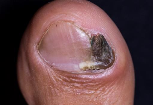
Male patient, 59 years old, with a 2-year course of hyperkeratosis, dystrophy and ungual hyperchromia of the left hallux (Figure 1).
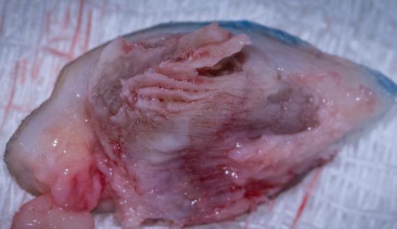
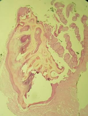
The patient was referred to the dermatology service for diagnostic and treatment, and the tumor was excised (Figure 2). During the surgical procedure, it was possible to visualize the classic structures for diagnosis and confirmed by histopathological examination, compatible with onychomatricoma (Figures 2 & 3).
Discussion
Onychomatricoma is a benign and rare fibroepithelial tumor of the nail matrix, first reported by Baran and Kint [1, 2]. It has brownish hyperkeratotic bands and deformities of the nail plate. The macroscopic picture of onychomatricoma mimics other tumors and conditions, and biopsy and histopathological analysis are essential for diagnostic confirmation.
Nail surgery is a painful procedure and often leaves permanent dystrophies on the nail plate. After careful diagnostic analysis of the lesion, an experienced dermatologist must perform the surgical procedure, as it is necessary to distinguish the nail pathology from other diseases, and thus schedule an adequate surgical margin. Fibrokeratoma, periungual fibroma, osteochondroma, onicopapilloma, squamous carcinoma of subungual exostosis, amelanotic melanoma, keratoacanthoma, common wart, onychogryphosis, Darier's disease, and nail lichen planus are differential diagnoses that should be questioned during the evaluation [3]. It is important to remember that despite the benign appearance, recurrence can occur and long-term follow-up is indicated because it is uncertain if the onychomatricoma will turn to malignant [4].
Following complete excision of the lesion, it is possible to visualize digitiform structures that are typical, and after that the diagnosis is confirmed with a histopathological examination. The characteristics are deep epithelial invaginations, fibrillar and fibrotic stroma in the proximal nail fold, and multiple projections in the distal zone.
References
-
Nakandakari S, Marques GF, Soares CT, Santos LSS, Sousa JMP (2014) Onicomatricoma: Tumor raro do aparelho ungueal − Relato de três casos. Surg Cosmet Dermatol 6(1): 902.
-
Baran R, Kint A (1992) Onychomatrixoma. Filamentous tufted tumour in the matrix of a funnel - shaped nail: a new entity (report of three cases). Br J Dermatol 126: 510-505.
-
Cinotti E, Veronesi G, Labeille B, Cambazard F, Piraccini BM, et al. (2018) Imaging technique for the diagnosis of onychomatricoma. J Eur Acad Dermatol Venereol 32(11): 1874-1878.
-
Durrant MN, Palla BA, Binder SW (2012) Onychomatricoma: a case report with literature review. Foot Ankle Spec 5(1): 41-44.
- Epithelioid Granuloma; 3cases with Different Clinical Features
- Advancing Representation in Dermatology Clinical Trials: Ethical, Scientific, and Regulatory Imperatives for Inclusion Across all Fitzpatrick Skin Types
- A Case of Atopic Dermatitis with Concurrent Psoriasis Vulgaris: Successful Treatment with Upadacitinib
- Innovation Lifting Eyeshadow: A Synthesis of Makeup and Optical Illusion
- Distinguishing Superficial Actinic Porokeratosis from Actinic Keratosis with UVF Dermoscopy: A Case Report
- High Mobility Group Box 1 (HMGB1) in Cutaneous Inflammation: An Immune Modulator Bridging Cellular Stress, Ferroptosis and Danger Signaling