Clinical Dermatology Open Access Journal
Research Article
1 min read
Nodules of Scalp in Infant: What is your Diagnosis
* Corresponding author
Keywords
Juvenile xanthogranuloma
Chilhood
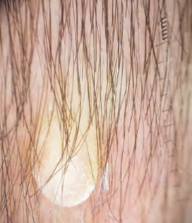
Dermoscopy
Abstract
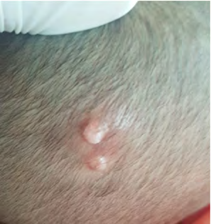
A 6 week-old boy, was evaluated for 2 asymptomatic,erythematous lesions on the right scalp vertex that was present 7 days after birth. He was otherwise healthy. Dermatological examination revealed two wellcircumscribed papule and nodule , erythematous in color,7- 10mm in diameter with firm consistency, located on the right scalp vertex. The rest of the physical examination was normal. An ophthalmologic examination was performed and was normal.
Conflict of Interest
The authors do not declare any conflict of interest.
Click to enlarge
Click to enlarge
Contributions of Authors
All authors contributed to the writing of this article. The authors also state that they have read and approved the final version.
References
-
Helwig EB, Hackney VC (1954) Juvenile xanthogranuloma (nevoxanthoendothelioma). Am J Pathol 30: 625-626.
-
Pajaziti L, Hapçiu S, Pajaziti A (2014) Juvenile xanthogranuloma: a case report and review of the literature. BMC Res Notes 7(1): 174.
-
Herbst AM, Laude TA (1999) Juvenile xanthogranuloma: further evidence of a reactive etiology. Pediatr Dermatol 16(2): 164.
-
Dehner LP (2003) Juvenile xanthogranulomas in the first two decades of life: a clinicopathologic study of 174 cases with cutaneous and extracutaneous manifestations. Am J Surg Pathol 27(5): 579-593.
-
Liang S, Liu YH, Fang K (2009) Juvenile xanthogranuloma with ocular involvement. Pediatr Dermatol 26(2): 232- 234.
-
Oliveira TE de, Tarlé RG, Mesquita LA de F (2018) Dermoscopy in the diagnosis of juvenile xanthogranuloma. Anais Brasileiros de Dermatologia 93(1): 138-140.
-
Litaiem N, Zeglaoui F (2018) Is the setting sun dermoscopic pattern specific to juvenile xanthogranuloma? J Am Acad Dermatol 78(2): 49.
← Previous Article
Reversal Reactions Observed in Two Cases of Subpolar Lepromatous Leprosy
Next Article →
When Trichoscopy Reveals Things to us
More from this journal
- Epithelioid Granuloma; 3cases with Different Clinical Features
- Advancing Representation in Dermatology Clinical Trials: Ethical, Scientific, and Regulatory Imperatives for Inclusion Across all Fitzpatrick Skin Types
- A Case of Atopic Dermatitis with Concurrent Psoriasis Vulgaris: Successful Treatment with Upadacitinib
- Innovation Lifting Eyeshadow: A Synthesis of Makeup and Optical Illusion
- Distinguishing Superficial Actinic Porokeratosis from Actinic Keratosis with UVF Dermoscopy: A Case Report
- High Mobility Group Box 1 (HMGB1) in Cutaneous Inflammation: An Immune Modulator Bridging Cellular Stress, Ferroptosis and Danger Signaling