Clinical Dermatology Open Access Journal
Research Article
1 min read
When Trichoscopy Reveals Things to us
* Corresponding author
Keywords
Trichoscopy
Abstract
A 40-year-old patient, with no notable pathological antecedents, followed in internal medicine for erythematous lupus with renal, immunological and cutaneous involvement retained on clinical, histological and immunological criteria using synthetic antimalarials, with oral corticotherapy
Figures
Click to enlarge
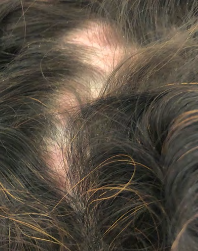
![Figure 2: green circles] linear sinuous arborescent vessels [Figure 2: gray arrow]. Corkscrew hairs [Figure 2: red circle] and scales [Figure 2: blue arrow]. Apart from dermoscopic signs of lupus, there have been signs in favor of a tinea capitis, The mycological samples taken were in favor of a trichophyton rubrum, the patient was put on oral and topical griseofulvin as well as hygienic rules adapted with good clinical progress.](/fulltextimages/4915/fig_2.png)
Click to enlarge
References
-
Francès C (2015) Manifestations cutanées des lupus érythémateux. EMC-Dermatologie 10: 1-14.
-
El-Khalawany M, Shaaban D, Hassan H, Abdalsalam F, Eassa B, et al. (2013) A multicenter clinicomycological study evaluating the spectrum of adult tinea capitis in Egypt. Acta Dermatovenerol Alp Pannonica Adriat 22(4):77-82.
-
Cervetti O, Albini P, Arese V, Ibba F, Novarino M, et al. (2014) Tinea capitis in adults. Adv Microbiol 4: 12.
← Previous Article
Nodules of Scalp in Infant: What is your Diagnosis
Next Article →
Treatment of Cutaneous Leishmaniasis with Photodynamic Therapy: The First Case Report from Khyber Pakhthunkhwa (KPK), Swat Pakistan
More from this journal
- Epithelioid Granuloma; 3cases with Different Clinical Features
- Advancing Representation in Dermatology Clinical Trials: Ethical, Scientific, and Regulatory Imperatives for Inclusion Across all Fitzpatrick Skin Types
- A Case of Atopic Dermatitis with Concurrent Psoriasis Vulgaris: Successful Treatment with Upadacitinib
- Innovation Lifting Eyeshadow: A Synthesis of Makeup and Optical Illusion
- Distinguishing Superficial Actinic Porokeratosis from Actinic Keratosis with UVF Dermoscopy: A Case Report
- High Mobility Group Box 1 (HMGB1) in Cutaneous Inflammation: An Immune Modulator Bridging Cellular Stress, Ferroptosis and Danger Signaling