Easy Diagnosis of Basal Cell Carcinoma by Demosocpie
The BCC is a cancer with low grade and rares métastases. Superficial basal cell carcinoma (SSBCC) comprise up to 25% of all histological sub-types. They are more likely to occur on the trunk and in the younger age groups especially in females. SSBCC appears as a scaly and well-defined area. It can resemble a patch of dermatitis and can be confused with eczema, psoriasis, lichen planus, or Bowen’s disease. Thus, the clinical features alone may not point to the appropriate diagnosis. The histopathology is the most reliable diagnostic modality for SSBCC. Surgical excision is the most commonly used treatment for BCC. Topical chemotherapy agents such as imiquimod or 5-fluorouracil may be various alternatives or adjuvants in the treatment of SSBCC.
Case Report
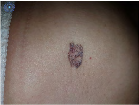
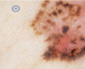
It is a patient of 53 years, without significant pathological antecedents, who presents for 2 years a lesion in the right hypochondrium, gradually increasing in size. Clinical examination found a slightly infiltrated 2 cm plaque with a pearled outline at the level of the right hypochondrium (Figure 1), dermoscopy showing ovoid nests, dots and globules, eccentric radial striations, and a maple leaf appearance (Figure 2).
The histopathology exam was in favor of a superficial basal cell carcinoma, with healthy margins, and the evolution was favorable without recurrence.
References
-
Singha J, Patel N (2016) Superficial Basal Cell Carcinoma on the Face is a Diagnostic Challenge Indian J Dermatol 61(2): 236.
-
Wong CSM, Strange RC, Lear JT (2003) Basal cell carcinoma. BMJ 327(7418): 794-798.
-
Bastiens MT, Hoefnagel JJ, Bruin JA, Westendorp RG, Vermeer BJ, et al. (1998) Differences in age, site distribution and sex between nodular and superficial basal cell carcinomas indicate different types of tumours. Jnl Invest Dermatol 110(6): 880-884.
-
Chen CC, Chen CL (2006) Clinical and histopathologic findings of superficial basal cell carcinoma: A comparison with other basal cell carcinoma subtypes. J Chin Med Assoc 69(8): 364-371.
-
Scalvenzi M, Lembo S, Francia MG, Balato A (2008) Dermoscopic patterns of superficial basal cell carcinoma. Int J Dermatol 47(10): 1015-1018.
- Epithelioid Granuloma; 3cases with Different Clinical Features
- Advancing Representation in Dermatology Clinical Trials: Ethical, Scientific, and Regulatory Imperatives for Inclusion Across all Fitzpatrick Skin Types
- A Case of Atopic Dermatitis with Concurrent Psoriasis Vulgaris: Successful Treatment with Upadacitinib
- Innovation Lifting Eyeshadow: A Synthesis of Makeup and Optical Illusion
- Distinguishing Superficial Actinic Porokeratosis from Actinic Keratosis with UVF Dermoscopy: A Case Report
- High Mobility Group Box 1 (HMGB1) in Cutaneous Inflammation: An Immune Modulator Bridging Cellular Stress, Ferroptosis and Danger Signaling