Ecthyma Gangrenosum in 5-Month-Old Long-Term Ventilated Infant: A Case Report
Rationale: Ecthyma gangrenosum (EG) is an aggressive cutaneous disease caused by local or systemic infection with Pseudomonas aeruginosa. EG is characterized by cutaneous manifestations ranging from nodule and papule to necrotic ulceration with surrounding erythema, especially with black eschar or central crust. EG presents with characteristic skin lesions which is important to establish diagnosis of sepsis caused by P aeruginosa, a serious condition that can be treated efficiently if diagnosed early. Patient concerns: A 5-month-old male infant, case of Severe HIE, Ventilatory dependent with Tracheostomy, NGT feeding, Epilepsy and dystonia admitted to Cambridge Medical and rehabilitation Center (CMRC)–AUH -UAE. Developed characteristic skin lesions of EG and developed sepsis. Diagnoses: Ecthyma gangrenosum and sepsis caused by Pseudomonas aeruginosa. Interventions: Meropenem was used in combination with Vancomycin at first and then continued only with Meropenem. Outcomes: Cure. Lessons: Early recognition of EG plays an important role in providing appropriate empiric antibiotic treatment at early stage and improves the prognosis. Close clinical follow up on skin lesion evolution is an alternative to possible invasive management.
Introduction
Ecthyma gangrenosum is a skin lesion that results from either primary skin infection or hematogenous seeding by a bacterium. EG gangrenous ulcer is characterized by a black eschar or central crust and may be surrounded by a red halo [1, 2, 3]. EG is mainly caused by infection with Pseudomonas aeruginosa (P aeruginosa), which has a high mortality. EG is clinically relevant not only because of its potentially fatal prognosis, but also because it may signal the presence of a predisposing condition [4]. EG has rarely been reported in Long term ventilated patients. Here, we report a case of severe P aeruginosa sepsis in 5-month-old male infant, case of Severe HIE, Ventilatory dependent with Tracheostomy, NGT feeding, Epilepsy and dystonia admitted to CMRC– AUH -UAE. Had characteristic skin lesions of EG and developed sepsis, treated empirically with broad spectrum antibiotics and improved.
Case presentation A 5-month-old male infant was admitted to our Long- term ventilated Unit as a case of: Severe HIE, Birth Asphyxia, Tracheostomy status, Ventilator dependent, Epilepsy, Dystonia, NGT feeding. Developed characteristic Lesions on nose and face of Erysipelas. Amoxicillin with Clavulanic acid via NGT initiated for 24 hours, because of the skin lesions become more necrotic, leading to a clinical suspicion of Ecthyma gangrenosum at that point patient appeared toxic, with mild fever, relative Sinus tachycardia with stable Other Vital signs.
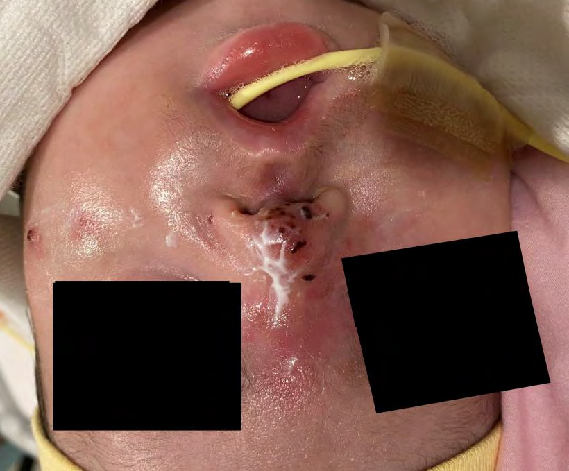
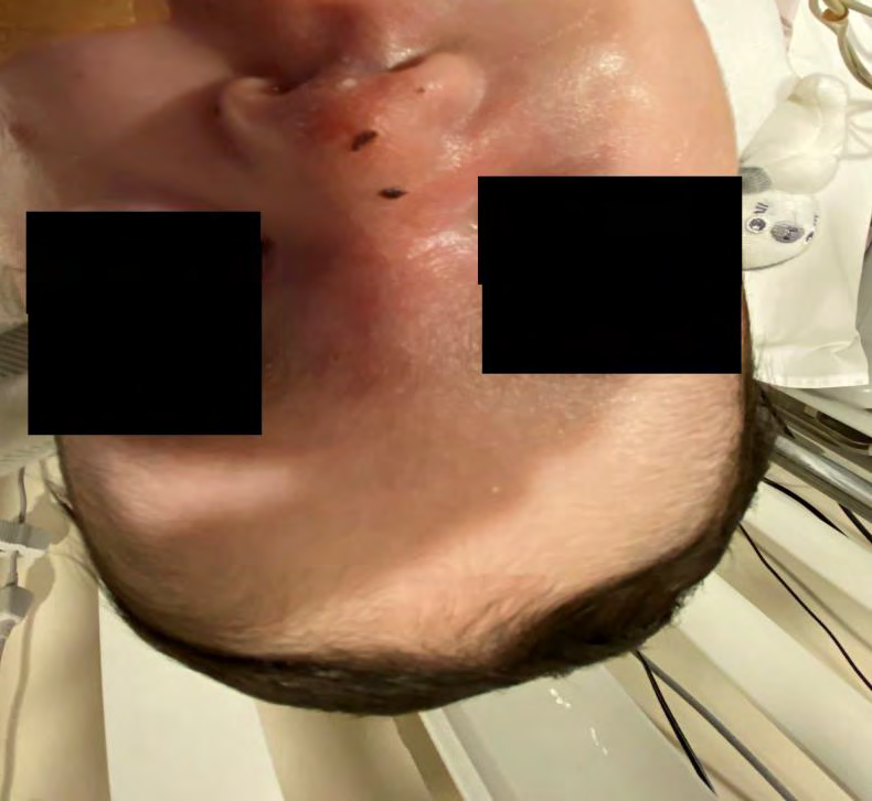
Physical examination highlighted multiple confluent skin lesions, non-blanchable, persistent, dark red to dark purple, hemorrhagic patches and plaques that occurred on the previously described skin pathology with characteristic branched configuration. Lesions relatively small (0.5 to 1 cm) in diameter, confluent and limited to Nose with peripheral erythema. Small solitary lesions with central necrosis and peripheral erythema noted on cheeks (Figure 1).
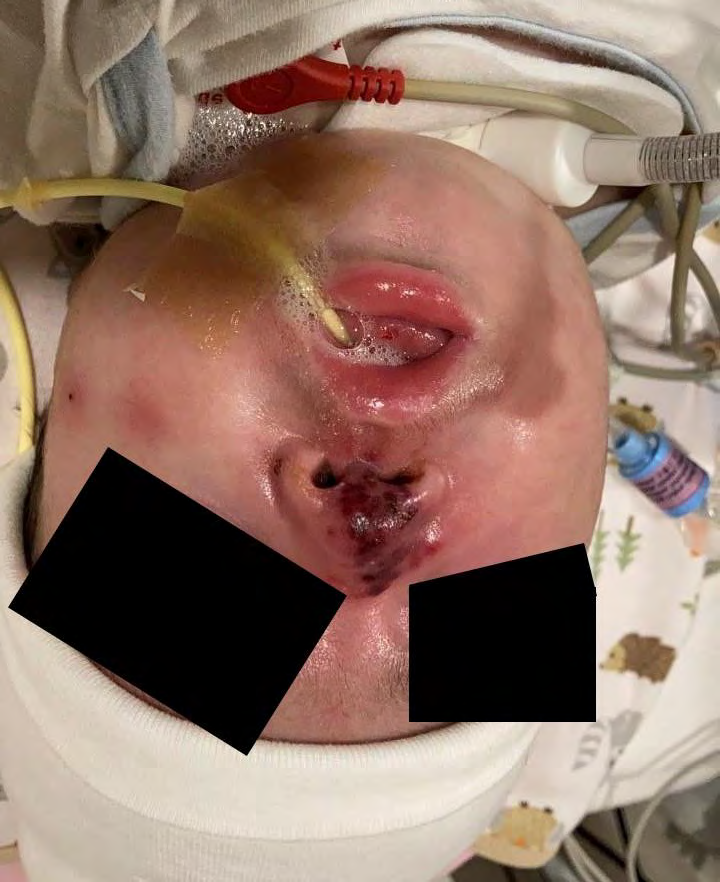
Figure 1: Multiple confluent skin lesions, non-blanchable, persistent, dark red to dark purple, hemorrhagic patches and plaques that occurred on the previously described skin pathology with characteristic branched configuration. Lesions relatively small (0.5 to 1 cm) in diameter, confluent and limited to Nose with peripheral erythema. Small solitary lesions with central necrosis and peripheral erythema noted on cheeks.
The laboratory results were as follows: hemoglobin, 9.4 g/dL; white blood cells, 4700/mm3; absolute neutrophil count, 366/mm3 (22%); platelets, 30,000/mm3; C-reactive Protein – 300 mg/L (normal up to 5 mg/L). Antibiotics shifted to intravenous meropenem (20 mg/kg per dose 8 hourly) and Vancomycin (15mg/kg per dose 8 hourly) after collecting blood, and skin lesion samples.
The patient’s general condition quickly improved, fever de- creased and Vital Signs improved. On day 2 of IV
antibiotics, the cultures of skin lesion grew P aeruginosa and blood culture remained sterile till day 7 of incubation. A repeated blood test performed on day 3 after initiation of IV antibiotics showed: hemoglobin, 9.75g/dL; white blood cells, 7,100/mm3; absolute neutrophil count, 1562 /mm3 (22%); normal red blood cells and Platelets, 66000/mm3.
Thus, the diagnosis of EG caused by P aeruginosa was not confirmed by Biopsy and as per the results of the drug susceptibility test, intravenous Meropenem continued for total of 14 days and Vancomycin was discontinued. Daily topical antibiotic therapy with iodine and Fusidic acid cream (20%) was instituted four times per day.
During treatment lesions gradually regressed (Figures 1-3) and disappeared without performing surgical intervention - excisions and without performing biopsy to confirm the diagnosis. Repeat blood counts were carried out every 3 days for 2 weeks, and consistently showed increased in absolute neutrophil counts to 3700/mm3 and normalization of Platelets count to 168000/mm 3 and nonreactive C- reactive Protein.
Discussion
Ecthyma gangrenosum is the cutaneous manifestation of pseudomonas infection in previously healthy patients with sepsis that can develop all over the body [1–3]. Patients with chronic diseases and immunodeficiency are typically at risk for developing EG. It was suggested that these patients may have risk factors for the development of EG or unrecognized underlying medical conditions [4].
Most previous reports mentioned that the skin lesions usually occur in the gluteal and perineal regions (57%), or in the extremities (30%) [5, 6, 7]. It was peculiar for our patient without previous signs of sepsis, or recent previous antibiotic therapy to suffer EG, with such an extremely rare distribution of the EG lesion. Two pathogenic mechanisms of EG have been well described, namely the bacteremic and the nonbacteremic form [8]. In the classic or bacteremic form, the skin lesions represent the hematogenous dissemination of the organism to the skin. Therefore, in such patients, blood cultures are positive. In the nonbacteremic form, the patients may tend to have a better prognosis than those who are septicemic [9]. However, secondary bacteremia may occur if treatment is delayed.
Our patient initially presented with skin lesions typical for erysipelas that evolved rapidly to characteristic lesions of EG. Sepsis developed later, as proven by the growth of P aeruginosa in the skin lesion. Some authors have reported P aeruginosa sepsis in previously healthy children without underlying medical problems. Different clinical manifestations were observed in these children, the most relevant being skin lesions, fever, diarrhea, pneumonia, and shock. The mortality rate was approximately 55% [10]. It is thus important to notice such manifestations early, so that appropriate antibiotic treatment can be instituted timely. This is especially true for patients such as ours, who had normal white blood cell count and was neutropenic at the time of suspected diagnosis. Qualitative neutrophil defects represent significant risk factors for sepsis. EG lesions usually present before the results of the blood and lesion cultures and help to administer appropriate antimicrobial therapy without delay [1]. In our patient, Localized EG lesions were characteristic of P aeruginosa infection. Antibiotic treatment is usually composed of cephalosporins, aminoglycosides, or penicillins, alone or in combination. For our patient, meropenem initially combined with Vancomycin resulted in a quick cure [10]. Additionally, although multiple confluent skin lesions, non-blanchable, persistent, dark red to dark purple, hemorrhagic patches and plaques with characteristic branched configuration were charcteristic lesions in our child, bacterial cellulitis, group A beta hemolytic deep impetigo, leukocytoclastic vasculitis-like vasculi-tis, and malignancy should also be included in the differential diagnosis. [7, 11].
EG treatment has 3 stages: initial empiric antibiotic therapy is administered as soon as infection is suspected; when the etiology is established, aggressive antibiotic or antifungal treatment is administered; finally, surgical excision is often necessary, because EG manifests as a necrotizing soft-tissue lesion [12]. The treatment plan of our patient was made according to the 2 stages. Based on the prognosis and outcome of our patient, surgical excision was not done and improvement was achieved with respect to the lesions in the process of conventional antibiotic treatment.
Conclusion
Early recognition of EG plays an important role in providing appropriate empiric antibiotic treatment early in the development of characteristic lesions and improves the outcome. Clinical awareness of P aeruginosa skin infection manifestation in Long Term Ventilated children should be increased. Early antibiotics treatment improve prognosis and might prevent the need for surgical excision and intervention with respect to the lesions in the process of conventional antibiotic treatment, since early antibiotic treatment was effective in our case.
References
-
Martín-Ancel A, Borque C, del Castillo F (1993) Pseudomonas sepsis in children without previous medical problems. Pediatr Infect Dis J 12: 258-260.
-
Reich HL, Williams Fadeyi D, Naik NS, Honig PJ, Yan AC (2004) Nonpseudomonal ecthyma gangrenosum. J Am Acad Dermatol 50(5): S114-117.
-
el Baze P, Thyss A, Vinti H (1991) A study of nineteen immunocompro- mised patients with extensive skin lesions caused by Pseudomonas aeruginosa with and without bacteremia. Acta Derm Venereol 71: 411-415.
-
Zomorrodi A, Wald ER (2002) Ecthyma gangrenosum: considerations in a previously healthy child. Pediatr Infect Dis J 21: 1161-1164.
-
Boxer LA, Blackwood RA (1996) Leukocyte disorders: quantitative and qualitative disorders of the neutrophil, Part 2. Pediatr Rev 17: 47-50.
-
Vaiman M, Lazarovitch T, Heller L (2015) Ecthyma gangrenosum and ecthyma-like lesions: review article. Eur J Clin Microbiol Infect Dis 34: 633-639.
-
Goolamali SI, Fogo A, Killian I (2009) Ecthyma gangrenosum: an important feature of pseudomonal sepsis in a previously well child. Clin Exp Dermatol 34: e180–182.
-
Boisseau AM, Sarlangue J, Perel Y (1992) Perineal ecthyma gangrenosum in infancy and early childhood: septicemic and non-septicemic forms. J Am Acad Dermatol 27: 415-418.
-
Huminer D, Siegman-Igra Y, Morduchowicz G (1987) Ecthyma gangrenosum without bacteremia. Report of six cases and review of the literature. Arch Intern Med 147(2): 299–301.
-
Viola L, Langer A, Pulitanò S (2006) Serious Pseudomonas aeruginosa infection in healthy children: case report and review of the literature. Pediatr Int 48: 330-333.
-
Biddeci G, Cutrone M, Mattei I (2015) Ecthyma gangrenosum of the cheek in a 6-month-old infant. Arch Dis Child 100(1): 55-56.
-
Pickard R, Llamas R (1970) Ecthyma gangrenosum complicating Pseudomonas bacteremia. Rare survival. J Fla Med Assoc 57: 34-35.
- Epithelioid Granuloma; 3cases with Different Clinical Features
- Advancing Representation in Dermatology Clinical Trials: Ethical, Scientific, and Regulatory Imperatives for Inclusion Across all Fitzpatrick Skin Types
- A Case of Atopic Dermatitis with Concurrent Psoriasis Vulgaris: Successful Treatment with Upadacitinib
- Innovation Lifting Eyeshadow: A Synthesis of Makeup and Optical Illusion
- Distinguishing Superficial Actinic Porokeratosis from Actinic Keratosis with UVF Dermoscopy: A Case Report
- High Mobility Group Box 1 (HMGB1) in Cutaneous Inflammation: An Immune Modulator Bridging Cellular Stress, Ferroptosis and Danger Signaling