Paraneoplastic Acanthosis Nigricans: Utility of Dermoscopy
This is 38-year-old woman who was followed up in oncogynecology for her neobreast, having benefited from a patey 2 years ago, presented with pigmented lesions on the left edge of the folds and opposite the patey scar, which became larger and more diffused throughout the trunk up to a few months ago. In addition to consulting several doctors, the patient was wrongly treated with pigmentogenic lichen, placed on undocumented treatment that did not improve his condition. On admission, the initial examination revealed multiple pigmented macular patches involving the neck, axillary and under mammary folds, trunk and back (Figures 1 & 2). Dermoscopy revealed a diffuse dark brown background, a cerebriform appearance, multiple crystals, furrows, milia cysts, hyperpigmented spots and striae.
Introduction
This is 38-year-old woman who was followed up in oncogynecology for her neobreast, having benefited from a patey 2 years ago, presented with pigmented lesions on the left edge of the folds and opposite the patey scar, which became larger and more diffused throughout the trunk up to a few months ago. In addition to consulting several doctors, the patient was wrongly treated with pigmentogenic lichen,
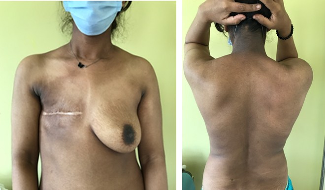
placed on undocumented treatment that did not improve his condition. On admission, the initial examination revealed multiple pigmented macular patches involving the neck, axillary and under mammary folds, trunk and back (Figures 1 & 2). Dermoscopy revealed a diffuse dark brown background, a cerebriform appearance, multiple crystals, furrows, milia cysts, hyperpigmented spots and striae (Figures 3a,b).
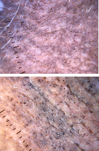
Figure 3a & b: Dermatoscopic examination showed multiple cristae and sulci, multiple white to brown exophytic papillary structures and black blotches .
The diagnosis of paraneoplastic acanthosis nigricans was retained, and the patient was put on topical retinoids and depigmenting creams. The patient was readmitted to the gynecology department for further evaluation.
Acanthosis nigricans (AN) is a common but often overlooked skin condition [1]. Affected areas include the neck, armpits, groin, popliteal fossa, ulnar fossa, and flexion creases. The skin is thickened and darkened in patches of velvety texture. In acanthosis nigricans, the exact mechanism of skin lesions development is unknown [2]. A more likely hypothesis is that a substance secreted by the tumor, or as a result of it, is the stimulating factor in the malignant form. A variety of disorders are associated with acanthosis nigricans, including diabetes mellitus, malignancies, HAIR-N syndrome, and many others. When acanthosis nigricans is diagnosed early and confirmed by dermoscopy, an invasive biopsy is not necessary. Dermatoscopic features reported for mild to moderate acanthosis nigricans include a diffuse dark brown background, cerebriform appearance, multiple crystals, furrows, milia-like cysts, hyperpigmented spots, and striae [3]. Due to the contrast with unaffected dark skin, the pattern of crystals and multiple furrows is particularly apparent in dark-skinned patients [4].
The use of dermoscopy in the diagnosis, prognosis, and follow-up of pigmentary disorders represents a potential noninvasive technique.
References
-
Das A, Datta D, Kassir M, Wollina U, Galadari H, et al. (2020) Acanthosis nigricans: A review. J Cosmet Dermatol 19(8): 1857-1865.
-
Pardeshi SS, Khemani UN, Kamath RR, Kura MM, Jafferany M (2020) Therapeutic implications of dermoscopic findings in acanthosis nigricans: A clinical and histopathological study. Dermatol Ther 33(6): e14521.
-
Elmas ÖF, Demirbaş A, Kutlu Ö, Kilitçi A, Atasoy M (2020) Utility of dermatoscopy in the diagnosis of acanthosis nigricans. J Cosmet Dermatol 19(12): 3426-3427.
-
Khopkar US, Bharti AM (2018) Advances in Dermoscopy of Pigmented Lesions. In: Kumarsinghe P (Eds.), Pigmentary Skin Disorders, 1st (Edn.), Gewebestrasse: Springer International Publication, pp: 79-93.
- Epithelioid Granuloma; 3cases with Different Clinical Features
- Advancing Representation in Dermatology Clinical Trials: Ethical, Scientific, and Regulatory Imperatives for Inclusion Across all Fitzpatrick Skin Types
- A Case of Atopic Dermatitis with Concurrent Psoriasis Vulgaris: Successful Treatment with Upadacitinib
- Innovation Lifting Eyeshadow: A Synthesis of Makeup and Optical Illusion
- Distinguishing Superficial Actinic Porokeratosis from Actinic Keratosis with UVF Dermoscopy: A Case Report
- High Mobility Group Box 1 (HMGB1) in Cutaneous Inflammation: An Immune Modulator Bridging Cellular Stress, Ferroptosis and Danger Signaling