Assessment of Anti-Hyperglycemic Effects and the Probable Mode of Action of Cynara scolymus Leaf and Flower Head Hydroethanolic Extracts in Type 2 Diabetic Wistar Rats
Introduction: The prevalence of type 2 diabetes mellitus has been increasing steadily all over the world. Because of the side effects of the conventional Antidiabetic chemical drugs, the development of alternative medicines is needed. Thus, this study was designed to assess the effect of Cynara scolymus (artichoke) leaf and flower head hydroethanolic extracts on oral glucose tolerance and to suggest their probable mode of actions in experimentally-induced type 2 diabetic Wistar rats. Materials and Methods: Type 2 diabetes mellitus was induced by a single intraperitoneal injection of streptozotocin (60 mg/kg body weight) dissolved in cold 0.01M citrate buffer (pH 4.5), 15 minutes after intraperitoneal injection of nicotinamide (60 mg/kg body weight). After induction of diabetes mellitus, the rats were allocated into four groups (each of six rats). Group 1 was kept as normal control. Group 2 was diabetic control. Groups 3 and 4were diabetic groups that were respectively treated with Cynara scolymus leaf and flower head extracts at dose of 100mg/kg body weight/day by oral administration for 28 days. Results: The treatment of the diabetic rats with leaf and flower head extracts resulted in a significant improvement of the impaired oral glucose tolerance, the lowered serum insulin and C-peptide levels, and the decreased β cell insulin content and HOMA-β cell function in association with amendment of the pancreatic islets histological architecture and integrity. The lowered visceral adipose tissue mRNA expression of adiponectin and the elevated resist in mRNA expression were significantly ameliorated in diabetic rats as a result of treatment with leaf and flower head extracts. The lowered HOMA-IS and raised HOMA-IR in diabetic rats was significantly alleviated due to treatment with flower head extract. Conclusion: The Cynarascolymus leaf and flower head hydroethanolic extracts successfully improved the oral glucose tolerance probably via amendment of pancreatic islets histological integrity, enhancement of the insulinogenic effects and attenuation of insulin resistance. The leaf extract had more potent antihyperglycemic effect than the flower head extract.
Introduction
Diabetes mellitus is a multi-factorial metabolic disorder characterized by chronic hyperglycemia with modificationsin carbohydrate, fat and proteins metabolism resulting from dysfunction of pancreatic β- cells and/or insulin resistance [1, 2]. With the increasing prevalence of diabetes, development of effective pre- clinical modelshas become crucialas a very useful toolfor studying possible drug discovery approaches for both diabetic prevention and treatment. Streptozotocin (STZ) has been reported to induce both type 1 diabetes mellitus (T1DM) and type 2 diabetes mellitus (T2DM) [3, 4, 5] depending on itsdose, single or multiple injection, the use of high-fat diet and its concomitant injection with or without nicotinamide (NA). In this regard, it was reported that the injection of NA 15 minutes before STZ in rats leads to T2DM [6, 7]. The major pathophysiologic abnormalities of T2DM include insulin resistance and subsequent loss of β-cell mass and β-cell dysfunction [8, 9, 10, 11]. Three key defects implicated in the onset of hyperglycemia in T2DM that are (1) increased hepatic glucose production, (2) diminished insulin secretion, and (3) impaired insulin action [12, 13]. T2DM accounts for _>_90% of diabetes and is associated with metabolic disorder of carbohydrate, lipid and protein [7, 14]. The sub-classes of lipids responsible for the pathogenesis of insulin resistance are intracellularly-accumulated free fatty acids (FFAs), diacylglycerol (DAG) and triacylglycerol (TG) moieties [15]. Despite the progress of modern medicine, traditional medicine remains an important pillar in the treatment of many chronic diseases including diabetes mellitus [16, 17]. Medicinal plants continue to provide valuable therapeutic agents and they have always been the sources of drugs that treat many diseases including diabetes mellitus [18, 19]. In this regard, the World Health Organization (WHO) has recommended the evaluation of traditional plant treatments for diabetes as they are effective, non-toxic, with less or no side effects and are considered to be excellent candidates for oral therapy [20].
The importance of the plant C. scolymus which is called artichoke or globe artichoke steamed from used as edible material for nutrition and from its content of phenolic acid constituent in particular cynarin and chlorogenic acid [21]. Previous studies found that after eating of an artichoke food supplement for three consecutive months reduced fasting and post-prandial glycaemia in T2DM diabetic patients [22]. Among the vegetables, the globe artichoke (Cynara cardunculus var. scolymus [L.] Fiori) plays an important economic role in Mediterranean agriculture [23]. Cynareae tribes present in Egypt are known for their efficacy in relieving some liver disorders [24]. The leaves of C. scolymus L. have been long-used in traditional medicine and now included in British and European Pharmacopeia (BP/EP), the British Herbal pharmacopeia (BHP) and the Complete German Commission E Monographs [21]. Artichokeleaf extracts decreased serum lipids, as well as hepatic and cardiac oxidative stress in rats fed on high cholesterol diet [55]. Several reports from folk medicine as well as preliminary data from scientifically designed pre- clinical and clinical studies have suggested that artichokes may have cholerectic, hypocholesterolemic and hypolipidemic properties [25, 26, 27, 28, 29, 30, 31]. The pharmacologic properties of artichoke flower heads are well documented in several in vivo and in vitro studies for the treatment of hepatobiliary dysfunction, dyspeptic syndromes, gastric diseases, as well as for inhibition of cholesterol biosynthesis and low density lipoproteins (LDL) oxidation-induced arteriosclerosis and coronary heart disease [31, 32, 33]. Little is known about the effects of C. scolymus leaf and flower head extracts on histopathological, ultrastructural, immunohistochemical and biochemical changes in diabetic rats. The present study aims to investigate the effects of C. scolymus on type 2 diabetic rats. This investigation will be monitored by histopathological, ultrastructural, immunohistochemical and biochemical parameters including serum glucose (fasting and post-prandial), insulin and C-peptide to assess the anti-diabetic efficacy of C. scolymus leaf and flower head extracts and to suggest their probable mechanisms of action.
Material and Methods
Chemicals
STZ and NA were obtained from Sigma Chemical Company (USA). Proteinase K was obtained from Bioline USA Inc., Taunton, MA, USA.All other used chemicals are of analytical grade.
Preparation of Plant Extraction
C. scolymus was collected from Beheira (Egypt) and was authenticated by Dr. Mohamed A. AbdallahFadl, Associate Professor of Taxonomy, Botany Department, Faculty of Science, Beni-Suef University, Egypt. The leaves and flower heads were washed, air dried at room temperature for 3 weeks in good aerated and shaded area, and then powderedby an electrical grinder. The powder of C. scolymus leaves and flower heads were separately soaked in 70% ethanol for 72 hours at room temperature. The mixtures were then filtered through man filter paper and evaporated under reduced pressure using rotary evaporator to a yield of crude extracts. The hydroethanolic extracts were kept at - 20˚C until used.
Experimental Animals
Healthy adult male Wistar rats weighing 120±20 g were purchased from Helwan Station of Experimental Animals, Egyptian Holding Company for Biological Products and Vaccines (Cairo, Egypt). All animals were kept under observation for two weeks before the onset of the experiment to exclude any intercurrent infections. Animals were maintained under standard conditions of light, ventilation, temperature (22±2 ˚C), and humidity (55±5%).The animals were allowed to access tap water and standard pellet diet ad libitum. All animals' procedures were in accordance with the ethical guidelines for the use and care of animal of the Experimental Animal Ethics Committee, Faculty of Science, Beni-Suef University, Egypt (Ethical Approval Number 2015/2). All efforts were done to reduce the number and suffering of animals. 4.4. Induction of T2DM T2DM was experimentally induced in rats deprived of food and water for 16 hours by intraperitoneal (IP) injection of 120 mg/kg b.wt dissolved in 0.9% NaCl solution 15 minutes before IP injection of STZ-NA at a dose of 60mg/kg/b.wt dissolved in citrate buffer at pH 4.5 [34, 35]. Ten days after STZ injection, rats were screened for blood glucose levels. Animals, deprived from food and water overnight (10-12 hours), were given glucose (3 g/kg b. wt.) by oral gavage. After 2 hours of oral administration, blood samples were taken from lateral tail vein and blood glucose concentration was measured by glucometerobtained from Life Scan Corporation, Canada. Rats having serum glucose ≥ 200 mg/dl, after 2 hours of glucose intake, were considered diabetic and selected for further biochemical, physiological and histological studies.
Experimental Design
After acclimation period and induction of T2DM, rats were randomly divided into 4 groups (6 rats for each). Group 1 was considered as normalcontrol group and was administered the equivalent volume of a vehicle, 1% CMC, daily by oral gavage for 28 days. Group 2 was diabeticcontrol group that consisted of diabetic rats administered the equivalent volume of 1% CMC (as vehicle) daily by oral gavage for 28 days. Group 3 was composed of diabetic rats treated with C. scolymus leaf hydroethanolic extract at a dose of 100mg/kg/b.wt, dissolved in 1% CMC, by oral gavage daily for 28 days. Group 4 consisted of diabetic rats treated with C. scolymus flower head hydroethanolic extract at a dose of 100mg/kg/b.wt, dissolved in 1% CMC, by oral gavage daily for 28 day.
Oral Glucose Tolerance Test (OGTT)
OGTT was performed tonormal control, diabetic control and diabetic treated rats at the day before sacrifice by oral administration of glucose solution (3 g/kg b.wt) to overnight (10-12 h) fasted animals then successive blood samples were then taken at 0,30, 60, 90 and 120 minutes. Blood samples were obtained from lateral tail vein and blood glucose concentration was measured by glucometer obtained from Life Scan Corporation, Canada.
Blood and Tissue Sampling
At the end of the experiment, animalswere overnight fasted, then blood was collected from jugular vein underdiethyl etheranesthesia and left to coagulate at room temperature. Serum was separated by centrifugation at 3000 rpm for 15 min and stored at - 20oC pending its use for the detection of insulin and C- peptide levels. The rats were rapidly dissected and visceral adipose tissue was excised and kept at -70oC until its use for RNA extraction and detection of mRNA of adiponectin and resistin by reverse transcriptase (RT)-PCR reaction. Pancreas was excised and fixed in 10% formalin pending histological and Immunohistochemical investigations. In addition, pieces of pancreas measuring 1 mm3 were immediately fixed in fresh 3% glutaraldehyde-formaldehyde for electron microscopic examination.
Detection of Serum Insulin and C-Peptide Levels
Serum was used for the determination ofinsulin and C-peptide levelsusing ELISA kit purchased from Linco Research, USA, according manufacturer's instruction.
Calculation of Homeostatic Model Assessment (HOMA) Indices
HOMA-Insulin resistance (IR), HOMA-insulin sensitivity (IS), and HOMA-β cell function were calculated according to Hsing_et al_. [36], Park et al. [37]and Aref_et al_. [38] from the following formulas. HOMA-𝛽 cell function = (20 × fasting insulin [µIU/ml])/(fasting glucose [mg/dl]- 3.5) HOMA-IS =10000/(fasting insulin[µIU/ml] × fasting glucose[mg/dl]) HOMA-IR = (fasting insulin [µIU/ml] × fasting glucose [mg/dl])/405
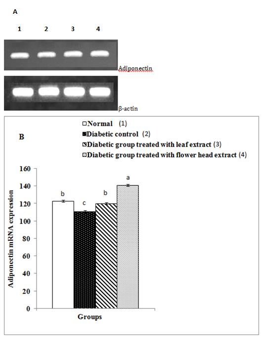
Detection of mRNA Expression of Adiponectinand Resistin
Total RNA was isolated from visceral adipose tissue according to the method of Chomzynski and Sacchi [39], using Thermo Scientific Gene JET RNA purification kit obtained from Thermo Fisher Scientific Inc., Rochester, New York, USA. Reverse transcription (RT) of RNA into cDNA and the PCR amplification in the presence of specific primers of adiponectin and resistin was performed using Thermo Scientific Verso 1-Step RT- PCR Reddy Mix kit (Thermo Fisher Scientific Inc., Rochester, New York, USA) and Thermal cycler Techne 312(Fisher Scientific, Leicestershire, LE11 5RG). The sense and anti-sense specific primers of adiponectin and resistin were obtained from Bioresearch Technologies, South McDowell Blud, Petaluma, CA, USA. The RT-PCR productswere loaded and electrophoresedat 90 volts on 1.5% agarose gel stained with ethidium bromide in 1X Tris Borate EDTA buffer (TBE) (pH 8.3-8.5). The bands on the agarose gel were viewed by UV transilluminator in a dark chamber and photographed by a camera using Gel Documentation System obtained from Raya for the Scientific Services, Giza, Egypt. The bands were analyzed by Raya Gel Docu Advanced Program accessed from Raya for the Scientific Services, Giza, Egypt. The mRNA levels of adiponectin and resistin were normalized to β-actin.
Histological Investigation
The pancreasfrom each rat wasrapidly excised after dissection andthen fixed in 10% neutral buffered formalin for 24 hours; the organs were routinely processed and sectioned at 4–5 µm thickness. Sections of pancreas were stained with haematoxylin and eosin [40, 41].
Immunohistochemistry of Pancreatic Islets
Immunolocalization technique for anti-insulin was performed on 5–6 mµ thickness sections and stained with the streptavidin–biotin–peroxidase staining method [42]. Paraffin sections were deparaffinized in xylene, rehydrated in descending grades of alcohol. Endogenous peroxidase and non-specific binding sites for antibodies were suppressed by treating the sections with 0.3% hydrogen peroxide for 20 min and 5% normal bovine serum (1:5 diluted TRIS) for 20 min at room temperature, respectively. The sections were washed in phosphate buffered saline and 10% normal goat serum was applied for 30 min to reduce non- specific binding. The sections were incubated for 1 h with anti-sera containing primary antibodies for rat insulin (polyclonal antibody) supplied by Bio Genex Cat. No. AR. 295-R and anti-TNF-α (Santa Cruz, CA, USA). The sections were incubated with biotinylated secondary antibody (Dako-K0690; Dako Universal LSAB Kit) and streptavidin horseradish peroxidase (Dako- K0690) for 30 min, and then 3,30-diam- inobenzidinetetrahydrochloride (Sigma-D5905; Sigma– Aldrich Company Ltd., Gillingham, UK) substrate kit for 10 min to obtain immunolabelling. Finally, (i) dehydrated in graded alcohol, (ii) cleared in xylene, and then (iii) mounted in DPX. The binding of antibodies was evaluated by examination under high-power light microscopy. All sections were incubated under the same conditions with the same concentration of antibodies and at the same time, so the immunostaining was comparable among the different experimental groups.
Ultra Structural Preparations
The specimens of pancreas at day 30 were cut into small pieces measuring about 1 mm3 and immediately fixed in fresh 3% glutaraldehyde-formaldehyde at 4ºC for 18-24 hours. The specimens were then washed in phosphate buffer (pH 7.4) and post-fixed in isotonic 1% osmium tetroxide for 1 h at 4ºC and then processed. Semithin sections (1 µm) were stained with toluidine blue. Ultrathin sections (70–80 nm) were stained with uranyl acetate and lead citrate [43] and examined on a Joel CX 100 transmission electron microscope operated at an accelerating voltage of 60 kV.
Statistical Analysis
Results were expressed as mean ± standard error (SE). The data are analyzed by one-way analysis of variance (ANOVA) using PC-STAT, University of Georgia, followed by LSD analysis to compare various groups with each other [44]. Values of P>0.05 were considered non-significantly different, while values of P<0.05 and P<0.01 were significant and highly significant different respectively.
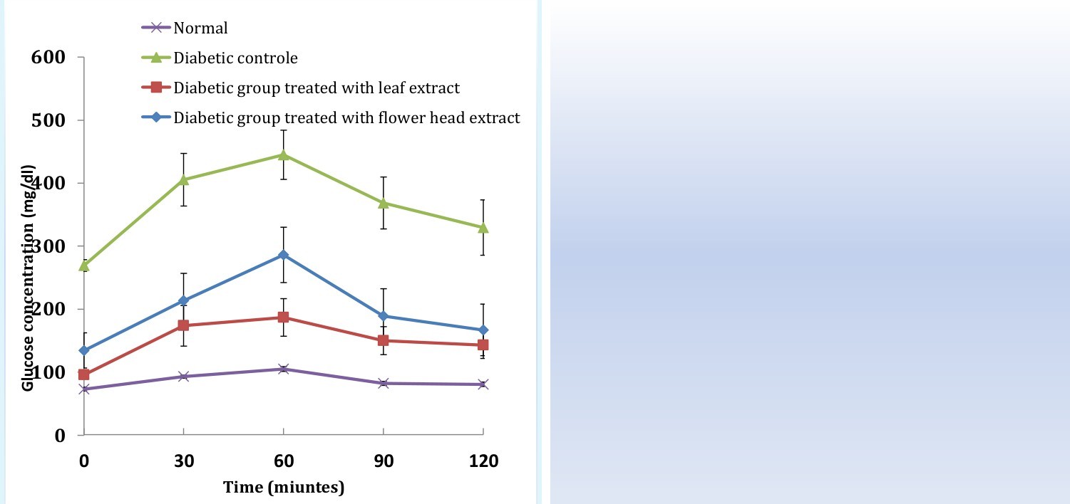
rats as compared to normal ones. The treatment of diabetic rats for 28 day with C. scolymus leaf and flower head extract successfully improved (p<0.01; LSD) the impaired glucose tolerance of diabetic rats. The leaf extract seemed to be more effective in improving the glucose tolerance than the flower head extract. F- probability of one-way ANOVA revealed that the effect on OGTT between groups was very highly significant throughout the experiment.
Results
Effect on Oral Glucose Tolerance
Oral glucose tolerance curves of normal, diabetic control and diabetic treated rats are illustrated in Figure 1. The serum glucose levels at all intervals (0, 30, 60, 90, 120 minutes) of glucose tolerance test were highly significantly (p<0.01; LSD) elevated in diabetic
The serum insulin and C-peptide concentrations at fasting statewere highly significantly (p<0.01; LSD) decreased in diabetic rats in comparison with those of normal control rats; the recorded percentage changes were -60.03and -74.03% respectively. The treatment of diabetic rats with C.scolymus leaf and flower head
| Insulin (μIU / ml) | Percentage change | C-peptide (pg/ml) | Percentage change | |
|---|---|---|---|---|
| Normal | 9.45±0.55a | 3.62±0.31a | ||
| Diabetic control | 3.75±0.46c | -60.03 | 0.94±0.15d | -74.03 |
| Diabetic group treated with leaf extract | 9.45±0.72a | 152.00 | 3.10±0.13ab | 229.78 |
| Diabetic group treated with fruit extract | 6.21 ±0.74b | 65.60 | 2.92±0.10bc | 210.64 |
| F-probability | P<0.001 | P<0.001 | ||
| LSD at the 5% level | 1.86 | 0.56 | ||
| LSD at the 1% level | 2.53 | 0.76 |
Table 1: Effect of C. scolymus leaf and fruit extracts on insulin and C-peptide levels in NA/STZ-induced diabetic rats.
-Data are expressed as mean ±SE. Number of animals in each group is six. -Means, which share the same superscript symbol(s), are not significantly different -Percentage changes were calculated by comparing diabetic treated groups with diabetic control.
Data describing the effect of treatment of diabetic rats
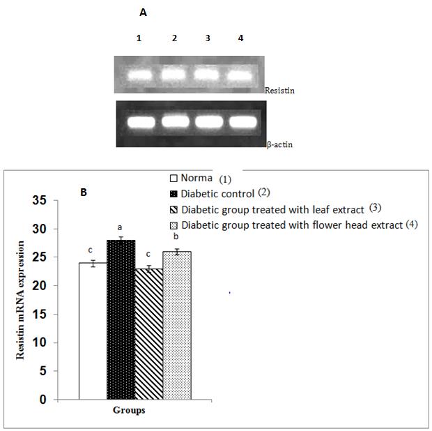
potent in decreasing the elevated resistin mRNA expression.
Effect on Homeostatic Assessment (HOMA) Indices
HOMA-β cell function, HOMA-IS and HOMA-IR of normal, diabetic control and diabetic rats treated with C. scolymus leaf and flower head extracts are represented in Table 2.
| Group | H | OMA-β cel | l | Percentage | HOMA-IS | Percentage | HOMA-IR | Percentage | ||||||
|---|---|---|---|---|---|---|---|---|---|---|---|---|---|---|
| function | change | change | change | |||||||||||
| Normal | 2.76±0.10a | 14.67±0.97a | 1.72±0.12b | |||||||||||
| Diabetic control | 0.27±0.04c | -90.22 | 10.58±1.84b | -27.88 | 2.66±0.25a | 54.65 | ||||||||
| Diabetic group treated with leaf extract | 2.08±0.12a | 670.37 | 11.56±1.20ab | 9.26 | 2.23±0.19ab | -16.17 | ||||||||
| Diabetic group treated with flower head extract | 1.26±0.22b | 366.66 | 17.48±2.04a | 65.22 | 2.03±0.23b | -23.68 | ||||||||
| F-probability | P<0.001 | P<0.05 | P<0.05 | |||||||||||
| LSD at the 5% level | 0.53 | 4.65 | 0.61 | |||||||||||
| LSD at the 1% level | 0.72 | 6.34 | 0.83 |
Table 2: Effect of C. scolymus leaf and flower head extracts on HOMA-β cell function, HOMA-IS and HOMA-IR in HOMA-β cell function
Table 2: Effect of C. scolymus leaf and flower head extracts on HOMA-β cell function, HOMA-IS and HOMA-IR in HOMA-β cell function and HOMA-IS were highly significantly (p<0.01; -90.22%)and significantly (p<0.05; -27.88%) decreased respectively in diabetic rats while HOMA-IR was highly significantly (p<0.01; 54.65%) increased. The treatment with C. scolymus leaf and flower head extracts induced a highly significant increase in HOMA-β cell function; the leaf extract seemed to be more effective in improving the impaired HOMA-β cell function than the flower head extract. The HOMA-IS was also detectably increased as a result of treatments with C. scolymus leaf and flower head extracts. While the effect of leaf extract was non- significant (p>0.05), the effect of flower head extract highly significant (p<0.01). In contrast, HOMA-IR was remarkably decreased after the treatments with C. scolymus leaf and flower head extracts; the decrease was significant only as a result of flower head extract.
HistopathologicalResults
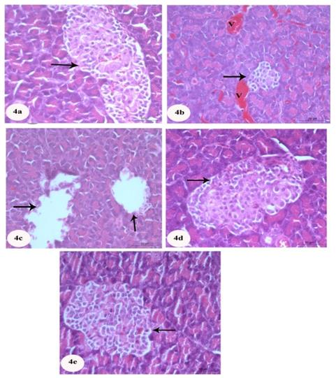
The histopathological examination of normal control pancreas sections showed closely packed lobules of pancreatic acini. Islets of Langerhans were embedded within the exocrine portions and alpha cells (Figure 4a). Diabetic control revealed histopathological changes of endocrine portions represented by marked decrease of β-cells and dissolution of some islets in other parts(Figures 4b and c). Congested stromal blood vessels was also seen(Figure 4b). Pancreas of rats treated with C. scolymus leaf (Figure 4d) and flower (Fig. 4e) showed nearly normal structure of islets of Langerhans.
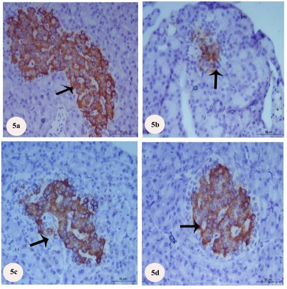
ImmunohistochemicalStaining of Insulin
Immunohistochemically, β-cells of the normal group stained with strong positive reaction for anti-insulin antibodies as brown granules in the cytoplasm of great number of the β-cells (Figure 5a). In diabetic group, the immune reactivity for anti-insulin antibodies was obviously decreased (Figure 5b). After treatment with C.
scolymus leaf extract, positive immunoreactions of β- cells for anti-insulin antibodies were obviously increased (Figure 5c). Treatment with C. scolymus flower head extract revealedmarkedly increased positive immunoreactions of β-cells for anti-insulin antibodies (Figure 5d).
Immunohistochemical Staining of TNF-alpha
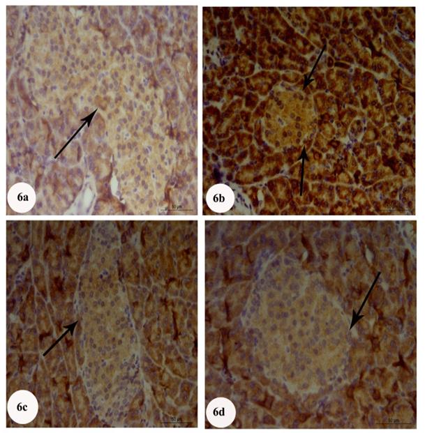
Immunohistochemical sections of control group showed no reaction for TNF-alpha (Figure 6a), while diabetic group revealed marked increase in intensity of TNF-alpha of pancreatic islets compared to normal group (Figure 6b). Treatment with C. scolymus leaf and flower head extract showed an obvious reduction in intensity of TNF-alpha of pancreatic islets (Figures6c and d) reflecting decrease in the expression of this cytokine.
Ultra Structure Results
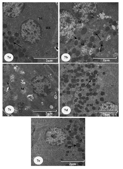
Acinar Cells: Ultra structure of control pancreas showed acinar cells with euchromatic nuclei, well- developed cisternae of rough endoplasmic reticulum, mitochondria and numerous electron dense secretory granules of variable sizes in the apical part (Figure 7a). Diabetic control showed marked changes in pancreatic acini represented by damaged swelling mitochondria, cytoplasmic vacuolation and an obvious decrease of zymogen granules. Nucleus with fragmented chromatin was also observed (Figures 7b and c). Diabetic rat pancreas treated with C. scolymus leaf and flower head extracts showed marked improvement represented by increase in zymogen granules, regular contours of nuclei and well-flattened rough endoplasmic except few vacuoles (Figures 7d-e).
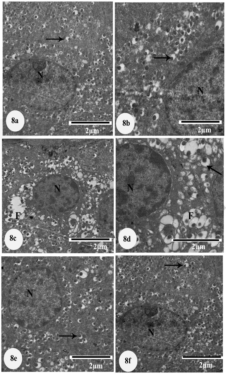
decrease of secretory granules, fusion of some secretory granules and pyknotic nuclei (Figures 8c-d). Treatment with C. scolymus leaf and flower head extract revealed euchromatic nucleus, few vacuoles in β-cells and increase of secretory granules compared to the diabetic ones (Figures 8e-f).
Discussion
Traditional herbal medicines have been used for a long time to treat diabetes, and many controlled trials have been done to investigate their efficacy.In the present study, the hydroethanolic extracts of C. scolymus leaves and flower heads were chosen to assess their anti-hyperglycemic efficacies and to suggest their possible mechanism(s) of action in experimentally T2DM induced by intraperitoneal injection of STZ following NA administration [45]. To explain the mode of action of NA and STZ in this animal model of T2DM, Novelli_et al_. [46] suggested that NA injection before STZ injection resulted in a partial loss of β-cell mass by necrotic actions. In addition, STZ-NA caused a significant reduction in the number of insulin receptors on many insulin target tissues reflecting insulin resistance in such tissues [45]. In the present study, the treatment of the diabetic rats with hydroethanolic extract of C. scolymus leaf and flower head resulted in a significant amelioration of the impaired glucose tolerance. The C. scolymus leaf extract had higher antidiabetic potency and improvement effect on OGT in NA/STZ- induced diabetic rats than C. scolymus flower head extract. This finding is in accordance with the data of Fantini_et al_. [47] who found that the hydroethanolic extract of C. scolymus flowering heads has hypoglycemic effects in both normal and obese rats and Nazni_et al_. [22] who stated that C. scolymus powder produced marked decrease of fasting and post-prandial blood glucose levels in diabetic subjects. In addition, Abdel Magied et al. [31] found that the treatment of diabetic rats with aqueous extracts of leaves and flower heads of two varieties of artichoke induced a significant decrease of the elevated serum glucose level. In the same way, Kuczmannová et al. [48] reported that daily oral administration of the Cynara cardunculus extract for 1 and 5 weeks significantly decreased glycemia after streptozotocin (STZ) administration. The hypoglycemic effects C. scolymus extract may be attributed to its effect to reduce hepatic glucose output by inhibiting hepatic glucose-6- phosphatase activity as indicated byEid and Haddad [49]. In disagreement with the present study, Fallah Huseini, et al. [50] found that C. scolymus hydroethanolic extract had no significant effects on the blood levels of fasting glucose, postprandial glucose, glycosylated hemoglobin in type 2 diabetic patients. The improvement of OGT due to the treatment of diabetic rats with extract of C. scolymus leaf and flower head hydroethanolic extracts was associated with a significant increase in the lowered serum insulin and C- peptide levels as well as the calculated HOMA-β cell function and also concomitant with a remarkable amendment of pancreatic islets' histological architecture as well as the increase in the islets' size and number of β-cells within the islets. In the present study, the adiponectin mRNA expressionin visceral adipose tissue was significantly decreased in NA/STZ-induced type 2 diabetic rats as compared with the normal, and the treatment with extract of C. scolymus leaf and flower head resulted in a significant amelioration. In agreement with these results, Szkudelska_et_ al. [51] reported that adipocytesisolated from epididymal fat tissue of STZ- NA-induced diabetic rats exhibited impaired secretion of adiponectin. In the same way, Ahmed et al. [52] found that serum adiponectin level was significantly depleted in HFD/STZ-induced type 2 diabetic ratswhile it was significantly alleviated as a result of the treatment of these diabetic rats with hesperidin and naringin. Moreover, it has been shown that mice lacking adiponectin expression have reduced insulin sensitivity or are more likely to suffer from insulin resistance [53]. In contrast to adiponectin in the current study, the resistin mRNA expression exhibited a significant elevation in adipose tissue of diabetic rats as compared with the normal. The treatment of diabetic rats with C. scolymus leaf and flower head extracts resulted in a marked improvement.This result goes parallel with that of Ahmed, et al. [52]Adel Abdel-Moneim_et al_. [54] who found that serum resistin level and adipose tissue resistin mRNA expression were significantly increased in high fat diet/streptozotocin-induced type 2 diabetic rats as compared with normal group, and the treatment of HFD/STZ diabetic rats with hesperidin and naringin induced a significant amelioration. It is worth mentioning here that resistin, a low molecular weight protein secreted from adipose tissue, acutely induces insulin resistance and glucose intolerance [55, 56, 57]. In addition, down-regulation of resist in expression and release from adipose tissue has been implicated in the insulin-sensitizing actions of many anti-hyperglycemic agents [58]. The depletion inadipose tissue adiponectinm RNA expression and the increase in resistin mRNA expression in NA/STZ-induced type 2 diabetes mellitus in association with reduced calculated HOMA-IS and raised calculated HOMA-IR reflects the role of these two adipokines in inducing insulin resistance and thereby impaired glucose tolerance in such animal model of diabetes mellitus.The treatment of NA/STZ-induced type 2 diabetic rats with C. scolymus leaf and flower head extract produced a marked increase in HOMA-IS and a decrease in HOMA-IR. However, while the effect of flower head extract was significant, the effect of leaf extract was not. In the present study, the NA/STZ-induced diabetic rats showed marked changes of the islets of Langerhans represented by few small pancreatic islets compared to the normal rats. These changes are similar to the results of Al-Hariri et al. [59] who observed few islets of Langerhans in diabetic group compared with normal group. The C. scolymus leaf and flower head extract treated rats exhibited marked increase in the number and size of islets of Langerhans as well as the number of islets cells within each islet. The improved pancreatic islet histological architecture and integrity attributed the increase in serum insulin and C-peptide levels as well as the enhanced HOMA-β cell function. In NA/STZ-induced diabetic rats of the current study, the immune reactivity for anti-insulin antibodies was markedly decreased in number of insulin cells. The C. scolymus leaf and flower head extract treated diabetic groups exhibited a high immunoreactivity evidenced by increase in the density and areas stained yellowish brown colour. These results are in accordance with Coskun_et al_. [60], Mendez and Haro Hernandez [61], Ao_et al_. [62], Simsek_et al_. [63] and Abdul-Hamid and Moustafa [64].
In the present study, pancreatic islets of NA/STZ- induced diabetic rats showed a significant increase of TNF-alpha expression.The treatment of diabetic rats with C. scolymus leaf and flower head extract showed a remarkable reduction in the TNF-alpha. These results are consistent with Ferreira et al. [65] who observed an increase the intensity of TNF-alpha immune stained cell in pancreas of diabetes group, while treatment with minocyclineor glibenclamide daily for 30 days resulted in marked reduction in the intensity of TNF-alpha immune stained cell. Since plasma TNF-alpha is associated to insulin resistance, one can assume that this cytokine plays a significant role in the pathogenesis of chronic insulin resistance in humans [66]. Moreover, TNF-alpha is a proinflammatory cytokine that may havea role in the mild damage of islets cells by inflammation in NA/STZ-induced diabetes. Its decrease in the treated diabetic rats may be in turn of importance in the improvement of insulin resistance and islets histological architecture and integrity. The present ultra-structural study of the diabetic group showed marked changes in pancreatic acini represented by decrease of secretory granules, cytoplasmic vacuolation and damaged mitochondria. Moreover, β-cells showed obvious vacuolation and decrease of secretory granules. On the other hand, the β cells of C. scolymus leaf and flower head extract treated diabetic groups showed an increase of secretory granules number and less vacuolated cytoplasm in comparison with the diabetic control group. These evidences were concomitant with the present immunohistochemical results which indicated a high immunoreactivity of anti-insulin antibodies reflecting increased insulin expression in the β cells of treated diabetic rats as compared with the diabetic control group. The present finding is in agreement with the result of Ahmed et al. [67] and Abdul-Hamid and Moustafa [68] who observed an increase of insulin secretory granules in STZ-induced diabetic rats treated with various plant constituents as compared with the corresponding diabetic control. In conclusion, the present study suggested that the C. scolymus leaf and flower head hydroethanolic extract improved the oral glucose tolerance via enhancement of the insulinogenic effects and attenuation of insulin resistance. The leaf extract was more potent in improving the oral glucose tolerance. Moreover, while the leaf extract seemed to have more potent insulinogenic effect, the flower head extract had stronger activity in improving insulin sensitivity and in suppressing insulin resistance.
References
-
Safdar M, Khattak MM, Siddique M (2004) Effect of diabetic individuals. Pak J Nutr 3: 268-227.
-
Rydén L, Grant PJ, Anker SD, Berne C, Cosentino F, et al. (2013) ESC Guidelines on diabetes, pre- diabetes, and cardiovascular diseases developed in collaboration with the EASD. European heart journal 34(39): 3035-3087.
-
Kwon NS, Lee SH, Choi CS, Kho T, Lee HS (1994) Nitric oxide generation from Streptozotocin. FASEB 8(8): 529-533.
-
Akbarzadeh A, Norouzian D, Mehrabi MR, Jamshidi SH, Farhangi A, et al. (2007) Induction of diabetes by streptozotocin in rats. Indian J Clin Biochem 22(2): 60-64.
-
Goud BJ, Dwarakanath V, Chikkaswamy BK (2015) Streptozotocin- A Diabetogenic Agent in Animal Models. International Journal of pharmacy and pharmaceutical research 3(1): 253-269.
-
Ahmed OM (2010) Antihyperglycemic effects of water extract of _Ulva_ _lactuca_ and its polysaccharides in nicotinamide-streptozotocin- induced diabetic rats. Egypt J Zool 54: 273-297.
-
Ahmed OM (2010) Anti-hyperlipidemic, antioxidant and cardiac improving effects of water extract of _Ulva_ _lactuca_ and its polysaccharides in nicotinamide-streptozotocin-induced diabetic rats. Egypt J Zool 54: 253-272.
-
Shaw JE, Sicree RA, Zimmet PZ (2010) Global estimates of the prevalence of diabetes for 2010 and 2030. Diabetes Res Clin Pract 87(1): 4-14.
-
Farag YM, Gaballa MR (2011) Diabesity: an overview of a rising epidemic. Nephrology Dial Transplan 26(1): 28-35.
-
Ashcroft FM, Rorsman P (2012) Diabetes mellitus and the beta cell: the last ten years. Cell 148(6): 1160-1171.
-
Halban PA, Polonsky KS, Bowden DW, Hawkins MA, Ling C, et al. (2014) β-cell failure in type 2 diabetes: postulated mechanisms and prospects for prevention and treatment. J Clin Endocrinol Metab 99(6): 1983-1992.
-
DeFronzo RA, Bonadonna RC, Ferrannini E (1992) Pathogenesis of NIDDM. A balanced overview. Diabetes Care 15(3): 318-368,
-
Stumvoll M, Goldstein BJ, van Haeften TW (2005) Type 2 diabetes principles of pathogenesis and therapy. Lancet 365(9467): 1333-1346.
-
Parillo M, Riccardi G (2004) Diet composition and the risk of type 2 diabetes: epidemiological and clinical evidence. British J Nutr 92(1): 7-19.
-
Kurek K, Garbowska M, Ziembicka DM, Łukaszuk B, Rogowski J, et al. (2016) Myriocin treatment affects lipid metabolism in skeletal muscles of rats with streptozotocin-induced type 1 diabetes. Adv Med Sci 62(1): 65-73.
-
Othman BB, Ibrahim H, Mankai A, Abid N, Othmani N, et al. (2013) Use of hypoglycemic plants by Tunisian diabetic patients. Alexandria Journal of Medicine 49(3): 261-264.
-
AbouZid SF, Ahmed OM, Ahmed RR, Mahmoud A, Abdella E, et al. (2014) Antihyperglycemic effect of crude extracts of some Egyptian plants and algae. J Med Food 17(3): 400-406.
-
Ahmed OM, Abdel-Reheim ES, Ashour MB,Abdel- Tawab SM (2007) Evaluation of anti-hyperglycemic effects of some medicinal plant ingredients and their probable mechanisms on streptozotocin- induced diabetic rats. J Egypt Ger Soc Zool 53A: 53- 100.
-
Ahmed OM, Abdel Hamid H, Bastawi MA, Hasona NA (2006) Antihyperglycemic effects of _Plantago_ _ispaghula_ seeds aqueous extract in diabetic and hypercholesterolemic rats. J Egypt Ger Soc Zool 51A: 371-393.
-
Shokeen P, Anand P, Murali YK, Tandon V (2008) Antidiabetic activity of 50% ethanolic extract of Ricinuscommunis and its purified fractions. Food Chem Toxicol 46(11): 3458-3466.
-
Evans WC, Evans D, Trease GE (2009) Trease and Evans pharmacognosy 16th(Ed) Saunders/Elsevier, Edinburgh, pp. 184.
-
Nazni P, PoongodiVT, Alagianambi P, Amirthaveni M (2006) Hypoglycemic and Hypolipidemic Effect of _Cynara scolymus_ among Selected Type 2 Diabetic Individuals. Pakistan J Nutr 5(2): 147-151.
-
Mauromicale G, Ierna A (2005) Effects of gibberellic acid and sowing date on harvest time and yields of seed-grown globe artichoke (_Cynara scolymus_ L.). Agronomie 15(9-10): 527-538.
-
El-Sohafy SM, Alqasoumi SI, Metwally AM, Omar AA, Amer MM, et al. (2013) Evaluation of the hepatoprotective activity of some plants belonging to the tribe Cynareae growing in Egypt. J Med Plants Res 7(17): 324-328.
-
Kucukgergin C, Aydin AF, Ozdemirler GO, Mehmetcik G, Kocar-Toker N, et al. (2010) Effect of artichoke leaf extract on hepatic and cardiac oxidative stress in rats fed on high cholesterol diet. Biol Trace Elem Res 135(1-3): 264-274.
-
Gebhardt R (1998) Inhibition of cholesterol biosynthesis in primary cultured rat hepatocytes by artichoke (_Cynara_ _scolymus_ L.) extracts. J Pharmacolo Exp Ther 286(3): 1122-1128.
-
Englisch W, Beckers C, Unkauf M, Ruepp M, Zinserling V (2000) Efficacy of artichoke dry extract in patients with hyperlipoproteinemia. Arzneimittelforschung 50(3): 260-265.
-
Bundy R, Walzer AF, Middelton RW, Wallis C, SimpsonHC (2008) Artichoke (_Cynara scolymus_) reduces plasma cholesterol in otherwise healthy hypercholesterolemic adults: a randomized, double blind placebo controlled trial. Phytomedicine 15(9): 668-675.
-
Wider B, Pittler MH, Thompson-Coon J, Ernst E (2009) Artichoke leaf extract for treating hypercholesterolaemia. Cochrane Database Syst Rev 4: CD003335.
-
Küskü-Kiraz Z, Mehmetçik G, Dogru-Abbasglu S, Uysal M (2010) Artichoke leaf extract reduces oxidative stress and lipoprotein dyshomeostasis in rats fed on high cholesterol diet. Phytother Res 24(4): 565-570.
-
Abdel Magied MM, Hussien SE, Zaki SM, EL Said RM (2016) Artichoke (_Cynara scolymus_ L.) leaves and heads extracts as hypoglycemic and hypocholesterolemic in rats. Journal of Food and Nutrition Research 4(1): 60-68.
-
Lattanzio V, Kroon PA, Linsalata V, CardinaliA (2009) Globe artichoke: A functional food and a source of nutraceuticalsingredients. J Functional Foods 1(2): 131-144.
-
Ceccarelli N, Curadi M, Picciarelli P, Martelloni L, Sbrana C, et al. (2010) Globe artichoke as functional food. Mediterr J Nutr Metab 3(3): 197-201.
-
Aboonabi A, Rahmat A, Othman F (2014) Effect of Pomegranate on Histopathology of Liver and Kidney on Generated Oxidative Stress Diabetic Induced Rats. J Cytol Histol 6(1): 1-5.
-
Shirwaikar A, Rajendran K, Barik R (2006) Effect of aqueous bark extract of Garugapinnata Roxb. Instreptozotocin-nicotinamide induced type-II diabetes mellitus. J Ethnopharmacol 107(2): 285- 290.
-
Hsing AW, Gao YT, Chua Jr S, Deng J, Stanczyk FZ (2003) Insulin resistance and prostate cancer risk. J Natl Cancer Inst 95(1): 67-71.
-
Park J, Bong HY, Jeong HN, Kim YK, Kim JY, et al. (2009) postprandial hypoglycemic effect of mulberry leaf in Goto-Kakizaki rats and counterpart control Wistar rats. Nutrition Research and Practice 3(4): 272-278.
-
Aref AM, Ahmed OM, Ali LA, Semmler M (2013) Maternal rat diabetes mellitus deleteriously affects insulin sensitivity and beta-cell function in the offspring. Journal of Diabetes Research ID 429154: 1-10.
-
Chomzynski P, Sacchi N (1987) Single-step method of RNA isolation by acid guanidinium thiocyanate- phenol-chloroform extraction. Anal Biochem 162(1): 156-159.
-
Bancroft J, Gamble M (2002) Theory and Practice of Histological techniques 5th (Edn.) Churchill Livingstone pup, Edinburg pp. 172-175.
-
Suvarna SK, Layton C, Bancroft JD (2013) Bancroft’s Theory and Practice of Histological Techniques7th (Edn.) Elsevier, Churchilli Livingstone, England.
-
Cemek M, Kaga S, Simsek N, Buyukokuroglu ME, Konuk M (2008) Antihyperglycemic and antioxidative potential of Matricaria chamomilla L. in streptozotocin-induced diabetic rats. J Nat Med 62(3):284-293.
-
Bozzola JJ, Russell LD (1999) Electron Microscopy: principles and Techniques for Biologists 2nd (Edn.) Jones & Bartlett publishers, Sudbury, MA, USA.
-
Rao M, Blane K, Zonneberg M (1985) One Way Analysis of Variance, Version 1A (C), PC-STAT, University of Georgia, Athens, USA.
-
Murugan P, Pari L, Rao CA (2008) Effect of tetrahydrocurcumin on insulin receptor status in type 2 diabetic rats: studies on insulin binding to erythrocytes. J Biosci 33(1): 63-72.
-
Novelli M, Fabregat ME, Fernandez-Alvarez J, Gomis R, Masiello P (2001) Metabolic and functional studies on isolated islets in a new rat model of type 2 diabetes. Mol Cell Endocrinol 175(1-2):57-66.
-
Fantini N,ColomboG, Giori A, Riva A, Morazzoni P, Bombardelli E, Carai MAM (2011) Evidence of glycemia-lowering effect by _Cynara scolymus_ L. extract in normal and obese rats. Phytother Res 25(3): 463-466.
-
Kuczmannová A, Balažová A, Raˇcanská E, Kameníková M, Fialová S, et al.(2016) _Agrimoniaeupatoria_ L. and _Cynara cardunculus_ L. water Infusions: Comparison of anti-diabetic activities. Molecules 21(5): 1-12.
-
Eid HM, Haddad PS (2013) _Cynara Scolymus_leaf extract stimulates adipogenesis in 3T3-L1 preadipocytes and suppresses hepatic glucose output in H4IIE hepatocytes. Abstracts / Can J Diabetes 37(4): S59.
-
Fallah Huseini H, Kianbakht S, Heshmat R (2012) _Cynara_ _scolymus_ L. in treatment of hypercholesterolemic type 2 diabetic patients: a randomized double-blind placebo-controlled clinical trial. Journal of Medicinal Plants Journal of Medicinal Plants 11(41).
-
Szkudelska K, NogowskiL, Szkudelski T (2014) Adipocyte dysfunction in rats with streptozotocin– nicotinamide-induced diabetes. Int J Exp Path 95(2): 86-94.
-
Ahmed OM, Mahmoud AM, Abdel-Moneim A, Ashour MB (2011) Antihyperglycemic and antihyperlipidemic effects of hesperidin and naringin in high fat diet/streptozotocin type 2 diabetic rats. Life Science J 8(4): 91-101.
-
Nawrocki AR, Rajala MW, Tomas E, Pajvani UB, Saha AK, et al. (2006) Mice lacking adiponectin show decreased hepatic insulin sensitivity and reduced responsiveness to peroxisome proliferator activated receptor gama agonists. J BiolChem 281(15):2654-2660.
-
Abdel-Moneim A, Ashour MB, Mahmoud AM, Ahmed OM (2011) Insulin Sensitizing Effects of Hesperidin and Naringin in Experimental Model of Induced Type 2 Diabetes in Rats: Focus on Tumor Necrosis Factor-Alpha and Resistin. Nature and Science 9(10):134-141.
-
Steppan CM, Bailey ST, Bhat S, Brown EJ, Banerjee RR, et al. (2001) The hormone resistin links obesity to diabetes. Nature 409:307-312.
-
Gerber M, Boettner A, Seidel B, Lammert A, Bar J,et al. (2005) Serum resistin levels of obese and lean children and adolescents: biochemical analysis and clinical relevance. J Clin Endocrinol Metab 90(8):4503-4509.
-
Jamaluddin MS, Weakley SM, Yao Q, Chen C (2012) Resistin: functional roles and therapeutic considerations for cardiovascular disease. Br J Pharmacol 165(3): 622-632.
-
PatelL, BuckelsAC, Kinghorn IJ, Murdock PR, Holbrook JD, et al. (2003) Resistin is expressed in human macrophages and directly regulated by PPARγ activators. Biochemical and biophysical research communications 300(2): 472-476.
-
Al-Hariri MT, Eldin TAG, Al-Harb MM (2016) Protective effect and potential mechanisms of propolis on streptozotocin-induced diabetic rats. J Taibah University Medical Sciences 11(1): 7-12.
-
Coskun O, Kanter M, Korkmaz A, Oter S (2005) Quercetin, a flavonoid antioxidant, prevents and protects streptozotocin-induced oxidative stress and β-cell damage in rat pancreas. Pharmacol Res 51(2): 117-123.
-
Mendez JD, Hernandez RH (2005) L-Arginine and polyamine administration protect beta-cells against alloxandiabetogenic effect in Sprague–Dawley rats. Biomed Pharmacotherapy 59(6): 283-289.
-
Ao Y, Chen J, Yue J, Ren-Xiu P (2008) Effects of 18aglycyrrhizin on the pharmacodynamics and pharmacokinetics of glibenclamide in alloxan- induced diabetic rats. Eur J Pharmacol 587(1-3): 330-335.
-
Simsek N, Kaya M, Kara A, Can I, Karadeniz A, et al. (2012) Effects of melatonin on islet neogenesis and beta cell apoptosis in streptozotocin induced diabetic rats: an immunohistochemical study. Domestic Anim. Endocrinol 43(1):47-57.
-
Abdul-Hamid M, Moustafa N (2013) Protective effect of curcuminon histopathology and ultrastructure of pancreas in the alloxan treated rats for induction of diabetes. Journal of Basic and Applied Zoology 66(4): 169-179.
-
Ferreira PLT, Brito GAC, Menezes SMS, Garcia FAO, VianaGSB, et al. (2014) Minocycline decreases blood glucose and triglyceride levels and reverses histological and immunohistochemical alterations in pancreas, liver and kidney of alloxan-induced diabetic rats. J Diabetes and Endocrinol 5(4): 29-40.
-
Plomgaard P, Nielsen AR, Fischer CP, Mortensen OH, Broholm C, et al. (2007) Associations between insulin resistance and TNF-alpha in plasma, skeletal muscle and adipose tissue in humans with and without type 2 diabetes. Diabetologia 50(12): 2562- 2571.
-
Ahmed OM, Abdel-Reheim ES, Ashour MB, Abdel- Tawab SM (2007) Evaluation of anti-hyperglycemic effect of some medicinal plant ingredients and their probable mechanisms on streptozotocin-induced diabetic rats. J Egyptian German Society of Zoology 53(A):53-100.
- Investigation of Polymorphisms in PPAR-Ɣ and TRHR Genes and their Impact on Turkish Diabetic and Obese Individuals
- The Impact of Aircraft Noise Exposure on the Efficacy of Empagliflozin Therapy in an Animal Model of Obesity
- Rooibos Mitigates Metabolic and Inflammatory Dysfunctions in Mice Fed a High-Carbohydrate Diet
- Synergistic Effect of Combined Leaf Extract of Vernonia amygdalina, Ocimum gratissimum, and Zingiber officinale Tuber on Phytochemical Profile, Antioxidant Activity, Serum Insulin, and Biochemical Parameters in Streptozotocin-Induced Diabetic Rats
- Investigation of Cardiovascular Responses to Aerobic Exercise in Obese University Students
- A Look at the Phase Angle Obtained by Electrical Bioimpedance