The Effect of Fabric Type and Laundering Conditions on the Detection of Semen Stains
There has been little research into the effect of fabric type and different laundering conditions on the ability to detect semen stains on washed fabrics. This study aimed to investigate three potential factors affecting semen identification on laundered clothing: fabric type, water temperature during washing, and whether the stain was dry at the time of washing. Following laundering, semen stains on four fabric types (cotton, polyester, denim, and wool) were examined and tested with three common methods used to detect semen; screening with an alternate light source, acid phosphatase press test, and histological staining of spermatazoa. It was determined that semen was difficult to detect if it was still wet when the semen-stained article was washed. There did not appear to be any difference based on the temperature of the wash cycle. It was also determined that synthetic fabrics such as polyester may not effectively retain the components of semen during laundering, making detection more difficult.
Introduction
In forensic investigations, determining the types of body fluids present on items of evidence can assist in establishing if a crime was committed and can provide information for reconstructions of the sequence of events [1]. The identification of semen in sexual assault cases is just one example of body fluid identification establishing if a crime has occurred. Semen stains suspected to be from a sexual assault can be found on clothing and bedding, resulting in a large array of potential fabrics for stains to be present on [2]. However, these items may be washed in an attempt to destroy any biological evidence present before they are seized by police [2]. There have only been a small number of published studies investigating the detection and identification of semen/spermatazoa after washing/laundering [2, 3, 4, 5, 6]. In previous studies however, in terms of fabrics, cotton appears to be the most commonly used fabric in studies focusing on semen identification on laundered fabrics [2, 3, 4, 6, 7]. One study compared the effects of laundering on semen identification between cotton and nylon [3]. However, there appears to be little research using multiple fabric types, different temperatures, and whether the stain is dry or wet when washing as variables.
The Alternate Light Source (ALS) is a simple, non- destructive, and easy to use screening tool for locating possible semen stains at crime scenes [8]. Different wavelengths of light can be selected between approximately 300-900nm with most handheld ALS devices. Semen typically fluoresces at an excitation wavelength of approximately 455nm. The ALS is not specific to semen however, and a large number of other biological and non-biological stains will also fluoresce [8, 9, 10]. As the ALS is not a specific test, further presumptive and confirmatory tests are employed. The most common and long standing method used for the presumptive testing of semen is the acid phosphatase (AP) test [11, 12, 13, 14]. Acid phosphatases are a water soluble class of enzymes found in various living tissues, with seminal acid phosphatase (SAP) present in semen at approximately 50X higher than in other body fluids. It is considered a presumptive test as it does cause false positives with other substances [11, 15]. The most commonly utilized method for the confirmatory identification of semen is through microscopic examination of spermatozoa [16, 17]. Areas which produce a positive AP reaction are typically extracted to isolate the cells from the questioned stain. A variety of histological stains can be used to facilitate microscopic examination, such as picroindigocarmine and nuclear fast red (aka. Christmas tree stain), and the Haemtotoxylin and Eosin stains (aka. H&E) [17]. In cases where there are no spermatozoa present and if the suspected donor of the fluid may have had a vasectomy or be azoospermic, a further test to detect the Prostate Specific Antigen may be employed. This study aimed to investigate the effect of fabric type on semen stain identification using four common fabrics found in clothing. Different washing conditions were also investigated, including the temperature of the wash cycle used and if the stain was wet or dry at the time of laundering. Semen stain identification was evaluated using two screening methods, alternate light source (ALS) examination and Acid Phosphatase press test, and one confirmatory method, microscopic examination of spermatozoa using Christmas tree staining.
Materials and Methods
Sample Collection and Preparation
Following Institutional Review Board approval, semen samples were collected from volunteers with informed consent. 150μL of semen was deposited onto 1 of 4 different fabric types; cotton, polyester, denim, and wool. This was replicated to create 4 groups based on the washing conditions; hot water with dried stains, hot water with wet stains, cold water with dried stains, and cold water with wet stains. Unwashed positive controls were included and each sample was prepared in triplicate.
| Cotton | Polyester | Denim | Wool | |
| Hot/Dry | n=3 | n=3 | n=3 | n=3 |
| Hot/Wet | n=3 | n=3 | n=3 | n=3 |
| Cold/Dry | n=3 | n=3 | n=3 | n=3 |
| Cold/Wet | n=3 | n=3 | n=3 | n=3 |
| Unwashed | n=3 | n=3 | n=3 | n=3 |
Table 1: Sample set-up.
Table 1: Sample set-up. All samples were washed in the hot standard cycle (~60°C) or cold standard cycle (~30°C). Wet stains were washed within 30 minutes of the semen being deposited. No detergent was used during any of the washing cycles, and samples were air-dried after washing to prevent exposing the samples to the high temperatures associated with using a dryer.
Alternate Light Source Examinations
All of the samples were examined with a Mini-Crime Scope 400 from Spex Forensics at a 455 nm wavelength setting. The fluorescence of the stains was recorded as either strong, moderate, weak, or undetected.
Acid Phosphatase Testing
All of the samples were tested using the acid phosphatase press test method. The samples were first lightly sprayed with sterile ddH2O. A large Grade 1 filter paper was then pressed to the fabric. The filter paper was removed to the fume hood and sprayed with the combined alpha-naphthol phosphate and brentamine fast blue B reagents, aka AP test reagent, which was freshly prepared. A positive reaction was recorded if a purple color reaction occurred within two minutes, and the time of the initial color change was noted. If no color reaction occurred within two minutes, the sample was deemed negative.
Christmas Tree Staining
Following the AP testing, samples were taken from the fabric to extract the sperm cells from the fabric. Sections of one square centimeter were cut from the fabric based on the results of the AP press test. For samples that were AP-positive, the section was taken from the center of the area where the strongest AP positive reaction was recorded. For the AP-negative samples, sections were taken from the center of the swatch in approximately the area where the stain was originally deposited. The samples were extracted using standard protocols by adding sterile ddH2O, macerating the stain, placing the fabric into a spin basket, and spinning the sample down to form a pellet. The pellet was then re-suspended in 50µl of ddH2O. After being extracted, the samples were pipetted onto a slide, dried, and stained with Nuclear Fast Red for fifteen minutes. After the primary stain was rinsed, the picro-indigocarmine counter stain was applied for 15
| Sperm Density | Score |
|---|---|
| No Sperm Visible | Negative |
| Sperm Hard to Find | 1+ |
| Some Sperm in Some Fields, Easy to Find | 2+ |
| Many or Some Sperm in Most Fields | 3+ |
| Many Sperm in Every Field | 4+ |
Table 2: Table 2: Scoring system used to evaluate slides made using Christmas tree staining.
seconds and then rinsed off. Once the slides were dried completely, coverslips were mounted using Permount®. The slides were observed under Köhler illumination and scored based on the number of spermatozoa present using Table 2.
Results
denim was very difficult to detect. The polyester showed no fluorescence at all. For the washed stains, only the stains that were dry at the time of washing were observed, and only consistently on the cotton. The wool showed a small amount of fluorescence when the stain was dry and washed in cold water. None of the stains that were wet at the time of washing showed fluorescence.
Alternate Light Source Examination
For the ALS examination, the unwashed controls exhibited a range of variability based on the type of fabric the stain was present on (Table 3). While the semen stain on the cotton fluoresced strongly, the stain on the wool showed less fluorescence, while any fluorescence in the
| Cotton | Polyester | Denim | Wool | |
| Hot/Dry | Weak | Negative | Negative | Negative |
| Hot/Wet | Negative | Negative | Negative | Negative |
| Cold/Dry | Weak | Negative | Negative | Weak |
| Cold/Wet | Negative | Negative | Negative | Negative |
| Unwashed | Strong | Negative | Weak | Moderate |
Table 3: Results of ALS examination of samples.
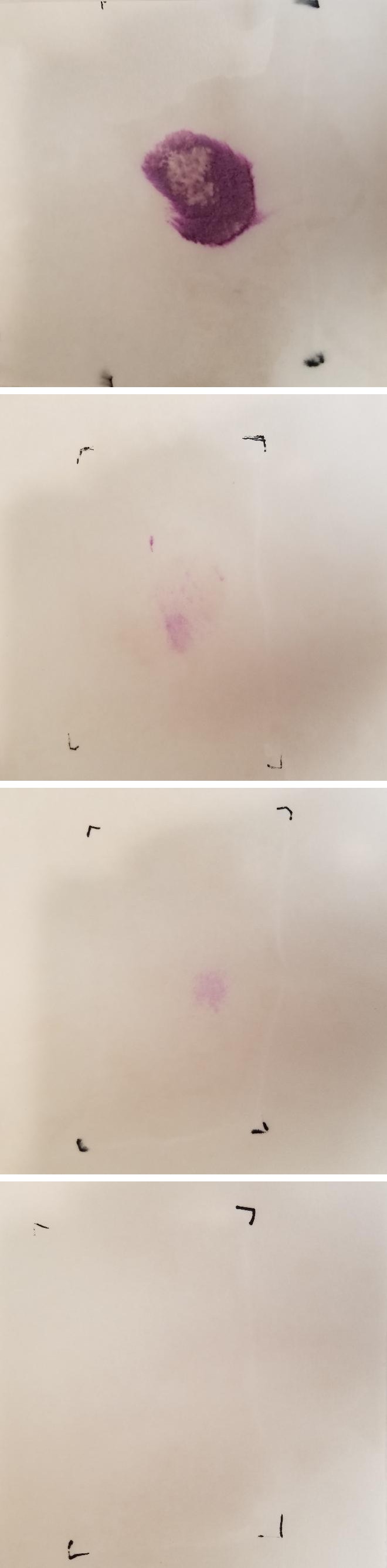
Acid Phosphatase Press Test
For the AP press test, all controls showed initial color development in less than ten seconds after the AP reagent was applied. None of the stains that were wet at the time of washing tested positive for the presumptive presence of semen. Of the stains that were dry, only polyester showed variable results, with each temperature setting resulting in two negatives and one presumptive positive out of the triplicates (Table 4).
| Cotton | Polyester | Denim | Wool | |||||||||
| Hot/Dry | + | + | + | - | - | + | + | + | + | + | + | + |
| Hot/Wet | - | - | - | - | - | - | - | - | - | - | - | - |
| Cold/Dry | + | + | + | - | - | + | + | + | + | + | + | + |
| Cold/Wet | - | - | - | - | - | - | - | - | - | - | - | - |
| Unwashed | + | + | + | + | + | + | + | + | + | + | + | + |
Table 4: Results of AP press test. “+” denotes positive, “-“denotes negative.
A.
C.
B.
D.
Christmas Tree Staining
For the Christmas tree staining, all the controls tested positive for the presence of spermatazoa, and were given scores of 2+ or 3+. The rest of the samples showed variable results, with none of the washed samples receiving a score above 1+ (Table 5). The denim samples showed to retain the most spermatazoa during washing, as there was only one sample from the denim that tested negative for the presence of spermatazoa.
| Cotton | Polyester | Denim | Wool | |||||||||
| Hot/Dry | 1+ | 1+ | - | 1+ | - | 1+ | 1+ | 1+ | 1+ | - | 1+ | 1+ |
| Hot/Wet | - | 1+ | - | - | - | - | 1+ | 1+ | 1+ | 1+ | 1+ | - |
| Cold/Dry | - | - | - | 1+ | 1+ | 1+ | 1+ | - | 1+ | 1+ | - | - |
| Cold/Wet | - | - | - | - | - | 1+ | 1+ | 1+ | 1+ | 1+ | - | - |
| Unwashed | 2+ | 2+ | 2+ | 3+ | 2+ | 3+ | 2+ | 2+ | 2+ | 2+ | 3+ | 2+ |
Table 5: Results of microscopic examination of Christmas tree stains.
Discussion
The failure to observe fluorescence on both the unwashed controls and washed samples appears to have been affected by the type of fabric the stain is present on. Even in the controls, the semen stains present on denim were difficult to observe due to the dark coloration of the substrate, as well as the lack of uniform coloration. The dark coloration of the polyester may have created a similar problem. While there are published studies investigating the impact of substrates when using ALS, these studies are limited to include substrates such as tile, concrete, wood, and fabric, which almost always is cotton [18, 19, 20, 21]. There is very little research investigating a variety of different fabric types. There have been studies to develop methods around substrate interference in ALS examination of biological stains to avoid problems such as color [18]. However, these methods require an image to be captured and analyzed in computer software, limiting how quickly ALS examinations can be performed. Fluorescence in the washed stains was observed only in cotton for the hot water/dried stains and in the cotton and wool for the cold water/dried stains. The observed fluorescence in the washed samples was less than that in the controls, demonstrating that the washing did reduce the ability of ALS to detect washed semen stains. However, the lack of fluorescence in some of the controls suggests that ALS is not a useful method for detecting semen stains on certain fabrics due to interference from the substrate. The AP testing demonstrated that washing the stains while they were wet would reduce the amount of the acid phosphatase enzyme present enough to cause a negative result for the test. This is most likely due to the semen not having time to fully soak into the fabric while drying, allowing it to be more easily washed away. The polyester samples also showed some inconsistencies for the samples that were washed once the stains were dried. However, because polyester is a synthetic fabric and possesses more uniformity, it may not have retained as much of the semen stains during the wash cycles as the other fabrics, which were composed of natural fibers. The Christmas tree staining showed much less consistency in each of the groups. The controls all worked as expected, meaning the inconsistencies may be coming from somewhere other than the methodology. The variability between samples in the same groups may be due to the swatches experiencing different washing conditions during the same cycle. Some of the samples may have bunched together in the washer, while the others remained separate. The clumping of some of the swatches may have allowed some of those swatches to retain more sperm cells than others. This could mean that if whole items were washed together, transfer could occur not only between items, but from one area of an item to another, delocalizing the original semen stain. The only fabric that gave consistent results for the Christmas tree staining was the denim. This is most likely due to it being a thicker weave fabric composed of natural fibers, which may have trapped more sperm cells during the wash cycles. There is also the potential for transfer between samples in the same wash cycle, as previous studies have shown is possible in standard wash cycles [5]. The other three fabrics may have shown less consistency and more negative results due to being thinner fabrics, allowing for the spermatazoa to be washed away more easily.
Conclusion
This study has highlighted some of the limitations of the current methodologies for semen identification for stains present on fabrics that have been washed/laundered. The use of ALS to locate potential biological stains may be severely inhibited by washing, regardless of water temperature or if the stain was wet or dry at the time it was washed. The acid phosphatase test can presumptively detect the presence of semen regardless of the water temperature when washed; however, if the stain is still wet at the time of washing, it may severely inhibit the detection of acid phosphatase. Further, the type of fabric may have an impact, as the AP test was inconsistent when attempting to detect washed semen stains on polyester. However, those inconsistencies were not seen in the other, natural fabrics used in this study. During washing, sperm may be transferred to other items, or lost entirely. The retention of sperm during washing could be affected by several factors: the type of fabric(s) the semen is deposited on, the water temperature, whether the stain is wet or dry, and other factors. The result of this study provide a valuable contribution to the forensic science field and its investigators, as it highlights the importance of choice of method and considerations to be taken when interpreting results for the detection of semen, particularly on items that may have been laundered
Acknowledgement
We would like to thank the Forensic Science Department at the University of New Haven for the use of their resources and facilities throughout the completion of this project.
References
-
Virkler K, Lednev IK (2009) Analysis of body fluids for forensic purposes: from laboratory testing to non- destructive rapid confirmatory identification at a crime scene. Forensic Sci Int 188(1-3): 1-17.
-
Crowe G, Moss D, Elliot D (2000) The effect of laundering on the detection of acid phosphatase and spermatazoa on cotton t-shirts. Canadian Society of Forensic Science Journal 33(1): 1-5.
-
Jobin R, De Gouffe M (2003) The Persistence of Seminal Constituents on Panties After Laundering. Significance to Investigations of Sexual Assault. Canadian Society of Forensic Science Journal 36(1): 1- 10.
-
Spector J, Von Gemmingen D (1971) The effects of washing on the detection of blood and seminal stains. Canadian Society of Forensic Science Journal 4: 3-9.
-
Kafarowski E, Lyon AM, Sloan MM (1996) The retention and transfer of spermatazoa in clothing by machine washing. Canadian Society of Forensic Science Journal 29(1): 7-11.
-
Farmen R, Cortez P, Skårland Frøyland E (2008) Spermatazoa recovered on laundered clothing. Forensic Science International: Genetics Supplement Series1(1): 418-200.
-
Joshi UN, Subhedar SK, Saraf DK (1981) Effect of water immersion on seminal stains on cotton cloth. Forensic Sci Int 17(1): 9-11.
-
Nelson DG, Santucci KA (2002) An alternate light source to detect semen. Acad Emerg Med 9(10): 1045-1048.
-
Stoilovic M (1991) Detection of semen and blood stains using polilight as a light source. Forensic Sci Int 51(2): 289-296.
-
Kobus HJ, Silenieks E, Scharnberg J (2002) Improving the effectiveness of fluorescence for the detection of semen stains on fabrics. J Forensic Sci 47(4): 819-823.
-
Kind SS (1957) The use of acid phosphatase test in searching for seminal stains. Journal of Criminal Law and Criminology 47(5).
-
Kaye S (1949) Acid phosphatase test for identification of seminal stains. J Lab Clin Med 34(5): 728-732.
-
Schiff AF (1978) Reliability of the acid phosphatase test for the identification of seminal fluid. J Forensic Sci 23(4): 833-844.
-
Schiff AF (1975) Sperm identification--acid phosphatase test. Med Trial Tech Q 21(4): 467-474.
-
Ablett PJ (1983) The identification of the precise conditions for seminal acid phosphatase (SAP) and vaginal acid phosphatase (VAP) separation by isoelectric focusing patterns. J Forensic Sci Soc 23(3): 255-256.
-
Allery JP, Telmon N, Mieusset R, Blanc A, Rouge D (2001) Cytological detection of spermatozoa: comparison of three staining methods. J Forensic Sci 46(2): 349-351.
-
Leubitz SS, Savage RA (1984) Sensitivity of picroindigocarmine/nuclear fast red (PIC/NF) stain for detection of spermatozoa: a serial dilution study of human ejaculate. Am J Clin Pathol 81(1): 90-93.
-
Miranda GE, Prado FB, Delwing F, Daruge E (2014) Jr. Analysis of the fluorescence of body fluids on different surfaces and times. Sci Justice 54(6): 427- 431.
-
Lee W, Khoo BE (2010) Forensic light sources for detection of biological evidences in crime scene investigation: a review. Malaysian Journal of Forensic Sciences 1.
-
Vandenburg N, Van Oorschot R (2006) The use of Polilight in the detection of seminal fluid, saliva, and bloodstains and comparison with conventional chemical-based screening tests. Journal of Forensic Sciences 51(2).
-
Viner TC, Kagan RA, Johnson JL (2014) Using an alternate light source to detect electrically singed feathers and hair in a forensic setting. Forensic Sci Int 234: e25-29.
- Forensic Implications of Adverse Drug Reactions in Schizophrenia A Case Series
- Narcotics and Digital Forensics: Bridging Crimes in the Digital Age
- Ethics in Forensic Psychiatry: Principles, Dilemmas, and Human Rights
- Impact of Acute Stress on Attentional Orienting to Social Cues
- Head Injury and Intracranial Hemorrhage in Western Region of Libya
- A Forensic Study on Handedness: Examination of Handwriting Features in Right and Left Handed Writers