Approaching Sex Determination by Computed Tomography Study of the Maxillary Sinuses: Application in a Tunisian Population and Literature Review
Purpose: The present study was performed to investigate the reliability and accuracy of maxillary sinus dimensions as a method for sex determination in a Tunisian sample. Method: This was a prospective study of a cohort of 225 Tunisian individuals (120 men and 105 women), aged 20 to 90 years who underwent a Computed Tomography (CT) scan of the maxillary sinuses to investigate whether these parameters could be used to determine the sex for forensic identification. The used measurement parameters were: the caniocaudal diameter (CCD), the anteroposterior diameter (APD), the transverse diameter (TD), and the volume (V). The statistical analyses were performed by using the SPSS 18.0 package program. The significance rate (p) was set at 0.05. Results: The anteroposterior, transverse, craniocaudal diameters and volume of the maxillary sinuses of males were found to be statistically larger than those of females. The study of the applicability of such method in the sex determination showed a good correlation with TD, APD and volume of the right maxillary sinus. We concluded that the correct predictive accuracy was 63.3% in males and 64.8% in females, with a mean of 64%. Conclusions: Our results suggest that maxillary sinuses CT-Scan analysis can be used for sex determination in the Tunisian population where other anthropological methods are not conclusive.
Introduction
Sex determination is still to be a challenge both in mutilated corpses and skeletal remains. For this purpose, several methods have been developed over the years to enhance the science of forensic anthropology, particularly radiological methods which have become a powerful tool in forensic sciences as they have the advantage of being simple, rapid and not requiring a heavy preparation of the studied bone parts [1, 2].
Historically, the early application of radiology in medico-legal sciences was introduced in 1896 [3]. Standard radiographs previously used for identification purposes have lost their place due to the development of cutting-edge imaging techniques that are increasingly being used in this field. Today, the introduction of Computed Tomography (CT), MRI and new multi-detector machines with fine and reformatted sections has allowed a more precise evaluation of the bone structures studied [4].
Several bones of the skeleton such as parts of the skull, ribs, hip, teeth, sternum, the pelvis, the vertebrae, the clavicle and the bones of the hand and foot have been the subject of radiological studies for identification purposes [5, 6, 7, 8, 9, 10, 11]. It has been reported that the precision of sex determination is about 100% from the skeleton, 98% from the pelvis and the skull, 95% from the pelvis alone or from the pelvis and long bones, 90-95% from the skull and long bones, and 80-90% from long bones only [12, 13]. Despite the sensitivity and specificity of the study of these bones in the determination of sex, the forensic pathologist is sometimes confronted with situations in which he has only certain parts of the skeleton or human remains. Therefore, evaluation of the discrimination index of other parts of the skeleton may be necessary.
Maxillary sinuses are the first sinus cavity to develop and appear at the end of the second month of embryonic life. During fetal development, they come from the invaginations which the nasal mucosa undergoes in the maxillary bone and extends on the roof of the permanent teeth when the milk teeth fall. This development explains its big anatomical variations [14, 15]. They are characterized by their ability to remain intact while the skull and other bones can be disfigured in victims who are incinerated. Indeed, it has been demonstrated that the shape and size of the maxillary sinus differ between individuals and by sex. Therefore, it can be used for the identification of mutilated or dilapidated bodies [16]. In 1927, Culbert and Law described the first complete radiological identification using the sinuses of the face [17].
It is within this framework that we have initiated several research projects within our department of forensic medicine, with the aim of individualizing specific methods of medico-legal identification to the Tunisian population [18, 19]. The purpose of the present study was to determine the reliability and accuracy of maxillary sinus dimensions as a method for sex determination in a Tunisian sample and through a review of the literature.
Method
It is a cross sectional study of CT scans of the maxillary sinuses of 225 living Tunisian individuals. The radiological examination was performed for diagnostic purposes in response to a medical indication. Only Tunisian individuals aged over 20 were included in this study. Patients who have history of traumatic and / or iatrogenic maxillary sinus lesions, and those having sinus malformations or conditions that may affect bone growth and maturation (metabolic abnormalities, hereditary hyperostosis diseases, radiotherapy history, sinus tumor, bone metastases) were not included in this study.
CT scans were acquired using a 16-stip “General Electric” scanner, with a 16 x 0.75 mm collimator and a die of 512 x 512 pixels. The parameters of the acquisition were as follows: Field of view 15 cm, 200-380 mA, 140 kV, reconstruction interval of 0.75 mm, length of acquisition of 40 seconds.
Sagittal and coronal reconstructions were used for radiographic evaluation of the lower wall of the maxillary sinus. The acquired images were exported as DICOM files to an image post-processing console and visualized in bone density range (300-1500 gray scale range).
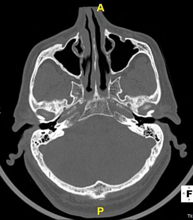
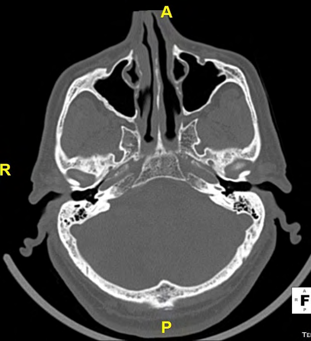
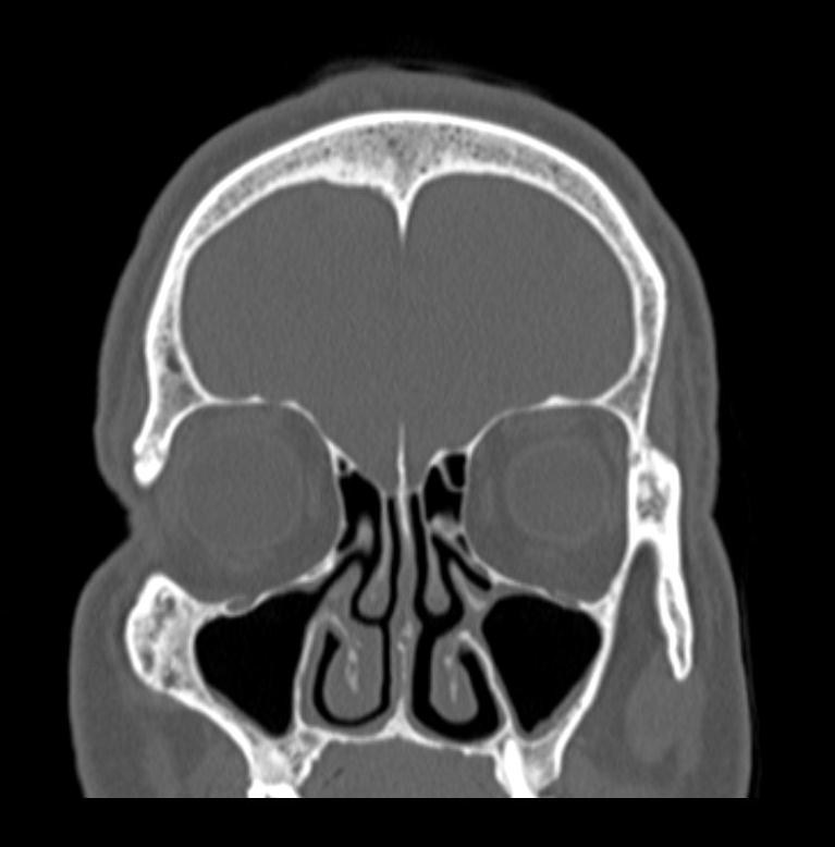
For each maxillary sinus (right and left), four measures were analyzed: - The anteroposterior diameter (APD) in axial section: this is the distance separating the anterior wall and the posterior wall of the sinus (Figure 1). - The transverse diameter (TD) in axial section: this is the distance separating the side walls of the sinus (Figure 2). - The craniocaudal diameter (CCD) in coronal section: this is the distance separating the lower wall and the upper wall of the sinus (Figure 3). - The volume (V): it was calculated by multiplying the three parameters mentioned above (V = CCD × APD × TD).
The greatest measurements were taken after going through different slices in axial and coronal sections.
All patients gave their informed consent for the additional use of the performed CT scans as part of a research protocol.
The data collected were analyzed using SPSS 18.0 on Microsoft Windows XP platform. The application of the Kolmogorov-Smirnov test made it possible to verify the normality of distribution of the sample studied. “P” value was considered significant at ≤ 0.05 and highly significant at ≤ 0.001.
The independent samples t-test was used to analyze differences regarding measurements among males and females. For sex determination, the student’s t-test was used to compare between means of the two groups. P value < 0.05 was considered statistically significant. A logistic regression analysis was used to estimate the relationship between sex and different measurements by calculating the odds ratio or / and the corresponding 95% confidence intervals (CIs). The final retained variables were those significant at the level of 5%. Different probabilities were evaluated from this analysis. Then, a ROC (Receiver Operator Characteristic) curve was performed to assess the validity of the tested variables. Discriminant value finally retained was based on the better specificity with the better sensitivity.
Results
A slight male predominance was noted with a sex ratio of 1.14 (H/F = 120/105). The mean age of the subjects included was 55.32 ± 18.15 years with extremes of 20 to 90 years and a median of 57 years.
For each maxillary sinus, four quantitative variables were studied. Table 1 illustrates the means of measurements of each variable for each side.
| N | Minimum | Maximum | Mean | Standard Deviation | |
|---|---|---|---|---|---|
| RTD | 225 | 9.6 | 40.7 | 26.4458 | 6.09895 |
| LTD | 225 | 10.7 | 44.2 | 26.0373 | 6.01163 |
| RAPD | 225 | 21.2 | 50.2 | 35.0573 | 4.16322 |
| LAPD | 225 | 22.1 | 45.9 | 34.9533 | 4.16862 |
| RCCD | 225 | 18.1 | 49.5 | 32.54 | 6.06747 |
| LCCD | 225 | 17.8 | 45.2 | 32.5951 | 5.70615 |
| RV (mm3) | 225 | 5260.99 | 100389.96 | 32098.214 | 14689.68625 |
| LV (mm3) | 225 | 6679.06 | 9149798 | 31449.061 | 14068.81716 |
Table 1: Means of measurements of variables (in millimeters). RTD: Right transverse diameter; LTD: Left transverse diameter; RAPD
Despite the variation of the measurements between the right and left sides of maxillary sinuses, we didn’t found statistical differences.
The different variables studied (APD, TD, CCD and Volume) varied significantly by sex. Measurements were on average significantly higher in male group than in female group. Metric data was summarized as mean, standard deviation and p values in Table 2.
| Variables | Minimum | Maximum | Mean | Standard Deviation | p | |
|---|---|---|---|---|---|---|
| RTD | M | 9,6 | 40,4 | 27,447 | 60,650 | 0,008 |
| F | 13,6 | 40,7 | 25,301 | 59,621 | ||
| LTD | M | 10,7 | 44,2 | 26,707 | 58,363 | 0,074 |
| F | 12,7 | 37,8 | 25,271 | 61,444 | ||
| RAPD | M | 21,2 | 50,2 | 35,827 | 42,914 | 0,003 |
| F | 22,3 | 44,5 | 34,178 | 38,465 | ||
| LAPD | M | 25,2 | 45,9 | 35,652 4 | 41,303 | 0,007 |
| F | 22,1 | 43,3 | 34,155 | 40,869 | ||
| RCCD | M | 18,1 | 49,5 | 34,495 | 60,705 | <10-3 |
| F | 18,4 | 41,7 | 30,306 | 52,608 | ||
| LCCD | M | 21,000 | 45,200 | 3,411,917 | 549,486 | <10-3 |
| F | 17,800 | 43,700 | 3,085,333 | 5,462,425 | ||
| RV | M | 5260,99 | 100390,0 | 35,937,264 | 15,564,077 | <10-3 |
| F | 6859,71 | 56787,90 | 27,710,728 | 12,294,382 | ||
| LV | M | 7931,95 | 91497,98 | 34,229,568 | 14,602,616 | 0,001 |
| F | 6679,06 | 66467,23 | 28,271,338 | 12,775,246 |
Table 2: Distribution of means for anthropometric measurements (in millimeters) according to sex. M: Male; F: female; The volume
Three variables selected using the stepwise statistics were retained (logistic regression analysis). Each step was statistically significant with p value < 0.05. These variables consist of the RTD, RAPD and RV. Discriminant function coefficients of these parameters are shown in the following table (Table 3).
| Discriminant Coefficients Function « B » | Confidence Interval of B (CI: 95%) | p | |
|---|---|---|---|
| RTD | 1.268 | 1.115 – 1.442 | 0.008 |
| RAPD | 1.149 | 1.008 – 1.309 | 0.003 |
| RV | 1 | 1.000 – 1.000 | < 10-3 |
| Constant | 0.003 |
Table 3: Discriminant function coefficients of the maxillary sinuses.
The discrimination function “DF” for sex determination established using these variables was as follows: “DF’’ = 0.003 + (1.268 x RTD) + (1.149 x RAPD) + (1 x RV).
We calculated the point of discrimination in males ‘DF (males)’ and in females ‘DF (females)’ using this formula: DF (males): 0.003 + (1.268 x 2.744) + (1.149 x 3.582) + (1 x 35.937) = 43.534. DF (women): 0.003 + (1.268 x 2.530) + (1.149 x 3.417) + (1x 27.710) = 34.847. (Measurements are in centimeters, volume in cm3). Cut point: DF (males) / DF (women) = 39.190.
So we can conclude that if “DF” is greater than 39.190, it is a male and if “DF” is less than 39.190, it is a female.
This formula was applied to the study sample. We represent in the following table the rates of sex determination from the variables measured (Table 4). The correct predictive accuracy in sex determination was 63.3% in males and 64.8% in females with an overall accuracy rate of 64%.
| Real Sex | Determined Sex | |
|---|---|---|
| Man | Women | |
| Man(120) | 76(63.3%) | 44 |
| Women(105) | 37 | 68(64.8%) |
Table 4: Determination of sex from the measured variables.
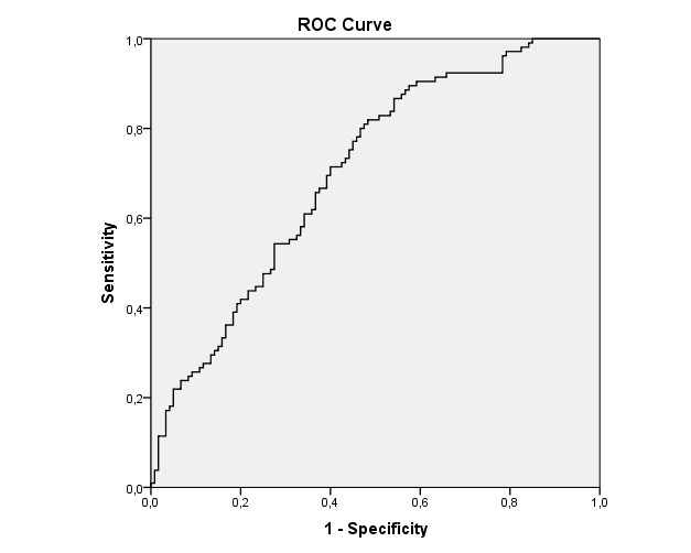
A ROC curve was performed for the sample (Figure 4). The area under the curve (AUC) is 0.703 which is considered “good”. The confidence interval of the 95% level is [0.636 – 0.770]. The sensitivity and specificity of this method in the sex determination, deduced by analyzing the ROC curve, were 67.2% and 60.7%, respectively.
Discussion
The identification of human bone remains a very important procedure in forensic practice. Although the methods of identification in Medico-legal Anthropology are similar, the results are never unequivocal. Indeed, different environmental, nutritional and ethnic conditions are likely to give distinct anthropometric features from one human group to another. Each population must, therefore, establish its own scores and formulas for sex determination and age estimation.
Sex determination is an important step in the forensic identification. Several bones of the skeleton have been the subject of investigations for gender determination with an accuracy rate of 100% when the skeleton exists completely, 98% in existence of pelvis and skull, 95% with only pelvis and long bones, and 80–90% with only long bones [12, 13].
Despite the higher accuracy rate of these bones in the sex determination, the anthropologist is sometimes confronted with situations in which the whole skeleton is not available, like in explosions and mass disasters. Therefore, evaluation of the discrimination index of other parts of the skeleton, like maxillary sinuses, may be necessary.
It has been demonstrated in various populations that the shape and size of the maxillary sinus differ between individuals and by sex. That’s why several teams attempted to establish population-specific equations to determine sex from maxillary sinus using CT-scan measurements [20, 21, 22, 23, 24, 25, 26]. Results varied from one population to another [15, 20, 21, 22, 23, 24, 25, 26, 27, 28, 29, 30, 31, 32, 33, 34]. The present study was conducted to evaluate the reliability and accuracy of maxillary sinus anthropometric measurement as a method for sex determination in the Tunisian population.
However, sex determination from maxillary sinuses measurements is not recommended on the bones of preadolescent individuals since the secondary sexual characteristics do not appear until the bones are transformed during puberty under the influence of estrogens and androgens. For this reason, in the present study, as well as in the majority of published studies, subjects less than 20 years of age were not included. In some studies, the lowest age limit was set at 18 years [4, 27].
In the present study, maxillary sinus measurements were studied using MDCT scan. For each side, four variables were evaluated. We found that the mean values of right side maxillary sinus measurements were higher than the left side, but these variations were not statistically significant. In the literature, the majority of published studies have reported that the left maxillary sinus had dimensions larger than those of the right side in both sex [22, 23, 24, 25, 26, 33, 34]. However, this dimensional variability between the two sides was not always significant. In the literature, no explanation has been proposed for the variation of maxillary sinus measurements from the side [4, 15, 20, 21, 22, 23, 24, 25, 26, 27, 28, 29, 30, 31, 32, 33, 34].
In addition, the measurements performed on the maxillary sinuses in males were on average significantly larger than those in females, and these findings were reported in the literature [22, 23, 24, 25, 26]. However, this result was not admitted in Zululand where maxillary sinuses were found to be narrower in males than in females [30]. Enlow [35] proposed two explanations for the variability of dimensions between the two sexes. First, men need to have larger lungs to support their relatively more massive muscles and organs. Second, men need more developed airways, starting with the nose and the nasopharynx. In other words, physiological changes in the size and shape of the nasal cavity occur as a direct result of breathing-related needs, such as warming and humidification of the inhaled air.
In the current study, the best discriminatory parameters for sex determination were the TD, APD and volume of the right maxillary sinus whereas for Amin and Hassan [20], only the CCD and volume of the left maxillary sinus were determinant. For Kiruba, et al. [22], the discriminatory formula was established from the TD, APD and CCD of the left maxillary sinus and the APD and CCD of the right side. For Vidya, et al. [29], only the left TD and the right sinus volume showed statistically significant values.
In the present study, we concluded that the correct predictive accuracy was 63.3% in males and 64.8% in females with an overall accuracy rate of 64%. Our results are similar to those reported in the literature with an average precision rate ranging from 63.6% to 73.9% [20, 21, 22, 23, 24, 25, 26] (Table 5).
| Our Study | A Study Population | Number | Precision Rate | ||
|---|---|---|---|---|---|
| Male 70,8% | Female 62,5% | Average 66,7% | |||
| Amin, et al. [20] | Egyptian | 96 | |||
| Attia,et al. [21] | Egyptian | 73 | 71,8% | 67,6% | 69,9% |
| Kiruba, et al. [22] | Indian | 200 | 69,5% | 55% | 63,6% |
| Sharma, et al. [23] | Indian | 102 | 65,16% | 68,9% | 67,03% |
| Uthma,n et al. [24] | Iraqi | 88 | 74,4% | 73,3% | 73,9% |
| Teke, et al. [25] | Turkish | 127 | 69,2% | 69,4% | 69,3% |
| Azhar Gh [26] | Iraqi | 119 | 56,1% | 71% | 63,9% |
| Ekizoglu, et al. [27] | Tunisian | 225 | 63,3% | 64,8% | 64% |
Table 5: Variation in sex determination from maxillary sinuses according to published studies.
Some authors have been particularly interested in the determination of sex by the volume of the maxillary sinus [4, 23, 32, 36, 37, 38]. Emirzeoglu, et al. [4] examined digitized CT images of 77 Turkish patients. They reported a significant difference in the mean volume of the maxillary sinus between men and women. Similar results were described by Sahlstrand-Johnson, et al. [32] and Karakas [38]. This variability of volume by sex was explained by the difference in the shape and morphology of the male and female face especially around the mid-face region.
According to the study conducted by Ekizoglu, et al. [27], the sinus volume was considerably smaller in females (P <0.001), with an overall accuracy rate of sex determination estimated at 77.15%. In an Indian study reported by Kanthem, et al. [39], the volume had a higher percentage of sexual dimorphism with an overall accuracy rate of 85.46% for the right side and 78.38% for the left side.
Recently, Italian researchers conducted an anthropometric study to validate the use of the software Dolphin in the analysis of the tomographic images and in the estimation of the volumes of the maxillary sinuses, and to specify whether the maxillary sinus volumes can be used to determine the sex of the unknown persons [40]. No statistically significant differences were observed between the operators, confirming the reliability of the technique. In addition, there were no statistically significant differences between right and left maxillary sinus volumes (p = 0.2) and by gender (p = 0.1). The authors had rejected the hypothesis that the morphology of the maxillary sinuses, especially volume, is crucial for determining sex, in contrary to what was reported in the literature [40].
Variations in anthropometric measurement of maxillary sinuses in most studies reported in the literature are likely due to the combination of many factors such as differences in ethnic and racial groups, genetic and environmental factors, sample size, measurement technics, and anatomical variations of the sinuses [23].
Our results suggest that maxillary sinuses CT-Scan analysis can be used for sex determination in the Tunisian population where other anthropological methods are not conclusive. The establishment of an equation specific to the Tunisian population to determine sex from maxillary sinuses with rates of specificity and sensitivity that exceed 60% represents a breakthrough in the field of Medico-legal Anthropology in Tunisia. This equation must be validated on other Tunisian samples to draw more strong conclusions.
References
-
Iscan MY (2005) Forensic anthropology of sex and body size. Forensic Sci Int 147(2-3): 107-112.
-
Cunha E, Baccino E, Martrille L, Ramsthaler F, Prieto J, et al. (2009) The problem of aging human remains and living individuals: A review. Forensic Sci Int 193(1-3): 1-13.
-
Eckert WG, Garland N (1984) The history of the forensic applications in radiology. Am J Forensic Med Pathol 5(1): 53-56.
-
Emirzeoglu M, Sahin B, Bilgic S, Celebi M, Uzun A (2007) Volumetric evaluation of the paranasal sinuses in normal subjects using computer tomography images: a stereological study. Auris Nasus Larynx 34(2): 191-195.
-
Bodey TE, Loushine RJ, West LA (2003) A retrospective study evaluating the use of the panoramic radiograph in endodontics. Mil Med 168(7): 528-529.
-
Ukoha U, Egwu OA, Okafor IJ, Anyabolu AE, Ndukwe GU, et al. (2011) Sexual dimorphism in the Foramen magnum of Nigerian adult. Int J Biol Med Res 2(4): 878-881.
-
Rejtarova O, Slizova D, Smoranc P, Rejtar P, Bukac J (2004) Costal cartilages—a clue for determination of sex. Biomed Pap 148(2): 241-243.
-
Varga M, Takacs P (1991) Radiographic personal identification with characteristic features in the hip joint. Am J Forensic Med Pathol 12(4): 328-331.
-
MacLaughlin SM, Oldale KN (1992) Vertebral body diameters and sex prediction. Ann Hum Biol 19(3): 285– 292.
-
McCormick WF, Stewart JH, Greene H (1991) Sexing of human clavicles using length and circumference measurements. Am J Forensic Med Pathol 12(1): 175- 181.
-
Smith SL (1997) Attribution of foot bones to sex and population groups. J Forensic Sci 42(2): 186-195.
-
Krogman WM, Isçan MY (1986) The human Skeleton in Forensic Medicine. 2nd (Edn.), Springfield: Charles Thomas Publisher.
-
Hauser G, Stefeno DGF (1989) Epigenetic variants of the human skull. E. Stuttgart: Schweizerbart’sche Verlagsbuchhandlung, pp: 38-40.
-
Champsaur P, Pascal T, Vidal V, Gaubert JY, Bartoli JM, et al. (2003) Radio anatomie des sinus de la face. J Radiol 84: 885-900.
-
Amusa YB, Eziyi JA, Akinlade O, Famurewa OC, Adewole SA, et al. (2011) Volumetric measurements and anatomical variants of paranasal sinuses of Africans (Nigerians) using dry crania. Int J Med Sci 3(10): 299- 303.
-
Cameriere R, Ferrante L, Mirtella D, Rollo UF, Cingolani M (2005) Frontal sinuses for identification: quality of classifications, possible error and potential corrections. J Forensic Sci 50(4): 770-773.
-
Culbert WL, Law FM (1928) Identification by comparison of roentgenograms of nasal accessory sinuses and mastoid processes. JAMA 88: 1634-1636.
-
Haj Salem N, Aissaoui A, Mesrati MA, Belhadj M, Quatrehomme G, et al. (2014) Age estimation from the sternal end of the fourth rib: a study of the validity of İşcan’s method in Tunisian male population. Legal Med 16(6): 385-389.
-
Haj Salem N, Saadi S, Ben Jomaa S, Othmani H, Hmida B, et al. (2019) Age estimation at death by the study of chest plate radiographs: establishing a Tunisian male score. Int J Legal Med 134: 775-782.
-
Amin MF, Hassan M, Eman I (2012) Sex identification in Egyptian population using Multidetector Computed Tomography of the maxillary sinus. J Forensic Leg Med 19(2): 65-69.
-
Attia AM, El-Badrawy AM, Shebel HM (2012) Gender identification from maxillary sinus using multi-detector computed tomography. J Forensic Med Clin Toxicol 10: 17-28.
-
Kiruba LN, Gupta C, Kumar S, D’Souza AS (2014) A study of morphometric evaluation of the maxillary sinuses in normal subjects using computer tomography images. Arch Med Health Sci 2: 12-215.
-
Sharma SK, Jehan M, Kumar A (2014) Measurements of maxillary sinus volume and dimensions by computed tomography scan for gender determination. J Anat Soc India 63(1): 36-42.
-
Uthman AT, Al-Rawi NH, Al-Naaimi AS, Al-Timimi JF (2011) Evaluation of maxillary sinus dimensions in gender determination using helical CT scanning. J Forensic Sci 56(2): 403-408.
-
Teke HY, Duran S, Canturk N, Canturk G (2007) Determination of gender by measuring the size of the maxillary sinuses in CT scans. Surg Radiol Anat 29(1): 9-13.
-
Azhar Gh, Ibrahim S, Salah M, Ghadah N (2015) CT scan images analysis of maxillary sinus dimensions as a forensic tool for sexual and racial detection in a sample of kurdish population. Eur Sci J 11: 272-281.
-
Ekizoglu O, Ince E, Hocaoglu E (2014) The use of maxillary sinus dimensions in gender determination: a Thin-slice Multidetector computed tomography Assisted morphometric study. J Craniofac Surg 25(3): 957-960.
-
Masri A, Asilah Yusof, Rozita Hassan (2013) A Three Dimensional Computed Tomography 3D-CT: A Study of Maxillary Sinus in Malays. CJBAS 01: 125-34.
-
Vidya CS, Shamasundar NM, Manjunatha B, Keshav R (2013) Evaluation of size and volume of maxillary sinus to determine gender by 3d computerized tomography scan method using dry skulls of south indian origin. Int J Cur Res Rev 5: 97-100.
-
Fernandes CL (2004) Volumetric analysis of maxillary sinuses of Zulu and Europen crania by helical, multislice computed tomography. J Laryngol Otol 118(11): 877- 881.
-
Kim HJ, Yoon HR, Kim KD, Kang MK, Kwak HH, et al. (2003) Personal computer based three dimensional reconstruction and simulation of maxillary sinus. Surg Radiol Anat 24: 393-399.
-
Sahlstrand-Johnson P, Jannert M, Strömbeck A, Abul- Kasim K (2011) Computed tomography measurements of different dimensions of maxillary and frontal sinuses. BMC Med Imaging 11: 8.
-
Jehan M, Bhadkaria V, Trivedi A, Sharma SK (2014) Sexual Dimorphism of Bizygomatic distance and Maxillary sinus using CT Scan. IOSR-J Dent Med Sci 13(3): 91-95.
-
Baweja S, Dixit A (2013) Study of age related changes of maxillary air sinus from its anteroposterior, transverse and vertical dimensions using Computerized Tomographic (CT) scan. IJBR 4(1): 21-25.
-
Enlow DH (1990) Facial Growth. 3rd (Edn.), Saunders Philadelphia, pp: 6-7.
-
Kawarai Y, Fukushima K, Ogawa T (1999) Volume quantification of healthy paranasal cavity by threedimensional CT imaging. Acta Oto-laryngologica 540: 45-49.
-
Chang-Hee P, Kim KD, Park CS (2000) Measurements of maxillary sinus volume using Computed Tomography. Korean J Oral Maxillofac Radiol 30: 63-70.
-
Karakas S, Kavakli A (2005) Morphometric examination of the paranasal sinuses and mastoid air cells using computed tomography. Ann Saudi Med 25(1): 41-45.
-
Kanthem RK, Guttikonda VR, Yeluri S, Kumari G (2015) Sex determination using maxillary sinus. J Forensic Dent Sci 7(2): 163-167.
-
Saccucci M, Cipriani F, Carderi S, Di Carlo G, D’Attilio M, et al. (2015) Gender assessment through three- dimensional analysis of maxillary sinuses by means of Cone Beam Computed Tomography. Eur Rev Med Pharmacol Sci 19(2): 185-193.
- Narcotics and Digital Forensics: Bridging Crimes in the Digital Age
- Ethics in Forensic Psychiatry: Principles, Dilemmas, and Human Rights
- Impact of Acute Stress on Attentional Orienting to Social Cues
- Head Injury and Intracranial Hemorrhage in Western Region of Libya
- A Forensic Study on Handedness: Examination of Handwriting Features in Right and Left Handed Writers
- Techniques for Latent Fingerprint Development Using Natural and Synthetic Powders: A Review