Imaging of Fingermark Chemistry via Spectroscopic Techniques
Fingermarks are one of the earliest forensic evidence linking a crime scene to a criminal and is based on the assumption that everyone has a unique set of patterns at their fingertips. In fact, fingermarks are much more than their formal appearance; they are our chemical identities. Most forensic scientists now consider not the chemicals produced by a person's body, but the chemicals they touched before leaving fingermarks; explosives, narcotics, drugs, etc. Therefore, there has long been a need in the forensic science community to explore new ways to analyze fingermarks. Spectroscopic methods are the leading methods that will allow the multi-faceted examination of the evidence detected at the crime scenes, the preservation of the structure of the evidence during the examination and the re-analysis of the evidence in the judicial proceedings. Today, with the developing technology, a large number of spectra can be collected by area scanning with spectroscopic techniques and these can be converted into images with various software. Spectroscopic imaging is an emerging technology that combines digital imaging and molecular spectroscopy, where high-quality spectral and spatial information is collected over a period of time. In terms of sensitivity, reproducibility, selectivity, reliability and finally applicability of each method; it has advantages and limitations to be used in routine forensic science applications or academic research studies. In this review, apart from the more commonly used mass spectrometry techniques, it is aimed to discuss chemically monitoring narcotic substances and gunshot residues in fingermark samples by some spectroscopic techniques. When the literature is examined, the limited number of studies on the analysis and chemical imaging of fingermark chemistry with spectroscopic techniques is the most unique aspect of this discussion.
Introduction
The value of fingermarks as evidence stems from the fact that the fingermarks left by the person when touching the surface can be detected even if their qualities are weak [1]. This has made fingermarking an identification method that has been used for more than a hundred years [2]. Fingermarks encountered in daily life are generally not easily visible to the naked eye, and therefore, various chemical, physical and optical methods are used to visualize fingermarks [3, 4, 5]. The fingermarks found at the crime scene show some differences according to the characteristics of the objects they are left on.
However, sometimes not all fingermarks on the scene are suitable for this purpose. For example, fingermarks taken from the crime scene may not be available in the database or may not be sufficient in terms of details [6]. Thanks to the rapid developments in the fight against crime and criminals, it has become possible to make comprehensive and versatile investigations on the findings collected from crime scenes. Today, as a result of developing technology and new researches, information is obtained not only about the visual properties of fingermarks, but also about their chemistry. This situation will make a great contribution to the solution of judicial events.
Various studies have been conducted to investigate the organic and inorganic composition of the body fluid formed by fingermarks using Gas Chromatography-Mass Spectrometry (GC-MS) [7, 8]. In addition, researchers working in this field include Desorption Electrospray Ionization Mass Spectrometry (DESI-MS) [9], Matrix Assisted Laser Desorption Ionization Mass Spectrometry (MALDI-MS) [10] and Secondary Ion Mass Spectrometry (SIMS). Bailey MJ, et al. [11], Muramoto S, et al. [12], Szynkowska MI, et al. [13], they were able to obtain both chemical compositions and chemical images of fingermarks by using techniques for this purpose.
Besides chromatographic and mass spectrometric
techniques, there are other techniques that can be used for the chemical identification of certain fingermark compounds. Spectroscopic imaging is an important tool in this regard. Spectroscopic imaging is an up-to-date and useful technology combining digital imaging and molecular spectroscopy where high quality spectral and spatial information are collected [14]. Wei, Zhang, Ogorevc and Zhang [15] reported that such techniques are also used in chemical imaging of fingermarks in a compilation study published.
Apart from mass spectrometric techniques, Raman Spectroscopy, Micro X-ray fluorescence (MXRF), Fourier Transform Infrared Spectroscopy (FTIR) and X-ray Photoelectron Spectroscopy (XPS) have also been used in recent years to image fingermark chemistry. Unlike other techniques, faster results can be obtained with these techniques. Additionally, these techniques do not require chemicals and special sample preparation [16, 17, 18, 19].
In this study, a brief literature review has been made regarding the chemical imaging and analysis of fingermark chemistry using spectroscopic techniques. In this context, a brief summary of how spectral imaging was acquired was also mentioned.
Composition and Properties of Fingermarks
Fingermarks have a complex chemical composition that may contain both natural secretions and contaminants. The chemicals present are not necessarily homogeneously distributed, both at a microscopic and macroscopic level. The chemical composition will change after deposition of the mark.
| Location | Constituents | |
|---|---|---|
| Eccrine sweat | Eccrine sweat glands found all over the body and particularly abundant on the palms of hands and fingertips | Water, urea, uric acid, creatinine, amino acids, ammonia, choline, glucose and other reducing sugars, lactic acid and lactate, sodium, chloride, potassium, calcium, trace metal ions, phosphate, sulphate, enzymes, peptides, proteins, vitamins |
| Sebum | Sebaceous glands on the face, head and other locations associated with hair follicles | Free fatty acids (C7-C22, saturated and unsaturated), cholesterol esters, mono-, di- and triacylglycerols, wax esters, cholesterol, squalene and other hydrocarbons |
| Apocrine sweat | Apocrine sweat glands found in the axillary regions of the body, namely, the armpits and genital area | Ammonia, androgenic steroids, cholesterol, glycogen (carbohydrates), iron, proteins and water |
| Epidermal (skin surface) lipids | From touching other areas of the body (the epidermis) and migration of material from the nonpalm side of hand | Free fatty acids, glycerides, proteins, sterols, sterol esters |
| External contaminants (exogenous substances) | Picked up as a consequence of touching other objects and surface | Illicit drugs, nicotine, cosmetics, explosives, foodstuffs, dust, grease |
Table 1: Summary of latent fingermark residue sources [16].
In addition to the chemical properties, fingermarks also have both physical and biological properties that can be exploited Fingermarks are formed as the result of an interaction between a finger and a surface. Several outcomes are possible from this interaction, including deposition of positive marks, removal of material to leave negative marks and formation of impressions on the surface. Latent fingermarks can consist of components from a range of sources as outlined in Table 1. The relative contribution of each of these sources varies for each fingermark. Consequently, no two fingermarks have the exact same chemical composition, although there can be similarities in the relative composition [16].
Chemical Imaging of Fingermarks by Spectroscopic Techniques
Chemical imaging is just another term for spectral imaging. In practice, there are no differences. Spectral imaging refers to a group of analytical techniques that collect spectroscopic information and imaging information at the same time. The spectroscopic information tells us about the chemical makeup at the individual points of the image (pixels, x and y) allowing a chemical map of the imaged area to be produced. Spectral imaging is a technology that integrates conventional imaging and spectroscopy to get both spatial and spectral information from an object [17].
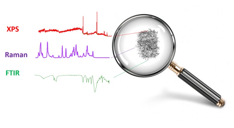
Nowadays, visualizing human fingermarks do not provide now enough information about the suspects and/ or the criminals. For this reason, researchers try to find more about the fingermarks; that is their chemistry. Figure 1 shows the concept of chemical imaging using different spectroscopic techniques. This type of information could be useful for reducing the pool of potential suspects in criminal investigations when latent fingermarks are unsuitable for comparison by traditional methods.
![Figure 1: Concept of chemical imaging (adopted from [18]).](/fulltextimages/6263/fig_1.jpeg)
In recent literature, we come across with some studies regarding spectroscopic imaging of human fingermarks due to their advantages. Most importantly, the techniques in question do not damage the sample. However, so far, very few attempts have been made on the chemical determination of human fingermarks by XPS [11, 19]. On the other hand, the first study on chemical imaging was carried out by Erdogan, Esen and Simpson [20] in 2020.
XPS is a surface analysis technique that provides both qualitative and quantitative information with a sampling depth of ≤ ~ 10 nm. All elements except hydrogen and helium can be detected with XPS. The limit of detection for this technique is reported as approximately 0.05% by mass for each element. In addition to the basic surface composition, XPS provides crucial information about the oxidation state and chemical bonding that cannot be achieved with other techniques. Despite its many advantages as a surface analytical technique, XPS is not yet widely used in forensic science [21]. Watts briefly talked about what kind of applications can be made in forensic science related to XPS in 2010. These include chemical identification of fingermarks, analysis of residues on surfaces, and examination of residues of explosives. In addition, Watts emphasized the shortcoming in this area and advocated the routine use of XPS like other techniques [22]. Another study on XPS Strohmeier, Bunker, McAllister, Marquis, Piasecki and McAllister [23] in 2011 mentions the analysis of firearm remains. In this study, lead (Pb), antimony (Sb) and barium (Ba) elements and their oxides were examined in the residues using both Scanning Electron Microscopy (SEM) and XPS, but no spectral imaging / mapping was performed. In later years Bailey, et al., on the other hand, XPS analysis for fingermark chemistry was done for the first time. In this study, the difference between fingermarks of two different donors could be reproducibly distinguished by XPS. Thus, it has been shown that XPS is not only useful for the detection of inorganic substances, but also a technique that can give an idea about the identification of some organic compounds. However, XPS alone may not be sufficient for the structure determination of organic compounds. Therefore, other complementary techniques like IR and Raman spectroscopy besides XPS are required for the identification and / or verification of more complex organic compounds, structural isomers, and similar structures. Figure 2 presents conceptually how different spectroscopic techniques complement each other for the chemical imaging of human fingermark.
In the same study, the researchers revealed that the difference in depth is important in their analysis with Attenuated Total Reflection-Fourier Transform Infrared Spectrometry (ATR-FTIR). While information on a surface chemistry up to 10 nm can be obtained with XPS, FTIR can reach micron levels. Infrared (IR) spectroscopy is based on the principle of absorbing the infrared radiation sent on the molecule under study and examining the corresponding vibrational wave numbers. In this method, a molecule can be easily distinguished from the others thanks to the molecular vibrations specific to each molecule. Therefore, the IR technique has a wide range of use in forensic applications. With this technique, the chemical components in the fingermark residues can be analyzed with the help of the microscope in the spectrometer, and the substance in the focus point can be easily analyzed. In addition, the IR technique provides significant advantages in that it does not damage the sample and generally does not require an additional sample preparation process after the sample is taken. With this technique, spectra can be collected from the scanned area (pixel-based) and images consisting of thousands of characters can be obtained microscopically and mapping and chemical imaging can be performed. As a result of this mapping, detailed information about the chemical structure in the studied microscopic region can be obtained. Bhargava et al. (2009), thanks to the vibrations of the C-H bond and the differences in other vibration modes, spectroscopic imaging was performed with IR to distinguish overlapping fingermarks [24]. The overlapping fingermarks could be distinguished due to the different ratios of chemical components from the two donors.
Apart from the IR spectroscopy technique, Raman spectroscopy is also widely used. In Raman Spectroscopy, the object to be examined is first imaged with the help of a microscope and laser radiation in the visible region at certain wavelengths is sent on the material and the radiation of different wavelengths scattered from the sample is examined. The difference of Raman spectroscopy from IR spectroscopy is that it is a scattering spectroscopy. With this technique, some wavelength values that cannot be obtained in the IR spectroscopy are also determined, and in this sense, the Raman spectroscopy plays a complementary role to the IR spectroscopy. This technique also does not damage the sample and requires almost no preparation of the sample [25].
Day, et al. reported that Raman spectroscopy was applied to detect illegal drugs and other residues in hidden fingermarks [26]. In another study conducted by Widjaja, mapping was performed again with the Raman technique and the remains found in fingermarks were detected using multivariate data analysis [27]. As with all other techniques, there are some limitations in FTIR and Raman spectroscopy. There may be some difficulties in identifying chemically similar compounds. FTIR is mostly used in determining polymers and organic molecules. There are some limitations in that it cannot be used to identify and quantify inorganic compounds, metals, metal oxides and composite materials. It is possible for chemicals or mixtures with similar structures to overlap or show serious similarity spectrally. Therefore, these techniques may not always give precise results and should be used as a complement to each other. On the other hand, the IR spectroscopy causes difficulties in liquid samples, especially since water bands make the spectra difficult to interpret, whereas in the Raman spectroscopy, liquid samples can be studied quite easily.
For example, since it is possible for the water vapor / moisture in the environment to accumulate in the sample, it is necessary to dry the samples in a vacuum oven before analysis to compensate for this. A disadvantage of Raman spectroscopy is that the wavelength of the radiation sent on the sample under study does not give the spectrum bands that can distinguish the chemical contained in the sample as a result of scattering from the sample. In this case, radiation at a wavelength that can distinguish that sample must be sent. For example, while there are no characteristic bands in the Raman spectrum for a banned substance in the sample, on which a 532 nm wavelength laser light is sent, when the sample is stimulated with 785 nm wavelength laser radiation, the detection of this banned substance can be made immediately. The use and widespread use of new techniques has led to significant improvements in chemical characterization of other substances in fingermarks. It is very important to detect components such as explosive residues and drugs deposited on fingertips for many reasons [13, 28].
Chemical analysis of fingermarks may provide insights into the suspect’s use of illicit drugs (heroin, cocaine, amphetamine, methamphetamine, codeine, morphine, etc.) or skin care products (including TiO2, SiO2) and smoking habits (such as nicotine) [29, 30, 31]. Apart from these, determining the remains of firearms in fingermarks can also provide very important information [23].
Conclusion
The identification of human fingermarks has long been a vital point of criminal investigation and forensic science. Forensic researchers are continually aiming to develop novel methods that are fast, precise, simple, low- cost, friendly, non-invasive, portable and compatible with conventional fingermark development for obtaining personal chemical information and identification. Accurate fingermark identification is of great significance in providing further chemical analysis about a fingermark donor’s age, race, gender, diet habit, etc. In general, extracting chemical information from a fingermark is far more important than just physically enhancing the fingermark. Herein, it has been reviewed that the recent developments in the chemical imaging of latent fingermarks by using some spectroscopic methods. As might be expected, all of these techniques have advantages and disadvantages depending on how the analysis needs to be performed and what or how the analytes can be detected.
This review highlights the considerable advances in the chemical imaging of human fingermarks that provide more chemical information. Although a number of techniques exist that allow for the detection of specific components in fingermarks, there is still a gap between forensic research in the lab and forensic practice at a crime scene. In future, methods that are reliable, portable, fast, low-cost, sensitive, selective and non-destructive are highly needed for visualizing latent fingermarks physically and for imaging them chemically.
In future, with the progress in the determination of fingermark chemistry via spectroscopic imaging, significant contributions to the institutions and organizations (Forensic Medicine Institutions and Police Criminal Laboratories) will be provided that will implement these results, by reaching more information about the suspects or criminals. Because nowadays fingermarks are no longer just a visual data, more information has been obtained thanks to existing technologies. In this context, new researches will contribute to a social problem globally.
Acknowledgement
Dr. Ayşegül Erdoğan (MC Substitute of COST Action 16101-MULTI-FORESEE-MULTI-modal Imaging of FOREnsic SciEnce Evidence tools for Forensic Science) appreciate the inspiration provided by Prof. Dr. Simona Francese (Chair of COST Action 1610).
Declaration of Conflicting Interests: The authors declared no conflicts of interest with respect to the research, authorship, and/or publication of this article.
Funding: This study was not supported financially by any of the organizations.
References
-
Lennard C (2020) Fingermark detection and identification: current research efforts. Australian Journal of Forensic Sciences 52(2): 125-145.
-
Girod A, Ramotowski R, Weyermann C (2012) Composition of fingermark residue: a qualitative and quantitative review. Forensic science international 223(1-3): 10-24.
-
Burns DT, Brown JK, Dinsmore A, Harvey KK (1998) Base- activated latent fingerprints fumed with a cyanoacrylate monomer (1998) A quantitative study using Fourier- transform infra-red spectroscopy. Analytica chimica acta 362(2-3): 171-176.
-
Choi MJ, Smoother T, Martin AA, McDonagh AM, Maynard PJ, et al. (2007) Fluorescent TiO2 powders prepared using a new perylene diimide dye: Applications in latent fingermark detection. Forensic science international 173(2-3): 154-160.
-
Qin G, Zhang M, Zhang Y, Zhu Y, Liu S, et al. (2013) Visualizing latent fingerprints by electrodeposition of metal nanoparticles. Journal of Electroanalytical Chemistry 693(15): 122-126.
-
Van Helmond W, Van Herwijnen AW, Van Riemsdijk JJ, Van Bochove MA, de Poot CJ, et al. (2019) Chemical profiling of fingerprints using mass spectrometry. Forensic Chemistry 16: 100183.
-
Cadd S, Mota L, Werkman D, Islam M, Zuidberg M, et al. (2015) Extraction of fatty compounds from fingerprints for GCMS analysis. Analytical Methods 7(3): 1123-1132.
-
Weyermann C, Roux C, Champod C (2011) Initial results on the composition of fingerprints and its evolution as a function of time by GC/MS analysis. Journal of forensic sciences 56(1): 102-108.
-
Ifa DR, Manicke NE, Dill AL, Cooks RG (2008) Latent fingerprint chemical imaging by mass spectrometry. Science 321(5890): 805-805.
-
Lauzon N, Dufresne M, Chauhan V, Chaurand P (2015) Development of laser desorption imaging mass spectrometry methods to investigate the molecular composition of latent fingermarks. Journal of the American Society for Mass Spectrometry 26(6): 878- 886.
-
Bailey MJ, Bright NJ, Croxton RS, Francese S, Ferguson LS, et al. (2012) Chemical characterization of latent fingerprints by matrix-assisted laser desorption ionization, time-of-flight secondary ion mass spectrometry, mega electron volt secondary mass spectrometry, gas chromatography/mass spectrometry, X-ray photoelectron spectroscopy, and attenuated total reflection Fourier transform infrared spectroscopic imaging: An intercomparison. Analytical chemistry 84(20): 8514-8523.
-
Muramoto S, Sisco E (2015) Strategies for potential age dating of fingerprints through the diffusion of sebum molecules on a nonporous surface analyzed using time- of-flight secondary ion mass spectrometry. Analytical chemistry 87(16): 8035-8038.
-
Szynkowska MI, Czerski K, Rogowski J, Paryjczak T, Parczewski A (2009) ToF-SIMS application in the visualization and analysis of fingerprints after contact with amphetamine drugs. Forensic Science International 184(1-3): e24-e26.
-
Exline DL, Wallace C, Roux C, Lennard C, Nelson MP, et al. (2003) Forensic applications of chemical imaging: latent fingerprint detection using visible absorption and luminescence. Journal of forensic sciences 48(5): 1047- 1053.
-
Wei Q, Zhang M, Ogorevc B, Zhang X (2016) Recent advances in the chemical imaging of human fingermarks (a review). Analyst 141(22): 6172-6189.
-
Dorakumbura BN, Boseley RE, Becker T, Martin DE, Richter A, et al. (2018) Revealing the spatial distribution of chemical species within latent fingermarks using vibrational spectroscopy. Analyst 143(17):4027-4039.
-
Boseley RE, Dorakumbura BN, Howard DL, de Jonge MD, Tobin MJ, et al. (2019) Revealing the elemental distribution within latent fingermarks using synchrotron sourced x-ray fluorescence microscopy. Anal Chem 91(16):10622-10630.
-
Ricci C, Bleay S, Kazarian SG (2007) Spectroscopic imaging of latent fingermarks collected with the aid of a gelatin tape. Anal Chem 79(15):5771-5776.
-
Worley CG, Wiltshire SS, Miller TC, Havrilla GJ, Majidi V (2006) Detection of visible and latent fingerprints using micro-x-ray fluorescence elemental imaging. J Forensic Sci 51(1):57-63.
-
Erdoğan A, Esen M, Simpson R (2020) Chemical Imaging of Human Fingermark by X‐ray Photoelectron Spectroscopy (XPS). Journal of forensic sciences 65(5): 1730-1735.
-
Wagner JM (2011) X-ray Photoelectron Spectroscopy (Chemical Engineering Methods and Technology). Nova Science Publishers Incorporated.
-
Watts JF (2010) The potential for the application of X‐ray photoelectron spectroscopy in forensic science. Surface and Interface Analysis: An International Journal devoted to the development and application of techniques for the analysis of surfaces, interfaces and thin films 42(5): 358- 362.
-
Strohmeier BR, Bunker KL, McAllister DR, Marquis JP, Piasecki JD, et al. (2011) Application of X-ray photoelectron spectroscopy (XPS) for the surface characterization of Gunshot Residue (GSR). Microscopy Today 19(2): 40-45.
-
Bhargava R, Perlman RS, Fernandez DC, Levin IW, Bartick EG (2009) Non-invasive detection of superimposed latent fingerprints and inter-ridge trace evidence by infrared spectroscopic imaging. Analytical and bioanalytical chemistry 394(8): 2069-2075.
-
Larkin P (2017) Infrared and Raman spectroscopy: principles and spectral interpretation. Elsevier.
-
Day JS, Edwards HG, Dobrowski SA, Voice AM (2004) The detection of drugs of abuse in fingerprints using Raman spectroscopy I: latent fingerprints. Spectrochimica Acta Part A: Molecular and Biomolecular Spectroscopy 60(3): 563-568.
-
Widjaja E (2009) Latent fingerprints analysis using tape- lift, Raman microscopy, and multivariate data analysis methods. Analyst 134(4): 769-775.
-
Rowell F, Seviour J, Lim AY, Elumbaring Salazar CG, Loke J, et al. (2012) Detection of nitro-organic and peroxide explosives in latent fingermarks by DART-and SALDI- TOF-mass spectrometry. Forensic science international 221(1-3): 84-91.
-
Benton M, Chua M, Gu F, Rowell F, Ma J (2010) Environmental nicotine contamination in latent fingermarks from smoker contacts and passive smoking. Forensic science international 200(1-3): 28-34.
-
Draelos ZD (2005) Cosmetic Formulation of Skin Care Products. CRC Press, pp: 25-26.
-
Morelato M, Beavis A, Tahtouh M, Ribaux O, Kirkbride P, et al. (2013) The use of forensic case data in intelligence- led policing: the example of drug profiling. Forensic science international 226(1-3): 1-9.
- Narcotics and Digital Forensics: Bridging Crimes in the Digital Age
- Ethics in Forensic Psychiatry: Principles, Dilemmas, and Human Rights
- Impact of Acute Stress on Attentional Orienting to Social Cues
- Head Injury and Intracranial Hemorrhage in Western Region of Libya
- A Forensic Study on Handedness: Examination of Handwriting Features in Right and Left Handed Writers
- Techniques for Latent Fingerprint Development Using Natural and Synthetic Powders: A Review