Possible Age and Sex Dependency of Costal Cartilage Calcification Degree Investigated by Computed Tomography: First Costal Cartilage Calcification is of Significance
Introduction: This study provides the costal cartilage calcification degree (CCCD) with more focus on first costal cartilage calcification (FCCC) in different sexes and age groups in an Iranian population. Materials and methods: This is a cross-sectional retrospective study including 400 chest computed tomography (CT) scans of the participants aging from 20 to 89 years old, from 2020 to 2021. The chest CT scans were categorized based on two factors of sex (n= 400; male and female), and age (n: 400; 30 years). Degree of calcification was categorized into five groups including: 1 (
Introduction
Rib cage, which mostly consists of twelve pairs of ribs, functions as a protective (protection of viscera), and supportive (supporting of muscle attachments helping in breathing and visceral protection) structure. Ribs are attached to sternum by means of cartilage in a pattern in which first four costal cartilages (CCs) connect to sternum directly, fifth to seventh CCs connect to sternum and costal cartilages above each other, and eighth to tenth CCs connect to sternum by means of upper CCs [1]. Costal cartilages are made up of a core hyaline cartilage and a peripheral perichondrium [1].
Costal cartilage calcification (CCC) or ossification occurs mostly in an age dependent manner [2, 3], but it can also occur due to pathological states (e.g. metabolic changes, or nutritional state) [3], corticosteroid medication [4, 5], and congenital diseases [4]. CCC is of significant importance from several clinical aspects. Cartilaginous structure of rib cage accounts for flexibility and better endurance against traumatic injury, but the process of CCC stiffens the rib cage which exerts stress loads on ribs putting them at risk of fracture [1, 6]. The other clinical importance of CC is they can be used for nasal, auricular, or temporomandibular joint grafts in which CC calcification can adversely affect the efficiency of grafts [7, 8, 9]. CCC studies have also been used in age and sex discrimination in forensic medicine [3, 4, 10].
Regarding the clinical significance of CCC in traumatic injuries, grafts, and forensic investigations, this study provides the degree of CCC with more focus on first costal cartilage calcification in different sexes and age groups in an Iranian population by means of multidetector computed tomography scan and 3-dimensional volume images. Since the CCC can be readily detected in CT and there is no sufficient data on the pattern of CCC in the literature – particularly FCCC which may be depended upon sex and age – this study was conducted to provide information on calcification of costal cartilages in different sexes and age limits.
Materials and Methods
Patients and Computed Tomography
A cross-sectional retrospective study was conducted including 400 chest CT scans of the participants aging from 20 to 89 years old, from 2020 to 2021 performed in Dr. Shariati Hospital, Mahdasht, Alborz, Iran. The chest CT scans were categorized based on two factors of sex (n= 400; male and female), and age (n: 400; <30 years and >30 years). Imaging device was multidetector computer tomography (Hitachi – Supria 16/32 with 51 kW power, 75 cm gantry bore, 180 cm scan range, 5 mega hit unit (MHU) X ray tube, and 0.675 mm minimum slice thickness). To avoid motion artifacts, patients had to stay still and hold their breath in deep inspiration. CT scans of the chest with 5mm thickness and 5mm interval were acquired. For analyzing costal cartilage calcification, the images were reconstructed with thickness and interval of 1.25mm and analyzed in 3-dimensional volume reconstruction.
Costal Cartilage Calcification Degree (CCCD)
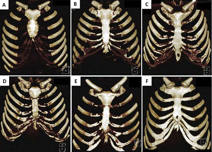
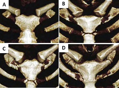
CCCD of first to eighth rib was investigated in 3-dimensional volume images. Degree of calcification was categorized into five groups including: 1 (<5%), 2 (5-25%), 3 (25-50%), 4 (50-75%), and 5 (75-100%) (Figure 1). First costal cartilage calcification (FCCC) was investigated meticulously by categorizing it into three groups including: 1 (complete fusion with sternum), 2 (partial fusion with sternum), and 3 (partial calcification) (Figure 2).
Statistical Analysis
Sex discrimination analysis in all the participants, and age groups was done by chi-square test. Then age groups were analyzed separately by chi-square test. Also logistic regression analysis was performed in separate sex groups, and age groups. All the analyses were done by SPSS software (version 16, Chicago, IL, USA), and P≤0.05 was considered significant.
Results
CCCD was reported based on sex (n=400, male= 223, female=177), and age (n=400, <30=91, >30=309) (Table 1). CCCD in both male and female demonstrated higher value in group 2 (5-25%) (male=65.1%, female=50.6%) compared to other groups, although CCCD did not show any significant difference between sex groups. FCCC did not show significant difference between male and female, either. However, complete fusion of first costal cartilage was more common (male= 70.9%, female=64.8%) (Table 1). Male and female differentiation was also investigated in two age groups. In <30 years group, 5-25% calcification (CCCD=2) is higher in male (52.4%) than female (19%) (Pearson Chi-square value (PCV): 5.65, P: 0.05). Also, in <30 years group, complete fusion is higher in female (80%) than male (18.2%) (PCV: 6.07, P: 0.05) (Table 2). Age-based analysis without considering sex demonstrated that <5% calcification (CCCD=1) is higher in age group <30 (61.9%) than >30 years (15.8%) (PCV: 37.50, P: 0.000), and complete fusion (FCCC=1) is higher in age group >30 (72.4%) than <30 years (37.5%) (PCV: 8.13, P: 0.01) (Table 3). Logistic regression results demonstrated no predicting value of CCCD, and FCCC in sex discrimination. But in age discrimination, FCCC can be considered a possible predictor with an overall correct classification of about 88.6% (Table 4).
| Sex | Rib calcification | ||||||||||
|---|---|---|---|---|---|---|---|---|---|---|---|
| CCCD | FCCC | ||||||||||
| 1 | 2 | 3 | 4 | 5 | T | 1 | 2 | 3 | T | ||
| Male | 45 (19.8%) | 145 (65.1%) | 23 (10.4%) | 6 (2.8%) | 4 (1.9%) | 223 (100%) | 166 (70.9%) | 34 (14%) | 36 (15.1%) | 236 (100%) | |
| Female | 60 (33.7%) | 89 (50.6%) | 11 (6%) | 11 (6%) | 6 (3.6%) | 177 (100%) | 96 (64.8%) | 21 (14.8%) | 30 (20.4%) | 147 (100%) | |
| Total | 105 (25.9%) | 234 (58.7%) | 34 (8.5%) | 17 (4.2%) | 10 (2.6%) | 400 (100%) | 262 (68.6%) | 55 (14.3%) | 66 (17.1%) | 383* (100%) | |
| Age | Rib calcification | ||||||||||
| CCCDa | FCCCb | ||||||||||
| 1 | 2 | 3 | 4 | 5 | T | 1 | 2 | 3 | T | ||
| <30 y | M | 22 (47.6%) | 24 (52.4%) | 0 (0%) | 0 | 0 | 46 (100%) | 5 (18.2%) | 13 (45.5%) | 10 (36.4%) | 28 (100%) |
| F | 34 (76.2%) | 8 (19%) | 3 (4.8%) | 0 | 0 | 45 (100%) | 12 (80%) | 0 | 3 (20%) | 15 (100%) | |
| T | 56 (61.9%) | 32 (35.7%) | 3 (2.4%) | 0 | 0 | 91 (100%) | 17 (37.5%) | 13 (31.2%) | 13 (31.2%) | 43 (100%) | |
| >30 y | M | 24 (12.9%) | 122 (68.2%) | 23 (12.9%) | 6 (3.5%) | 4 (2.4%) | 179 (100%) | 160 (78.7%) | 19 (9.3%) | 25 (12%) | 204 (100%) |
| F | 25 (19.4%) | 80 (61.3%) | 8 (6.5%) | 11 (8.1%) | 6 (4.8%) | 130 (100%) | 85 (63.3%) | 23 (16.3%) | 28 (20.4%) | 136 (100%) | |
| T | 49 (15.8%) | 202 (65.8%) | 31 (9.6%) | 17 (5.5%) | 10 (3.4%) | 309 (100%) | 245 (72.1%) | 42 (12.5%) | 53 (15.4%) | 340 (100%) | |
| Total | 105 (25.9%) | 234 (58.7%) | 34 (8.5%) | 17 (4.2%) | 10 (2.6%) | 400 (100%) | 262 (68.6%) | 55 (14.3%) | 66 (17.1%) | 383* (100%) |
Table 1: Costal cartilage calcification based on sex. CCCD: Costal cartilage calcification degree, 1: <5%, 2: 5-25%, 3: 25-50%, 4
Table 2: Costal cartilage calcification based on age and sex. a 5-25% calcification (CCCD=2) is higher in male (52.4%) than female (19%) in age group <30 years (Pearon Chi-square value: 5.65, P: 0.05); b Complete fusion is higher in female (80%) than male (18.2%) in age group <30 years (Pearon Chi-square value: 6.07, P: 0.05); CCCD: Costal cartilage calcification degree, 1: <5%, 2: 5-25%, 3: 25-50%, 4: 50-75%, 5: 75-100%, T: total; FCCC: First costal cartilage calcification, 1: complete fusion, 2: partial fusion, 3: partial calcification. * 17 cases had no calcification of first costal cartilage.
| Age | Rib calcification | |||||||||
|---|---|---|---|---|---|---|---|---|---|---|
| CCCDa | FCCCb | |||||||||
| 1 | 2 | 3 | 4 | 5 | T | 1 | 2 | 3 | T | |
| <30 y | 56 (61.9%) | 32 (35.7%) | 3 (2.4%) | 0 | 0 | 91 (100%) | 17 (37.5%) | 13 (31.2%) | 13 (31.2%) | 43 (100%) |
| >30 y | 49 (15.8%) | 202 (65.8%) | 31 (9.6%) | 17 (5.5%) | 10 (3.4%) | 309 (100%) | 245 (72.1%) | 42 (12.5%) | 53 (15.4%) | 340 (100%) |
| Total | 105 (25.9%) | 234 (58.7%) | 34 (8.5%) | 17 (4.2%) | 10 (2.6%) | 400 (100%) | 262 (68.6%) | 55 (14.3%) | 66 (17.1%) | 383* (100%) |
Table 2: Costal cartilage calcification based on age. a <5% calcification (CCCD=1) is higher in age group <30 (61.9%) than >30 ye
Table 3: Costal cartilage calcification based on age. a <5% calcification (CCCD=1) is higher in age group <30 (61.9%) than >30 years (15.8%) (Pearon Chi-square value: 37.50, P: 0.000); b complete fusion (FCCC=1) is higher in age group >30 (72.4%) than <30 years (37.5%) (Pearon Chi-square value: 8.13, P: 0.01); CCCD: costal cartilage calcification degree, 1: <5%, 2: 5-25%, 3: 25-50%, 4: 50-75%, 5: 75-100%, T: total; FCCC: First costal cartilage calcification, 1: complete fusion, 2: partial fusion, 3: partial calcification. * 17 cases had no calcification of first costal cartilage.
| Based on age | Variables in the equation | % of correctly grouped sternum | ||||||||
|---|---|---|---|---|---|---|---|---|---|---|
| B | S.E. | Wald | df | Sig. | Exp (B) | Observed age group | Predicted age group | |||
| <30 | >30 | Correct | ||||||||
| FCCC | -0.72 | 0.30 | 5.68 | 1 | 0.01 | 0.48 | <30 | 0 | 45 | 0 |
| Constant | 3.25 | 0.62 | 27.1 | 1 | 0.00 | 25.90 | >30 | 0 | 338 | 100 |
| Overall | 88.6 |
Table 3: Logistic regression results. FCCC: First costal cartilage calcification
Discussion
CCCD showed no significant difference between male and female, although when sex differentiation was considered in <30 years age group, there was a significant higher 5-25% calcification in male than female. And also in the same age group, FCCC showed significant higher complete fusion rate in female than male. Age-dependent differentiation of CCCD and FCCC was also evident in which lower <5% CCCD, and higher complete fusion rate was evident in >30 years than <30 years age group.
CCC process begins after puberty at both ends of costal cartilage in a marginal to central pattern making the central parts of the costal cartilages the last parts to calcify. This process of calcification or ossification can be put in neither of the two types of ossification – intramembranous or endochondral – making it a rather specific physiological age- related change [2].
Although some of the studies in the literature report occasional sex-related differences in costal cartilage calcification degree, it shows extensive variations and cannot be considered a reliable factor by itself [3, 9, 10]. In this study, general sex differentiation without considering the age of participants was not applicable, however, when considered in a specific age group (<30 years), sex discrimination was possible.
Age-related changes in CCC is emphasized in the literature [3, 9, 11]. It has been reported that CCC does not occur under the age of 20 and its presence is low in participants under 30 years [3, 11]. In our study, in <30 years age group, most of the CCC were <5%, and there was not any case of calcification with more than 50%. More frequent and extensive calcification can be seen in first costal cartilage [2, 12]. In our study, FCCC was seen in more than 90% of the participants showing at least partial calcification. Also significant higher rates of complete fusion of first rib to manubrium resulting from complete calcification (complete calcification of the outer layer of cartilage) of first costal cartilage was seen in >30 years age group.
In cases of lack of CCC [11], sternal parameters and the fusion of sternal components are considered assisting predictors for sex discrimination [13]. Limitation of this study includes the low number of participants with the age limit of below 30 years, and the fact that CCC does not demonstrate significant changes in age limit of above 30 years, for example a 40-year-old individual does not demonstrate a significant difference in CCC compared to a 50 or 60-year-old individual.
Conclusion
In this study, it was reported that CCC is rather an age- dependent than a sex-dependent feature. The pattern in FCCC can be of significant importance in age discrimination – FCCC demonstrates a significant higher rate in individuals above 30 years. For reliable sex discrimination it is suggested to accompany CCCD with age and patterns of calcification such as marginal, central, and mixed patterns. Since the pattern of CCC – specifically the FCCC – can be readily detected by CT and 3-dimensional volume reconstruction and it can provide a clue in sex and age estimation of mortal remains or deceased individuals, it is suggested to use medical imaging modalities such as CT in forensic sciences.
Acknowledgement
Our deep gratitude goes to the staff of medical imaging center of Dr. Shariati hospital, Mahdasht, Alborz, Iran.
References
-
Forman JL, Kent RW (2014) The effect of calcification on the structural mechanics of the costal cartilage. Comput Methods Biomech Biomed Engin 17(2): 94-107.
-
Kampen WU, Claassen H, Kirsch T (1995) Mineralization and osteogenesis in the human first rib cartilage. Ann Anat 177(2): 171-177.
-
Rejtarová O, Slízová D, Smoranc P, Rejtar P, Bukac J (2004) Costal cartilages--a clue for determination of sex. Biomed Pap Med Fac Univ Palacky Olomouc Czech Repub 148(2): 241-243.
-
Rhomberg W, Schuster A (2014) Premature calcifications of costal cartilages: a new perspective. Radiol Res Pract 2014: 523405.
-
Thomas L (1964) The effects of papain, vitamin a, and cortisone on cartilage matrix in vivo. Biophysical journal 4(1 Pt 2): 207-213.
-
Griffin MF, O’Toole G, Sabbagh W, Szarko M, Butler PE (2020) Comparison of the compressive mechanical properties of auricular and costal cartilage from patients with microtia. J Biomech 103: 109688.
-
Cochran CS (2016) Harvesting Rib Cartilage in Primary and Secondary Rhinoplasty. Clin Plast Surg 43(1): 195- 200.
-
Goerke D, Sampson DE, Tibesar RJ, Sidman JD (2013) Rib reconstruction of the absent mandibular condyle in children. Otolaryngol Head Neck Surg 149(3): 372-376.
-
Zhang S, Zhen J, Li H, Sun S, Wu H, et al. (2017) Characteristics of Chinese Costal Cartilage and Costa Calcification Using Dual-Energy Computed Tomography Imaging. Scientific Reports 7(1): 2923.
-
Inoi T (1997) Estimation of sex and age by calcification pattern of costal cartilage in Japanese. Nihon Hoigaku Zasshi 51(2): 89-94.
-
Middleham HP, Boyd LE, Mcdonald SW (2015) Sex determination from calcification of costal cartilages in a Scottish sample. Clinical Anatomy 28(7): 888-895.
-
Navani S, Shah JR, Levy PS (1970) Determination of sex by costal cartilage calcification. Am J Roentgenol Radium Ther Nucl Med 108(4): 771-774.
-
Ghorbanlou M, Moradi F, Asgari HR (2022) Morphometric study of sternum by computed tomography in an Iranian population: A method to discriminate between male and female. Forensic Imaging 28: 200501.
- Forensic Implications of Adverse Drug Reactions in Schizophrenia A Case Series
- Narcotics and Digital Forensics: Bridging Crimes in the Digital Age
- Ethics in Forensic Psychiatry: Principles, Dilemmas, and Human Rights
- Impact of Acute Stress on Attentional Orienting to Social Cues
- Head Injury and Intracranial Hemorrhage in Western Region of Libya
- A Forensic Study on Handedness: Examination of Handwriting Features in Right and Left Handed Writers