Cutaneous T Lymphoma: Complete Therapeutic Response after Treatment with Contact Brachytherapy: About a Case and Review of Literature
Cutaneous T lymphoma is a relatively common type of non-Hodgkin’s lymphoma (NHL). It originates in mature T lymphocytes in the skin and takes several anatomical and clinical forms which can be very aggressive. We report the therapeutic outcome of a case of cutaneous T lymphoma located in the left forearm treated by contact brachytherapy in the RadiotherapyBrachytherapy department of the HASSAN II university hospital center in FES. The patient remains without recurrence with more than 24 months of follow up. Radiotherapy is a therapeutic means for cutaneous T lymphomas of slow progression of early stage (stage I or II). Brachytherapy allows excellent local control with good results both carcinological and cosmetic.
Introduction
Cutaneous T-cell lymphomas (CTCL) are a heterogeneous group of neoplastic T-cell proliferations that should be distinguished from lymph node lymphomas. These are clonal overgrowths which infiltrate the skin primarily, reach the lymph nodes only at an advanced stage and infiltrate the bone marrow only exceptionally. They are rare, representing only 2% of all lymphomas with an annual incidence estimated at 1-2/100,000. They are distinguished from lymph node lymphoma by their biological and clinical behavior, as well as by their prognosis which is generally much better than that of lymph node lymphoma [1]. There is no treatment for CTCL that can be “strictu-sensu” considered curative at present. The treatments available aim to obtain complete and prolonged remissions.
They are numerous, but only a minority has been validated by controlled studies. This is due to the fact that the rarity of cutaneous lymphomas and their slow evolution (years) require large studies with prolonged follow-up, which is difficult to achieve in practice. It has nevertheless been clearly demonstrated that the survival of patients suffering from a CTCL is not prolonged by multidrug therapy [2]. “Softer” therapies, either local for cutaneous use or systemic, modulating the biological response is therefore preferred, and this especially at the early stages where the anti-tumor immune response is still preserved. Among the treatments suggested for this pathology we mention contact brachytherapy. Brachytherapy is the administration of radiation through flexible catheters linked to an external programmable source and has been used for treatment of multiple cutaneous malignancies including advanced basal cell and squamous cell carcinomas and Merkel cell carcinoma [3, 4]. The use of fully customized molds in brachytherapy allows fine control of the depth of radiation penetration over complex convex surfaces.
Case Report
We report here the case of a 53 years old patient, chronic smoker for 40 years, who has family history of bladder cancer in his brother. This patient presented for 4 years erythematous lesions progressively increasing in size, concerning the left forearm with on examination a erythematato-purple tumor making 14cms of major axis very limited, occupying the front face of the forearm and extending to the posterior surface (Figures 1). When examining the lymph node areas there was no lymphadenopathy. A biopsy was done which was in favor of a CD30 + T lymphoproliferation, lymphomatoid papulosis (LyP) type A.
Figures 1: Appearance of skin lesions upon patient admission.
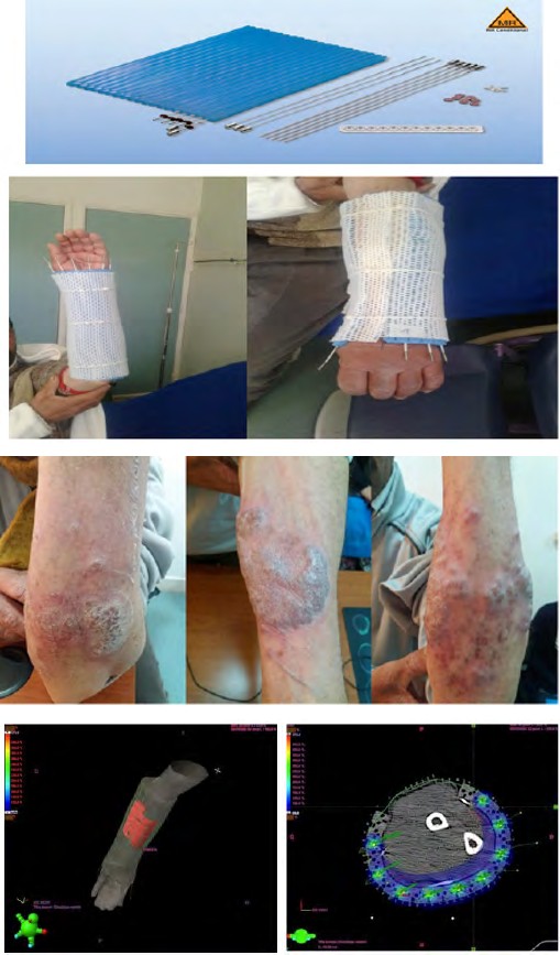
Laboratory evaluations including complete blood count, electrolytes and basic metabolic profile were normal. The patient was investigated with a computerized tomography (CT) scan of the chest and abdomen, with no evidence of distant metastasis. The tumor is classified T3N0M0, stage IIb according to the TNM System. The patient did not respond to systemic treatment with methotrexate, hence the indication for radiotherapy for him. The patient was hospitalized and received high dose rate brachytherapy (HDR), with a total dose of 30Gy in 5 fractions, 6Gy / fraction, 2 fractions per day at 6 hour intervals. The application was made by a Catheter Flap Set type applicator by the Gamma Medplus machine from VARIAN (Figures 2).
Figures 2: Application by a Catheter Flap Set.
The Catheter Flap Set is used for treating of superficial cancers. It is designed to create a defined space between the source and the tissue, and between the source channels. The highly flexible material easily adapts to complex anatomical regions. The catheter channels are 2mm in diameter and are positioned on the middle axis of the flap. The Catheter Flap is often affixed to a thermoplastic mesh (TM), commonly used in radiation therapy, to maintain a reproducible orientation relative to the patient’s anatomy. TM material was heated and formed around the patient’s left forearm. The Catheter Flap was attached to the TM. This TM design and position of the Catheter Flap enabled the Ir-192 HDR source to travel in close proximity to the cutaneous tissue to be treated.
A CT scan was performed. The images were imported into Eclipse (Varian Medical Systems, Palo Alto, Ca). The planning treatment volume (PTV) was outlined or ‘contoured’ on the CT images (Figures 3A & 3B). The patient received treatment according to plan and the tolerance was good. He was reviewed weekly during the first month after treatment by our team. There were no significant side effects observed except moderate pain. The patient was put on analgesic treatment.
Figures 3: Scanning sections showing dose distribution during brachytherapy.
Results
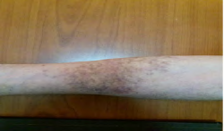
The patient was reviewed after 1 month. Skin lesions have significantly decreased. 2 months after the end of treatment, the lesions have completely disappeared (Figure 4). The patient did not present a local-regional recurrence or metastasis for more than 24 months of follow-up.
Discussion
Cutaneous T cell lymphomas (CTCLs) are a heterogeneous group of extranodal non-Hodgkin’s lymphomas that are characterized by a cutaneous infiltration of malignant monoclonal T lymphocytes. They typically afflict adults with a median age of 55 to 60 years, and the annual incidence is about 0.5 per 100,000. Mycosis fungoides (MF), Sézary syndrome (SS), and primary cutaneous peripheral T cell lymphomas not otherwise specified are the most important subtypes of CTCL. CTCL is a complicated concept in terms of etiopathogenesis, diagnosis, therapy, and prognosis [5].
Classification and Staging of Cutaneous T Lymphomas
A lymphoma is considered to be primarily cutaneous if it meets certain well-defined criteria for clinical pathological diagnosis and does not present any extra-cutaneous localization during the initial assessment of extension. The main types of CTCL are: fungus mycosis (44% of primary cutaneous lymphomas, LCP), Sézary syndrome (2% of LCP), cutaneous T cells with large CD30 + cells (9% of LCP), and lymphomas Skin cells with large CD30- cells (6% of LCP). Fungus mycosis (MF) is the first described of cutaneous lymphomas. It is characterized by a slow evolution in three clinical phases: erythema in non-infiltrated plaques, infiltrated plaques, nodules and tumors. Its five-year survival rate is high (87%).
Sézary syndrome is defined by the association of erythroderma due to diffuse infiltration of the skin by neoplastic T cells associated with a large number (1000/ mm3) of circulating neoplastic T cells and polyadenopathy. The prognosis of Sézary syndrome is more severe than that of MF, its five-year survival rate being 11% CD30 + large cell cutaneous T lymphoma often presents with a red skin tumor (s) and infiltrates immediately with a cytological appearance of high grade lymphoma (large anaplastic, immunoblastic or pleomorphic cells ). These worrying clinical and pathological aspects contrast with its favorable prognosis (> 90% five- year survival), hence the importance of recognizing and distinguishing it from secondary cutaneous involvement of lymph node T lymphoma and other cutaneous T lymphomas CD30- large cell cutaneous T lymphomas are fast-growing tumors that are clinically similar to CD30 + large cutaneous T cell lymphomas, but made up of large CD30- T cells. The absence of expression of the CD30 lymphocyte marker and a clearly poorer prognosis (15% five-year survival) distinguish this cutaneous lymphoma from cutaneous large T cell CD30 + lymphoma. It also demonstrates that CD30 is an immunophenotypic marker with high prognostic value in cutaneous T lymphomas.
Since 1997, a classification intended exclusively for cutaneous lymphomas has been developed by European cutaneous lymphoma experts within the framework of the European Organization for Research and Treatment of Cancers (EORTC) [6]. This EORTC classification, which is very close to the most recent WHO classification has the advantage of distinguishing the different types of cutaneous lymphomas according to their histopathological, clinical characteristics and their prognosis (indolent, intermediate or aggressive) [7]. This clinical pathological classification allows in most cases to quickly establish a prognosis, and therefore facilitates the choice of the optimal treatment [8].
As with lymph node lymphoma, clinical staging is done according to the TMN system. It includes a skin and systemic examination, biological investigations and a radiological assessment. The diagnosis of CTCLs is difficult at early stages because of the presence of multiple clinical presentations and lack of definitive diagnostic criteria [9, 10, 11, 12]. Hence, in most cases, it takes an average of 6 years from disease onset until confirmation of the diagnosis [9, 10, 11]. Recently, there have been advances in the accurate diagnosis of CTCLs. To diagnose the CTCLs, guidelines prepared by the National Comprehensive Cancer Network recommend biopsy of suspicious skin sites and subsequent assessment in terms of dermatopathology, immunohistochemistry, and molecular analysis (TCR gene rearrangement) [13]. Observation and palpation of the skin are mainstays in suspecting CTCLs. Palpation of lymph nodes remains the traditional approach for staging of these disorders [12, 13, 14]. Frequently, many biopsies are required to make the definitive diagnosis, as morphologic and phenotypic manifestations of CTCLs are variable and information derived from a single biopsy can lead to misdiagnosis [12, 13, 14, 15, 16]. Identifying malignant cells in the peripheral blood of patients with CTCL is invaluable for detecting SS in early stages and determining prognosis [17, 18]. However, blood analysis is of limited value because there is no precise marker in this analysis to detect the CTCLs in a sensitive way [13, 19]. Lactate dehydrogenase (LDH) is a non-specific marker of tumor burden and is related to poor prognosis of CTCLs [13, 19]. These studies provide a robust technique for assessing aberration of genes in the CTCLs [20, 21].
Detection of a malignant T cell clone is a critical marker for definite diagnosis of CTCLs [5]. T-cell-specific soluble IL-2 receptor (sIL-2r) is not specific for diagnosing CTCL but is a potential marker for activity, severity, and prognosis of this disorder. The association between increased sIL- 2r and either adnexal disease or advanced-stage MF has been reported. This factor has better specificity as a prognostic factor than does LDH [19]. Magnetic resonance imaging (MRI) or computed tomography (CT) scan is used to investigate nodal and systemic involvement [14, 15, 16, 17, 18, 19]. Fluorine-18 fluorodeoxyglucose positron emission tomography-CT (18F-FDG PET-CT) can determine cutaneous and extracutaneous lesions in CTCLs, response to therapy, and disease recurrence. In comparison with CT scan, this modality is more sensitive and specific in detecting both cutaneous and extracutaneous involvement, particularly in determining lymph node involvement [14]. Among the therapeutic arsenal currently available, a distinction is made between local treatments for cutaneous use, systemic treatments that modulate the biological response, and systemic cytotoxic treatments. These are used alone or sometimes in combination, with the aim of maximizing the chances of complete remission while preserving immunity, since the anti-tumor immune response is a factor naturally limiting the progression of CTCL.
The choice of the type of therapy primarily takes into account the histopathological type and the stage of cutaneous lymphoma, the latter integrating both cutaneous and extra- cutaneous extension. However, you should know that the choice between treatments of comparable effectiveness is often based on availability and local habits. This is particularly the case for treatments aimed strictly at the skin in the early stages of fungus mycosis. Multiple treatments are available and can be used alone or in combination: a) Local skin treatments: topical corticosteroids, carmustine, mechlorethamine, radiotherapy, electron baths, phototherapy (PUVA). b) Modulators of the biological response: extracorporeal photopheresis, interferon alpha, interferon gamma, retinoids (acitretin Néotigason®, bexarotene Targretin®).
c) Cytostatics.
Radiotherapy is an effective skin-directed therapy for the treatment of CTCLs [5, 22, 23]. Lymphocytes are sensitive to radiation therapy. In more advanced cases, radiation therapy to local lesions or to the entire skin can control disease. For cases with a single lesion, this modality can be curative [5, 22]. Electron beam radiation therapy (EBRT) is effective in treating CTCLs [5, 24, 25, 26, 27] in stages I to III [5, 25]. Whole-body total skin electron beam is an appropriate modality for more advanced cases [5, 9, 25]. Complete response rate is lower in tumor-stage disease in comparison with plaque-stage cases (36% versus 98.3%) [24]. X-ray radiotherapy (30-40 Gy administered in fractions of 2-4 Gy, 3 to 4 x / week) is an effective therapeutic complement at the tumor stage (IIB), and lends itself particularly well to localized tumor lesions [28].
Neelis KJ, et al. [29] utilized low-dose external beam radiotherapy to treat mycosis fungoides lesions refractory to PUVA and topical steroids. They reported that 65 lesions in 24 patients treated with 8 Gy in two fractions had a complete response rate of 92% with no skin toxicities noted [29]. Thomas et al. found a complete response rate of approximately 94.4% among 58 patients with CTCL treated with a single fraction of radiation therapy, the majority between 7 and 8 Gy, using either photons or electrons. The mean follow- up time was 41.3 months, and no significant long-term side effects were observed [30]. Low-dose total skin electron beam therapy has also shown satisfactory results with a good side effect profile for patients with more diffuse skin disease [31, 32]. Brachytherapy is a form of radiation therapy, by which a radioactive source is placed close to the tumor, either directly adjacent to it or inside the tumor itself. This procedure delivers a high dose of radiation to the target with only a minimal dose affecting the surrounding tissues.
One such applicator is the Catheter Flap, which is designed to allow the HDR Ir-192 source to travel approximately 5 mm from the skin surface. This method is noninvasive and ideal for delivering tumoricidal doses of radiotherapy to superficial lesions while limiting unfavorable delivery of radiation to healthy tissues due to rapid dose fall-off at the periphery of the lesions [33]. This is especially desirable when treating anatomic sites that are near tissues vulnerable to irradiation or that present significant cosmetic challenges to surgical excision such as the scalp, face, and hands [33, 34]. There are few reports utilizing brachytherapy for CTCL [33]. DeSimone, et al. reported on 10 patients with facial mycosis fungoides lesions that were successfully treated with HDR brachytherapy doses of 8 Gy in 2 fractions of 4 Gy. There were no recurrences in the median 6-month follow-up period [35]. Goddard AL, et al. [23] presented a case series utilizing HDR brachytherapy for the treatment of acral CTCL
skin lesions on six patients with eight lesions also treated with 8 Gy in 2 fractions. They reported an 88% control rate with only one lesion recurring locally within a mean follow- up period of 15.8 months [23]. Tao J, et al. [33] reported on a patient with multiple CTCL on the bilateral feet. His lesions on both feet were successfully treated with a total of 8 Gy in two fractions via high-dose-rate surface brachytherapy using the Freiburg Flap applicator, followed by 20 Gy in 10 fractions of 6 MeV external beam electron treatments to the bulky dorsal lesions. Both feet were still in remission at his most recent follow-up 21 months and 19 months after completing his left and right foot treatments, respectively [33].
Concerning the prognosis, CTCLs are lifelong disorders that recur after discontinuation of therapy, even in cases that do not progress [36]. In spite of the introduction of several therapeutic options for CTCLs, as they progress and become refractory to treatment, the malignant cells have the propensity to infiltrate lymph nodes and peripheral blood vessels, resulting in debilitating states. Progression to tumor stage where the neoplastic cells spread to the lymph nodes and internal organs has been reported in less than 5% of cases with CTCL [9]. Patients with MF have a chronic course lasting from years to decades; many of them die from unrelated disorders, whereas about 25% of them die of lymphoma [19]. Immunosuppression and opportunistic infections are the most common causes of disease-related death [37]. The prognosis of SS is poor. Its median survival rate is from 2 to 4 years 1, and its 5-year survival rate is approximately 18% to 20% [38].
Conclusion
Cutaneous T-cell lymphoma (CTCL) is a chronic, debilitating disease that has a severe impact on quality of life. This case report demonstrates that HDR brachytherapy provides excellent results for local control of CTCL lesions, offering homogeneous, controlled dosing for complex topographic sites with minimal to no cutaneous toxic effects.
References
-
Bekkenk MW, Geelen FA, Vader PC, Heule F, Geerts ML, et al. (2000) Primary and secondary cutaneous CD30 (+) lymphoproliferative disorders: A report from the dutch cutaneous lymphoma group on the long-term follow-up data of 219 patients and guidelines for diagnosis and treatment. Blood 95(12): 3653-3661.
-
Kaye FJ, Bunn PA Jr, Steinberg SM, Stocker JL, Ihde DC, et al. (1989) A randomized trial comparing combination electron-beam radiation and chemotherapy with topical therapy in the initial treatment of mycosis fungoides. N Engl J Med 321(26): 1784-1790.
-
Salah HB, Bahri M, Turki H, Abdelmoula M, Frikha M, et al. (2011) Radiotherapy for cutaneous cancers with xeroderma pigmentosum [in French]. Cancer Radiother 15(5): 400-403.
-
Cotter SE, Devlin PM, Sahni D, Hansen JL, O’Farrell DA, et al. (2010) Treatment of cutaneous metastases of Merkel cell carcinoma with surface-mold computer-optimized high-dose-rate brachytherapy. J Clin Oncol 28(27): 464- 466.
-
Bagherani N, Smoller BR (2016) An overview of cutaneous T cell lymphomas. F1000Res 5: 1882.
-
Willemze R, Kerl H, Sterry W, Berti E, Cerroni L, et al. (1997) EORTC classification for primary cutaneous lymphomas: A proposal from the cutaneous lymphoma study group of the European Organisation for Reasearch and Treatment of Cancer. Blood 90(1): 354-371.
-
Jaffe ES, Harris NL, Diebold J, Muller-Hermelink HK (1999) World Health Organization classification of neoplastic diseases of the hematopoietic and lymphoid tissues. A progress report. Am J Clin Pathol 111(1 S1): 8-12.
-
Willemze R, Meijer CJ (1999) EORTC classification for primary cutaneous lymphomas: The best guide to good clinical management. European Organization for Research and Treatment of Cancer. Am J Dermatopathol 21(3): 265-273.
-
Rodd AL, Ververis K, Karagiannis TC (2012) Current and Emerging Therapeutics for Cutaneous T-Cell Lymphoma: Histone Deacetylase Inhibitors. Lymphoma, pp: 1-10.
-
Sidiropoulos KG, Martinez-Escala ME, Yelamos O, Guitart J, Sidiropoulos M (2015) Primary cutaneous T-cell lymphomas: a review. J Clin Pathol 68(12): 1003-1010.
-
Kirsch IR, Watanabe R, O’Malley JT, Williamson DW, Scott LL, et al. (2015) TCR sequencing facilitates diagnosis and identifies mature T cells as the cell of origin in CTCL. Sci Transl Med 7(308): 308ra158.
-
Hughes CF, Newland K, McCormack C, Lade S, Prince HM (2015) Mycosis fungoides and Sézary syndrome: Current challenges in assessment, management and prognostic markers. Australas J Dermatol 57(3): 182-191.
-
Benjamin Chase A, Markel K, Tawa MC (2015) Optimizing Care and Compliance for the Treatment of Mycosis Fungoides Cutaneous T-Cell Lymphoma with Mechlorethamine Gel. Clin J Oncol Nurs 19(6): 131-139.
-
Alanteri E, Usmani S, Marafi F, Esmail A, Ali A, et al. (2015) The role of fluorine-18 fluorodeoxyglucose positron emission tomography in patients with mycosis fungoides. Indian J Nucl Med 30(3): 199-203.
-
Smoller BR, Bishop K, Glusac E, Kim YH, Hendrickson M (1995) Reassessment of histologic parameters in the diagnosis of mycosis fungoides. Am J Surg Pathol 19(12): 1423-1430.
-
Kash N, Massone C, Fink-Puches R, Cerroni L (2016) Phenotypic Variation in Different Lesions of Mycosis Fungoides Biopsied Within a Short Period of Time From the Same Patient. Am J Dermatopathol 38(7): 541-545.
-
Gibson JF, Huang J, Liu KJ, Carlson KR, Foss F, et al. (2016) Cutaneous T-cell lymphoma (CTCL): Current practices in blood assessment and the utility of T-cell receptor (TCR)-Vβ chain restriction. J Am Acad Dermatol 74(5): 870-877.
-
Aggarwal S, Topaloglu H, Kumar S (2015) Systematic Review of Burden of Cutaneous T-Cell Lymphoma. Value Health 18(7): 38.
-
Eklund Y, Aronsson A, Schmidtchen A, Relander T (2016) Mycosis Fungoides: A Retrospective Study of 44 Swedish Cases. Acta Derm Venereol 96(5): 669-673.
-
Lauenborg B, Christensen L, Ralfkiaer U, Kopp KL, Jønson L, et al. (2015) Malignant T cells express lymphotoxin α and drive endothelial activation in cutaneous T cell lymphoma. Oncotarget 6(17): 15235-15249.
-
Katona TM, O’Malley DP, Cheng L, Hiatt KM, Wang M, et al. (2007) Loss of heterozygosity analysis identifies genetic abnormalities in mycosis fungoides and specific loci associated with disease progression. Am J Surg Pathol 31(10): 1552-1556.
-
Tandberg DJ, Craciunescu O, Kelsey CR (2015) Radiation Therapy for Cutaneous T-Cell Lymphomas. Dermatol Clin 33(4): 703-713.
-
Goddard AL, Vleugels RA, LeBoeuf NR, O’Farrell DA, Cormack RA, et al. (2015) Palliative Therapy for Recalcitrant Cutaneous T-Cell Lymphoma of the Hands and Feet With Low-Dose, High Dose-Rate Brachytherapy. JAMA Dermatol 151(12): 1354-1357.
-
Ahmed SK, Grams MP, Locher SE, McLemore LB, Sio TT, et al. (2016) Adaptation of the Stanford technique for treatment of bulky cutaneous T-cell lymphoma of the head. Pract Radiat Oncol 6(3): 183-186.
-
Chmielowska E, Studziński M, Giebel S, Krause A, Olejniczak M, et al. (2015) Follow-up of patients with mycosis fungoides after interferon α2b treatment failure. Postepy Dermatol Alergol 32(2): 67-72.
-
Elsayad K, Kriz J, Moustakis C, Scobioala S, Reinartz G, et al. (2015) Total Skin Electron Beam for Primary Cutaneous T-cell Lymphoma. Int J Radiat Oncol Biol Phys 93(5): 1077-1086.
-
Gamsiz H, Beyzadeoglu M, Sager O, Dincoglan F, Uysal B, et al. (2015) Evaluation of mycosis fungoides management by total skin electron beam therapy with “translational technique”. J Buon 20(4): 1124-1131.
-
Micaily B, Miyamoto C, Kantor G, Lessin S, Rook A, et al. (1998) Radiotherapy for unilesional mycosis fungoides. Int J Radiat Oncol Biol Phys 42(2): 361-364.
-
Neelis KJ, Schimmel EC, Vermeer MH, Senff NJ, Willemze R, et al. (2009) Low-dose palliative radiotherapy for cutatneous B- and T-cell lymphomas. Int J Radiat Oncol Biol Phys 74(1): 154-158.
-
Thomas TO, Agrawal P, Guitart J, Rosen ST, Rademaker AW, et al. (2013) Outcome of patients treated with a single-fraction dose of palliative radiation for cutaneous T-cell lymphoma. Int J Radiat Oncol Biol Phys 85(3): 747- 753.
-
Kamstrup MR, Lindahl LM, Gniadecki R, Iversen L, Skov L, et al. (2012) Low-dose total skin electron beam therapy as a debulking agent for cutaneous T-cell lymphoma: An open-label prospective phase II study. Br J Dermatol 166(2): 399-404.
-
Kroeger K, Elsayad K, Moustakis C, Haverkamp U, Eich HT (2017) Low-dose total skin electron beam therapy for cutaneous lymphoma: Minimal risk of acute toxicities. Strahlenther Onkol 193(12): 1024-1030.
-
Tao J, Hentz C, Mysz M, Rashed I, Eilers D, et al. (2018) Extensive Cutaneous T-Cell Lymphoma of the Feet Treated with High-Dose-Rate Brachytherapy and External Beam Radiation. Case Rep Dermatol Med, pp: 1-6.
-
Alam M, Nanda S, Mittal BB, Kim NA, Yoo S (2011) The use of brachytherapy in the treatment of nonmelanoma skin cancer: a review. J Am Acad Dermatol 65(2): 377- 388.
-
DeSimone JA, Guenova E, Carter JB, Chaney KS, Aldridge JR, et al. (2013) Low-dose high-dose-rate brachytherapy in the treatment of facial lesions of cutaneous T-cell lymphoma. J Am Acad Dermatol 69(1): 61-65.
-
Rook AH, Gelfand JM, Wysocka M, Troxel AB, Benoit B, et al. (2015) Topical resiquimod can induce disease regression and enhance T-cell effector functions in cutaneous T-cell lymphoma. Blood 126(12): 1452-1461.
-
Moyal L, Feldbaum N, Goldfeiz N, Rephaeli A, Nudelman A, et al. (2016) The Therapeutic Potential of AN-7, a Novel Histone Deacetylase Inhibitor, for Treatment of Mycosis Fungoides/Sezary Syndrome Alone or with Doxorubicin. PLoS One 11(1): 0146115.
-
Väkevä L, Niittyvuopio R, Leppä S, Heiskanen J, Lindström V, et al. (2016) Allogeneic Haematopoietic Stem Cell Transplantation for Patients with Cutaneous T-cell Lymphoma. Acta Derm Venereol 96(6): 816-817.
- Contribution of 18FDG PET in Atypical HORTON Disease
- Living Conditions, Healthy Practice and State of Households of a Town Rural in Colombia
- Background to the Health and Safety Regulation at Work in Colombia
- Risk Factors Psychology Workers Sena (Center for the Petrochemical Industry) Regional Bolívar, Colombia
- Diffuse Intense Pleural FDG Uptake with Smooth Thickening: A MARKER of Tuberculosis in Isolated Pleural Effusion
- Hypermetabolic Splenomegaly with Infarct in FDG PET/CT: A Clue to Scrub Typhus in PUO