Osteoradionecrosis of the Base of the Skull after Radiotherapy for Nasopharyngeal Carcinoma
We present the case of a 50-year-old patient with a history of undifferentiated nasopharyngeal carcinoma treated 6 years ago with chemotherapy combined with radiation therapy with intensity modulation and who currently has asymptomatic osteoradionecrosis of the base of the skull diagnosticated thanks to the CT examination objectifying atypical bone lesions of moth-eaten appearance.
Case Report
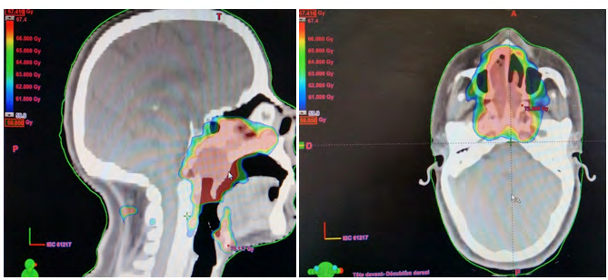
A 50 year old patient followed for a malignant tumor of the nasopharynx of undifferentiated carcinoma type, initially classified T2N2MO according to the eighth edition of the classification of the American joint committee on cancer for which he received a conformal radiotherapy with modulation of intensity with simultaneous integrated boost in 35 fractions of 1.6 Gy on the target low risk volume; 1.8 Gy on the target volume of intermediate risk and 2 Gy on the target volume of high risk thus obtaining three dose levels: 56, 63 and 70 Gy. The ballistics of the treatment consisted of fourteen beams of photons with an energy of 6 MeV. Radiation therapy was performed at a fraction per day and five fractions per week, in combination with concomitant Cisplatin chemotherapy administered weekly at a dose of 40 mg / m2 (Figure 1).
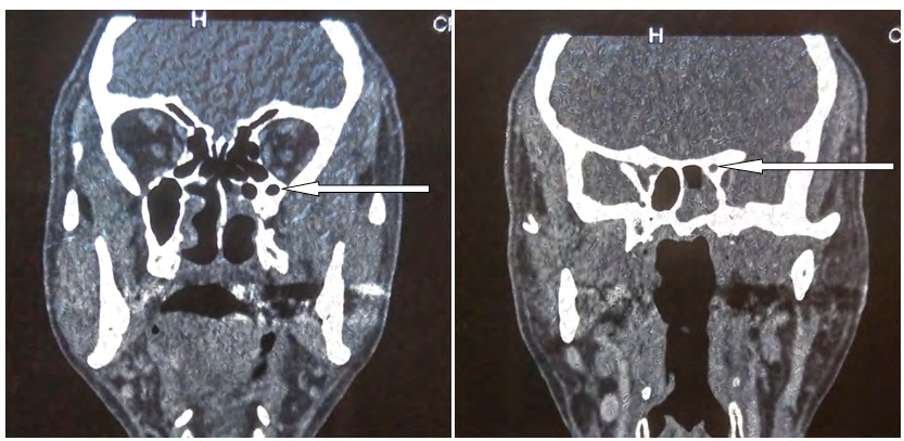
The evolution was characterized by the highlighting; after six years from the end of his treatment; by scanning CT imaging of a moth-eaten appearance at the level of the sphenoidal bone which was the site of rounded lytic lesions without contrast enhancement or infiltration of the soft tissues facing. The bone scintigraphy was without abnormalities. Clinically, the patient was asymptomatic (Figure 2).
Discussion
Osteoradionecrosis could result from several factors such as radio-induced damage to osteoblasts and osteoclasts or radio-induced vascular damage to bone tissue. Clinical studies have shown that it was associated with advanced tumors, high doses of nasopharynx radiation therapy and radiation fields that include the skull base [1]. Most osteoradionecrosis appears 1 to 3 years after irradiation [2]. It is often asymptomatic [3]. On CT images osteoradionecrosis appears in the form of a lytic zone within a demineralized bone. Pathological fracture, cortical erosions and bone loss can also be described. Osteoradionecrosis can sometimes simulate radiation-induced bone sarcoma. However, the absence of mass in the adjacent soft tissue points towards the diagnosis of radionecrosis rather than towards that of progressive tumor lesion [4].
Conclusion
Osteoradionecrosis of the base of the skull is a rare complication of radiotherapy for nasopahryngeal carcinomas. Its clinical and radiological presentation is variable. Radiological examinations make it possible to diagnose subclinical forms.
References
-
Han P, Wang X, Liang F, Liu Y, Qiu X, et al. (2018) Osteoradionecrosis of the skull base in nasopharyngeal carcinoma: incidence and risk factors. Int J Radiat Oncol Biol Phys 102(3): 552-555.
-
Resnick D (1995) Diagnosis of bone and joint disorders. 3rd (Edn.), Saunders, Philadelphia, pp: 3276-3308.
-
Mnejja W, Siala W, Boudawara T, Ghorbel A, Frikha M, et al. (2009) Ostéoradionécrose de la base du crane après radiothérapie pour cancer du nasopharynx. Cancer / Radiothérapie 13(6-7): 676-677.
-
Libschitz HI (1994) Radiation changes in bones. Seminars in Roentgenology 29(1): 15-37.
- Contribution of 18FDG PET in Atypical HORTON Disease
- Living Conditions, Healthy Practice and State of Households of a Town Rural in Colombia
- Background to the Health and Safety Regulation at Work in Colombia
- Risk Factors Psychology Workers Sena (Center for the Petrochemical Industry) Regional Bolívar, Colombia
- Diffuse Intense Pleural FDG Uptake with Smooth Thickening: A MARKER of Tuberculosis in Isolated Pleural Effusion
- Hypermetabolic Splenomegaly with Infarct in FDG PET/CT: A Clue to Scrub Typhus in PUO