Phytochemical Contents, Evaluation of Antiulcer, Antipyretic, Antinociceptive, and Anti-Inflammatory Activities of Artemisia absinthium L. (Asteraceae) Leaf Extract in Wistar Rats
Artemisia absinthium L.(wormwood) is popularly called ‘’Tazargaade’’ in northern Nigeria and is found in all parts of the country. The leaf is used to treat ulcers, fevers, pains and inflammations in Nigeria. The study was carried out aimed at analyzing the phytoconstituents of the leaf and evaluating the antiulcer, antipyretic, antinociceptive, and anti-inflammatory activities of A. absinthium extract in Wistar rats. Phytochemical analysis was carried out following standard procedures, the toxicity studies were carried out using the OECD methods, the antiulcer effect was evaluated using ethanol-induced, naproxeninduced and pyloric ligation-induced ulcer models, the antipyretic effect was evaluated using brewer’s yeast-induced pyrexia, the antinociceptive effect was evaluated using the acetic acid-induced and hot plate models, while the anti-inflammatory effect was determined using carrageenan-induced paw oedema in rats’ models at extract doses of 50-400 mg/kg body weight (p.o.). For the phytoconstituents, LC/MS and GC-MS were used to identify bioactive compounds. Various ulcer parameters were calculated from specific formulae for each model, while the abdominal writhing and volumes of paw oedema in rats were used to assess the potential of the extract in acetic acid and carrageenan-induced models respectively from 0 to 5 h. The phytochemical screening of leaf extract revealed the presence of saponins, flavonoids, carbohydrates, tannins, phenols and phytosterols while, the GC-MS and LC/MS analysis further confirmed three major bioactive compounds as cis-thujones, transsabinyl acetate, and β-thujone. An acute toxicity study of the extract showed that the plant was safe at a maximum dose of 2000 mg/kg b.w. (p.o.). There were no major histopathological defects observed on examination of vital organs, organ weights or biochemical changes in blood profiles, liver function parameters, and lipid profiles. There were no elevated values obtained as all the values fall within the acceptable ranges. Furthermore, the extract showed dose-dependent biological activities. The extract showed significant (p < 0.05) reductions in ulcer area, ulcer index, ulcer score, total acidity, and gastric volumes as well as increased percentage inhibition of ulceration (80.12 %) at the dose of 400 mg/kg b.w. when compared to the various control groups (p < 0.05). Similarly, significant antipyretic, antinociceptive and anti-inflammatory effects were observed in a dose-dependent fashion, with an average reduction in rectal temperature of 76.54 ±2.01℃ in pyrexia, 86.12 % inhibition of abdominal writhing in acetic acid-induced pain, and 82.14 % reduction in paw oedema within the study periods at the dose of 400 mg/kg b.w. (p.o.). There was a significant difference (p < 0.05) in the values obtained when compared to the standard drugs Our study showed that methanol leaf extract of A. absinthium possessed significant antiulcer, antipyretic, antinociceptive and anti-inflammatory activities in Wistar rats. These scientific findings further justify its use in Nigeria’s traditional medicine for the treatment of these ailments. However, there is the need to further investigate specific compound(s) responsible for these biological activities toward drug discovery.
Introduction
Most populations of under-developed and developing nations depend on medicinal plants as a source of primary healthcare [1]. These medicinal plants are easily accessible when looked for because of their potential to manage and treat various diseases such as ulcers, fevers, pains, inflammations, cancers, etc. thereby promoting the healing conditions of patients [2]. These medicinal plants are largely used in rural areas as a traditional healthcare [3]. Recent reports have shown that approximately 80 million indigenous people in Nigeria largely rely on medicinal plants for immediate treatment of many diseases due lack of accessibility to conventional medicine [4].
These plants have been screened for the presence of various types of metabolites called phytochemicals which are ingredients in plants’ parts showing different chemical structures. These phytoconstituents have been reported to have shown one or more biological activities in humans at various doses. These secondary metabolites include alkaloids, glycosides, terpenoids, flavonoids, saponins, tannins, phytosterols, etc. each playing critical roles in the human healthcare system [5]. Most of these metabolites have shown properties such as anti-ulcer, antipyretic, cardiac stimulants, anti-cancer, analgesic, anti-inflammatory, anti- diabetic, antioxidants, antihypertensive, anticonvulsant, antibacterial, and liver protection in various medical trials [6].
In the present research, ulcer, as an example, occur when there is a lack of balance between the digestive juice produced by the stomach, and the act of protecting the linings of the stomach in most cases involving bleeding in the linings of the stomach. In some cases, the stomach wall may be completely wear-out. For instance, a peptic ulcer occurs when there is an open wound in the stomach lining or intestine [7]. Peptic ulcer is divided into two types: gastric ulcer (when it occurs in the stomach), and duodenal ulcer (when occurs in the upper part of the small intestine). A microbe called Helicobacter pylori is known to be the major causative organism for stomach ulcers. Many treatment procedures for ulcers have mainly been carried out using conventional medicines such as omeprazole, cimetidine, antacids, etc. yet, these medicines have yielded little or no results due to the prevalence rate of ulcers in Nigeria and other African countries. The use of medicinal plants in the treatment of ulcers has yielded greater results in Nigeria’s traditional and herbal medicines [7, 8].
On the other hand, an antipyretic agent is an agent of pharmaceutical origin that alleviates or removes fever. Some of these agents either act indirectly by treating the origin of the fever or directly on the signs of the fever such as pains, inflammations and discomfort [9]. The most prevalent of the fevers is malaria. Malaria is a disease that is caused by a protozoan parasite of the genus Plasmodium, often widely distributed in the tropics where their vector (mosquitoes) exists in large populations [10]. In terms of the prevalence of malaria globally, Nigeria has the highest cases of malaria with about 51 million people diagnosed with about 207,000 deaths occurring sharing about 30 % of Africa’s malaria, especially among children who are less than five years of age. In the US, about 2,000 malaria cases per annum are reported.
The World Health Organization (WHO) has classified malaria as the deadliest disease with about 627,000 deaths per annum, mostly children from the continent of Africa and the Caribbeans. Various treatments against malaria especially the use of conventional drugs like artemisinin- based combination therapy (ATC) and chloroquine have not yielded much results due to the development of resistance by the parasite (Plasmodium) towards these chemotherapies [11]. From time immemorial, medicinal plants have been used to effectively treat malaria fever in Nigeria and most African countries. Some plants have been documented as being foremost for the treatment of malaria in Nigeria such as Azadirachta indica A. Juss. (neem), Chromolaena odorata (L.) R.M. King & H.Rob. (Awolowo weed), Cymbopogon citratus (DC.) Stapf. (lemon grass), Spondias mombin L. (African plume), Anacardium occidentale L. (cashew) and Mangifera indica L. (mango) [12].
Malaria often creates pains as one of the symptoms. Pain and inflammation are almost related always to each other. Pain has been defined as an unpleasant sensory and trauma witnessed by an individual due to certain defect or damage to tissues according to the International Association for the Study of Pain [13]. Pharmacologically, pains can be acute (short-lived) or chronic (long-lived). In most cases, both pain and inflammation can be treated using various medications such as NSAIDs, steroids and opiates, yet, there are many side effects noticed with the use of these chemotherapeutic agents [14]. Many medicinal plants have been reported to be used for treating and managing pains and inflammations, while much research is ongoing to discover many plants with antinociceptive (analgesics) and anti-inflammatory potentials [15]. One of the plants currently being used for the treatment of ulcers, malaria fever, pains and inflammations is Nigeria’s species of artemisia- Artemisia absinthium L.
Artemisia absinthium L. is a species found in Nigeria (especially southeastern and Northeastern zones). The name of this plant has been confirmed by ‘’World Flora Online’’ (www.worldfloraonline.org). The plant is native to Europe and is now distributed to North America, the northern United States, Canada, west African countries, and Madagascar [16]. It grows well on grasslands, marshlands and tropical rainforests of the eastern Nigerian states of Enugu, Imo and Anambra, where it can grow up to 50 m high. The plant is commonly called wormwood herb due to the presence of sesquiterpenoid compound absinthin which is bitter in taste and an excellent worm expeller, hence, the name wormwood [17]. The plant has been used in Nigeria for the treatment of various diseases such as ulcers, malaria fever, pains, worm expeller in children, inflammation and diabetes. In Nigeria, it is called ‘’Tarzargade’’ (Hausa language), and ‘’Onugbo-ohia’’ (Igbo language). Despite the uses of the plant, its toxicities have not been fully investigated, but it was considered very safe for use in traditional medicine [18].
This present study was carried out to determine the antiulcer, antipyretic, antinociceptive and anti-inflammatory activities of A. absinthium L. extract in the induced rats’ model.
Materials and Methods
Collection and Preparation of Plant Material
Fresh leaves of Artemisia absinthium L. were collected from a forest in Takum Taraba State, Nigeria in the evening hours in January 2022. The plant was authenticated by Dr. C.A. Ukwubile (a taxonomist) with a voucher specimen number of UMM/FPH/ASR/006 and deposited at the herbarium. The leaves were air-dried under shade for two weeks, reduced into a fine powder (weight:1000 g) using an electronic blender (M500, China), and then extracted with 5 L absolute methanol (Sigma Aldrich, St Louis Mo, USA). The dark-green extract (weight: 125 g; yield:12.5 %) was then stored in a refrigerator (LG, China) at 4℃ for further use.
Phytochemical Analysis
The preliminary phytochemical analysis of methanol leaf extract was carried out to detect the presence of some metabolites such as phenols, flavonoids, anthracenes, alkaloids, saponins, etc. following the procedures previously described [19].
Column Chromatography Isolation of Compounds: The methanol extract (ME) weighing 8.5 g was introduced into a long silica gel column (35 x 950 mm) and eluted by gradient elution technique using ethyl acetate: methanol in various volumes. In all, 30 fractions grouped into three subfractions (following their characteristics on TLC plates) were collected [20]. Further purification of the subfractions was done using a short glass column to obtain final purified isolates labelled as C1 (0.12 mg), C2 (2.8 mg) and C3 (1.6 mg). The dried isolates were then stored in ALS sample bottles for further analysis [4].
LC/MS and GC-MS Analysis of Compounds: The LC- MS analysis of isolated compounds (C1, C2 and C3) was performed using 6230B Time of Flight (TOF) LC/MS (Agilent Technologies, Inc., USA) while the GC-MS analysis was carried out in an Agilent 7890A coupled to a mass detector. The operating conditions for both apparatuses were maintained at optimal levels. The structures of compounds were compared with those in the NIST chemical database [21].
Experimental Animals
Wistar rats of both sexes weighing between 100 and 150 g were purchased from PJ Rats Farm Ltd, Jos Nigeria. The rats were housed in cages and allowed free access to food (Universal Feeds Ltd, Nigeria) and water for one week in the laboratory at room temperature. The ethical approval was given by the Animal Research Ethical Committee of PJ Rats Farm Ltd with approval number PJRF/RA0045/MAY/2022.
Toxicity Assessment of Extract
Acute (LD50) Oral Toxicity Study: The acute oral toxicity (LD50) of the extract was determined using the OECD up and down guidelines paragraph 425. Briefly, five Wistar rats comprising three males and two females were given a maximum oral dose of 2,000 mg/kg body weight A. absinthium extract (p.o.) and monitored for signs of toxicity such as restlessness, reddish eyes, inactiveness, itching, reduced feeding, and even death, especially in the first four hours. The rats were then observed further for 14 days for toxicity signs [22].
Subchronic Toxicity Study: Thirty (30) Wistar rats of
opposite sexes weighing 100-120 g were grouped into five groups of ten rats per group (i.e., 3 males plus 3 females). Group I was given 10 mL/kg distilled water, Groups II, III, IV and V were given A. absinthium extract (30 % LD50 dose) doses of 50, 100, 200, and 400 mg/kg (p.o.) respectively daily for 28 days [23]. The rats were observed three times weekly throughout the 28 days of the experiment for signs of toxicity such as mortality, removal of fur, changes in eyes, and behaviour. The animals were anesthetically sacrificed on the 29th day of the subchronic study. Fresh blood was collected from the rats by cardiac puncturing, while vital organs such as kidneys, lungs, heart, testes, ovaries and pancreas were collected by dissecting the rats and fixing each organ in 10 % formalin solution. Haematological parameters were analyzed using a Mindray auto haematology analyzer (Model: BC- 2800, UK), while histopathology examinations of the organs were carried out using the H & E staining procedure [24].
Antiulcer Evaluation of Extract
To determine the antiulcer activity of the extract, the following experiments were carried out for both acute and chronic ulcers.
Ethanol-induced model for acute ulcer: Rats were grouped as previously described with slight modification. The rats were denied access to food 24 hours before treatments. Group I was given 10 mL/kg distilled water (control), Group II was given 200 mg/kg standard ulcer drug sucralfate (Vixa Pharmaceutical Co. Ltd), while Groups III, IV and V were given 100, 200 and 400 mg/kg extract (p.o.) as prophylaxis doses. After 1 h of pretreatments, an ulcer was induced in the animals by giving them 5 mL/kg absolute ethanol orally (Sigma Aldrich, St. Louis Mo, USA). One hour after ulcer induction, the rats were sacrificed using the cervical dislocation method. The stomachs were carefully dissected, and observed under the compound microscope for ulceration using 40x magnification [25, 26, 27, 28]. Macroscopic Examinations of the Stomach: Physical examinations of the stomach for the presence of ulcers were carried out using the procedures previously described [29], and observed under 10x magnification. The severity scores used are 0 = no ulcer, 0.5 = red colouration, 1 = localized ulcers, 2 = deep ulcers, and 3 = stomach perforations. The ulcer index and the percentage of ulcer inhibition were determined using the formulae below:
( ) ( ) ( ) Ulcer index UI = NU + NS NA/10 [1]
where UI = ulcer index, NU = the average number of ulcers per rat, NS = the average number of severity scores, and NA = percentage of rats with ulcers.
UI of treated UI of control groups ulcer inhibition = 100 UI of control [2]
0 0 Naproxen-Induced Ulcer Model for Chronic Ulcer: Thirty (30) Wistar rats were given 30 mg/kg (p.o.) naproxen (Vixa Pharmaceutical Co. Ltd) for three consecutive days, thereafter, the rats were randomly grouped into five groups of six rats per group as described previously. Group I was given 10 mL/kg distilled (control), Group II was given 20 mg/kg omeprazole (Pharmaplus Nig., Ltd), Groups III, IV and V were given 100, 200 and 400 mg/kg extract (p.o.) after 24 h. The treatment was continued for eight weeks, with one rat sacrificed from each group until eight weeks. Their stomachs were carefully removed and dissected to expose the inner part, washed with normal saline and examined under the microscope [25, 28, 29, 30].
Pyloric Ligation-Induced Ulcer Model: The rats were randomly grouped into five groups of six as previously stated. The rats were starved for 24 hours and treated as described in section 2.5.2. Above. After 1 h of treatment, pyloric ligation was carried out in all the animals to promote peptic ulcer. Using a minor incision (2 cm below the xiphoid process), the abdomens were opened, and 10 mg/kg (i.p.) ketamine HCl (Alpha Pharmacy and Stores Ltd) was used to immobilize them. Then, the pyloric regions of the stomachs were carefully pushed out and ligated while ensuring that traction to the pylorus was avoided. The abdomen was then carefully returned, and its wall closed by interrupted suturing using the chromic catgut. The rats were euthanized using cervical dislocation after six hours of pyloric ligation, and their stomachs were removed. The stomach’s contents were then poured into the tubes and centrifuged for 15 min at 1500 rpm. Thereafter, the supernatants were used to evaluate the pH, acidity and gastric volume [29, 30, 31].
Antipyretic Evaluation
Brewer’s Yeast-Induced Pyrexia: This is the most common and reliable model used to evaluate the antipyretic activity of extracts. Briefly, thirty (30) Wistar rats of both sexes were randomly grouped into 5 groups of 6 rats per group. The rats were fasted overnight with free access to water only. The initial body temperatures of each rat were measured by inserting a well-lubricated thermistor probe digital thermometer (Model: 3800 Geokon, USA) 3 cm into the rectum. Before the induction of fever, the thermistor probe was quantified against the mercury thermometer, and their initial rectal temperatures were taken at 15-minute intervals for 1 h. Pyrexia (fever) was induced in the animals by subcutaneous injection of 30 % yeast extract (in 0.9 % normal saline) below the nape of their neck regions and their final temperatures were recorded. After 1 h, Group I
received 10 mg/mL distilled water, Group II received 20 mg/ kg aspirin (Biopharma Nigeria, Ltd), and Groups III, IV and V received 100, 200 and 400 mg/kg body weight extract orally respectively. The rectal temperature of the animals was then measured at 1, 2, 3, 4 and 5 h after treatment [9, 11, 32]. The percentage inhibition of pyrexia by the extract was then calculated using the formula below:
A – B inhibition of pyrexia= 100 A [3]
0 0 where, A = rectal temperature at 1 h after yeast injection, B = rectal temperature after treatments.
Antinociceptive Evaluation
Acetic Acid-Induced Writhing Test: In this test, thirty (30) Wistar rats of both sexes weighing 50-80 g were randomly grouped into five groups of six rats per group. Group I was given 10 mL/kg distilled water (orally), Group II was given 100 mg/kg diclofenac sodium (Kinapharm Nig., Ltd), Groups III, IV and V were given 100, 200 and 400 mg/kg body weight (b.w.) extract (p.o.) respectively. After 30 min, 0.6 % acetic acid (Sigma Aldrich, St Mo, USA) was injected into the rats (i.p.). The contraction of the abdominal muscles (writhing) was observed after a 5-minute cut-off time. Abdominal writhing in rats was observed using a magnifying hand lens at 5-, 10-, 20-, and 30-minute intervals [33, 34, 35]. The percentage of inhibition of analgesia was calculated from the formula below:
AWc-AWt inhibition= 100 AWc
[4]
0 0 where, AWc = mean number of abdominal writhing in the control group, and AWt = mean number of abdominal writhing in treatment groups.
Hot Plate Model for Antinociceptive Evaluation: Rats were selected based on their response latent times (5-7 s) when placed on Ugo Basile (Model 7280) hot plate (temperature 65.5 OC) before treatment. The rats were then grouped into five groups of five rats per group; Group I received 10 ml/ kg distilled water, Group II received 5 mg/kg morphine (Formulary Nigeria), Groups III, IV, and V received 100, 200, and 400 mg/kg b.w. extract (p.o.). The animals were then placed on a hot plate 30 min later. The latent time of response by the rats was immediately recorded at 0, 30, 60, 90, 120, 150, and 180 min. A delayed latency time of responses is an indication of the antinociceptive effect of the extract. A cut-off time of 20 s was observed before taking the readings [35, 36].
Anti-Inflammatory Evaluations
Carrageenan-Induced Paw Oedema in Rats: Thirty (30)
Wistar rats of both sexes (weight:100-150 g) were randomly grouped into five groups of six rats per group. Group I was given 10 ml/kg distilled water, Group II was given 200 mg/ kg aspirin, Group III, IV and V were given 100, 200 and 400 mg/kg b.w. extract (orally). After 30 min, 0.5 mL of 1 % carrageenan was injected via an intradermal route into the sub-plantar area of the rat’s right hind limb [37, 38]. The paw oedema volumes were then measured at 0,1, 2, 3, 4 and 5 h using the plethysmometer (Ugo Basile, model: 7280, Italy). The percentage of inhibition of paw oedema was calculated using the formula below:
0 0 Pc – Pt inhibition of paw oedema= 100 Pc [5]
where, Pc = average paw volumes of the control group, and Pt = average paw volumes of treatment groups.
Statistical Analysis
The data obtained were expressed as mean ± SD (n = 6). Statistically significant difference between control and treatment groups was analyzed at p < 0.05 (using one-way, two-way or split-plot ANOVA followed by Dunnett’s post hoc test) analyzed by SPSS statistical software version 23.
Results
Phytochemical Contents
The preliminary phytochemical screening of methanol leaf extract of Artemisia absinthium revealed the presence of some metabolites such as saponins, phytosterols, flavonoids, carbohydrates, tannins and phenols (Table 1).
| Phytoconstituents | Tests | Inferences |
|---|---|---|
| Alkaloids | Dragendorff’s | - |
| Wagner’s | - | |
| Meyer’s | - | |
| Saponins | Frothing | + |
| Hemolysis | + | |
| Phytosterols | Salkowski’s | + |
| Liebermann-Burchard | + | |
| Flavonoids | Shinoda’s | + |
| Carbohydrates | Molisch’s | + |
| Anthraquinones | Borntrager’s | - |
| Tannins | Acetic acid | + |
| Lead acetate | + | |
| Fats/oils | Spot | - |
| Phenols | FeCl 2 | + |
| Compounds | Peak area (%) | Retention time (min) |
| Cis-thujones* | 36.42 | 2.39 |
| Trans-sabinyl acetate** | 39.61 | 8.14 |
| Myrcene | 5.64 | 10.01 |
| β-pinene | 9.82 | 15.16 |
| Linalool | 4.81 | 5.52 |
| Trans-sabinol | 6.44 | 9.56 |
| 1,8-cineol | 6.71 | 12.14 |
| Trans-sabinene hydrate | 11.01 | 20.11 |
| β-thujone*** | 16.04 | 11.5 |
| Iso-3-thujanol | 4.14 | 8.12 |
| Estragole | 8.81 | 12.04 |
| Nerol | 5.32 | 15 |
| Geraniol | 8.1 | 16.21 |
| Eugenol | 6.42 | 10.14 |
| Methyl eugenol | 11.16 | 23.2 |
| Spathulenol | 5.43 | 16.72 |
| Herniarin | 6.64 | 12.42 |
Table 1: ** Phytochemical contents of R. absinthium methanol leaf extract.
+ = detected (present), - = not detected (absent). Table 1: Phytochemical contents of R. absinthium methanol leaf extract.
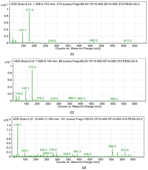
GC-MS Composition of Bioactive Compounds from Extract
The GC-MS analysis of isolated compounds C1, C2 and C3
showed the presence of cis-thujones, beta-thujone and trans- sabine acetate respectively which are the most abundance among other compounds in the extract (Table 2, Figure 1).
Acute (LD50) Oral Toxicity Determination
The result showed that the extract was tolerated by the animals at 2000 mg/kg b.w. dose (p.o.). However, signs such as reduced food and water intake, inactiveness for 5 min, reddish eyes which disappeared after 5 min, and short- lived itching in the eyes were observed within the first four hours. No mortality was witnessed after 14 days of acute intoxication of extract in the rats.
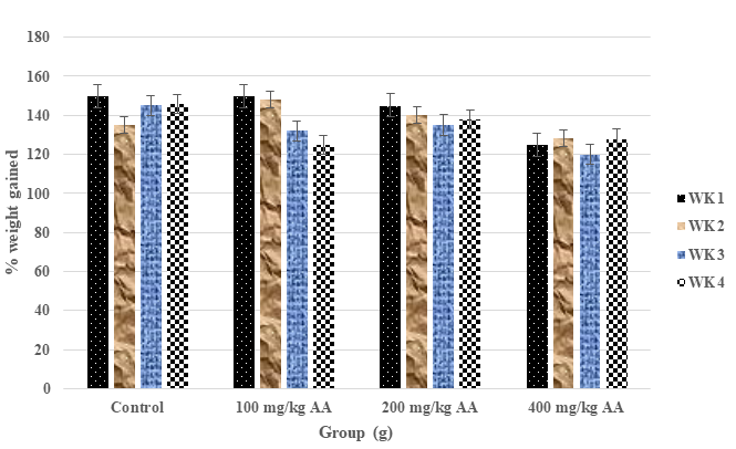
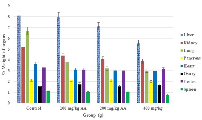
Effect of A. absinthium Extract on Body and Organ Weights of Rats in 28 Days
The results showed that there was a slight decrease in body weights of rats administered various doses (100, 200 and 400 mg/kg b.w.) of extract which were significantly different (p < 0.05) from the control (Figures 2 & 3).
Effects A. absinthium Extract on Blood Parameters of Rats
The results showed slight decreases in WBC, RBC, and MCV in a dose-dependent fashion. There were no significant reductions in the values of parameters such as MCH, MCHC, Hb and platelets at 400 mg/kg b.w. extract dose within 28 days of subchronic study. However, a significant increase in the values of monocytes and lymphocytes was witnessed (Table 3).
| Blood parameter | Control | 50 mg/g | 100 mg/kg | 200 mg/kg | 400 mg/kg |
|---|---|---|---|---|---|
| WBC (103/mm3) | 24.22±1.16* | 20.02±1.01 | 20.00±1.22 | 18.12±1.04 | 15.00±1.01* |
| Monocyte (%) | 3.02±0.01* | 3.88±0.01 | 4.24±0.01 | 4.92±0.01 | 8.12±0.02* |
| Lymphocytes (%) | 5.11±0.01 | 5.24±0.01 | 5.64±0.02 | 5.98±0.01 | 6.24±0.01 |
| Hb (g/dL) | 12.00±1.10* | 12.22±2.01 | 12.20±2.21 | 12.28±1.41 | 12.38±1.10* |
| RBC (106/mm3) | 25.18±2.32* | 25.12±1.01 | 25.02±1.11 | 22.16±1.04 | 18.22±2.14* |
| PCV (%) | 15.04±2.01* | 15.00±1.01 | 14.02±1.01 | 12.56±2.01 | 10.26±2.02* |
| MCV (fL) | 22.12±2.52 | 22.56±2.01 | 25.01±2.10 | 22.02±1.01 | 20.04±1.01 |
| MCH (Pg) | 8.24±0.01* | 8.98±0.01 | 12.02±0.02 | 12.34±1.01 | 14.28±2.01* |
| MCHC (g/dL) | 16.40±2.21* | 16.66±1.02 | 16.80±1.01 | 18.22±1.01 | 18.84±1.01* |
| Platelets (103/mm3) | 105.42±4.22 | 105.02±4.01 | 105.44±4.02 | 105.82±2.01 | 106.10±2.14 |
Table 2: Effects of A. absinthium leaf extract on blood parameters of Wistar rats (n = 6). Results are mean ± SD (n = 10). *Signi
Table 3: Effects of A. absinthium leaf extract on blood parameters of Wistar rats (n = 6). Results are mean ± SD (n = 10). *Significant difference (p < 0.05). WBC: white blood cells; Hb: hemoglobin content; RBC: red blood cells; PCV: packed cell volume; MCV: mean corpuscular volume; MCH: mean corpuscular hemoglobin; MCHC: mean corpuscular hemoglobin concentration.
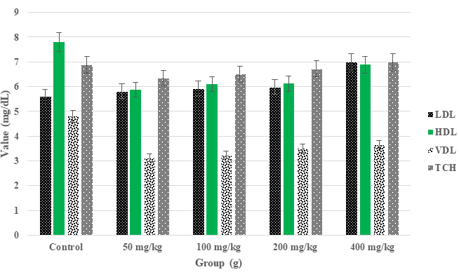
Effects A. absinthium Extract on Lipid Profiles and Ulceration of Rats
From the results obtained the extract showed a significant reduction in HDL cholesterol and total cholesterol levels of Wistar rats in a dose-dependent fashion while LDL and VDL increased likewise a dose-dependent ulceration. These reductions and increases were significantly different from the control group at 400 mg/kg b.w extract dose (Figure 4, Tables 4 & 5).
| Groups (g) | Ulcer score (US) | Reduction in US (%) | Ulcer index | % U inhibition |
|---|---|---|---|---|
| Control | 5.05 ± 0.01 | nd | 5.16 ± 1.01 | nd |
| Standard | 2.33 ± 0.02* | 53.86 ± 2.01 | 1.66 ± 0.01* | 67.83 ± 1.04 |
| 100 mg/kg AA | 2.55 ± 1.01 | 49.50 ± 1.02 | 2.30 ± 1.01 | 55.43 ± 1.02 |
| 200 mg/kg AA | 2.40 ± 0.01 | 52.48 ± 1.04 | 1.92 ± 0.01 | 62.79 ± 2.14 |
| 400 mg/kg AA | 2.02 ± 0.01* | 60.00 ± 2.02 | 1.01 ± 0.01* | 80.43 ± 4.08 |
Table 3: Effect of A. absinthium leaf extract on ethanol-induced ulcer in Wistar rats. Results are mean ± SD (n = 10). * Statisti
| Groups (g) | Ulcer area (mm2) | Ulcer index | % Ulcer inhibition |
|---|---|---|---|
| Control | 5.40 ± 1.01 | 0.54 ± 0.01 | nd |
| Standard | 3.14 ± 0.02* | 0.31 ± 1.01* | 42.60 ± 2.01* |
| 100 mg/kg AA | 4.22 ± 0.01 | 0.42 ± 1.01 | 22.22 ± 0.01 |
| 200 mg/kg AA | 4.12 ± 1.01 | 0.41 ± 0.01 | 24.07 ± 1.03* |
| 400 mg/kg AA | 3.01 ± 1.01* | 0.30 ± 0.02* | 44.44 ± 1.12* |
Table 4: Effect of A. absinthium leaf extract on ulcer area and ulcer index in naproxen-induced ulcer in rats. Results are mean ±
Table 5: Effect of A. absinthium leaf extract on ulcer area and ulcer index in naproxen-induced ulcer in rats. Results are mean ± SD (n = 10). * Statistically significant (p < 0.05) compared to control. AA: Artemisia absinthium extract, nd: not determined. 20 mg/kg Omeprazole (Pharmaplus Nig., Ltd) was the standard drug used. Ulcer index = 10/ulcer area (for 3 replicate readings).
Effects of A. absinthium Leaf Extract on Rat’s Liver Function Parameters
The results showed that there was no elevated level of the liver enzymes aspartate aminotransferase (AST) also known as serum glutamic oxaloacetic transaminase (SGOT) and alanine aminotransferase (ALT) also known as serum glutamic pyruvic transaminase (SGPT) as well as bilirubin in the animals within 28 days of subchronic study. All the parameters were within normal ranges (Figure 5).
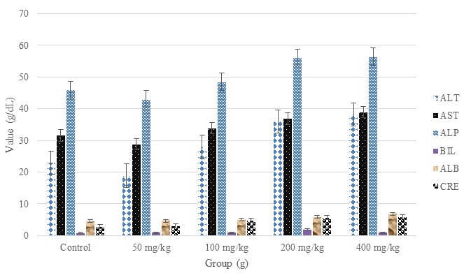
Figure 5: Effects of A. absinthium leaf extract on rat’s liver function parameters. ALT: alanine amino transferase (range: 7-56 g/dL), AST: aspartate aminotransferase (range: 5-40 g/dL), ALP: alkaline phosphatase (range: 45-115 U/L), BIL: bilirubin (range: 0.1-17 mg/dL), ALB: albumin (range: 3.5– 5.0 g/dL), CRE: creatinine (range: 0.1-7.5 g/dL); these values vary from laboratories. Results are mean ± SD (n = 10), p<0.05 using one-way ANOVA followed by Dunnett’s post hoc test).
Photomicrographic Examination of Some Vital Organs in Rats after Subchronic Study
The examination of some vital organs of the animals after 28 days of subchronic study with H & E stain showed slight changes in certain organs such as kidney, lung, ovary and testes at the highest dose of 400 mg/kg b.w. extract. The results equally showed that there was no significant change in these organs when compared to the control group at the highest dose (Figure 6). There was an indication of clear blood vessels with no vascular congestion seen in the liver, heart, pancreas, and spleen in the treated groups.
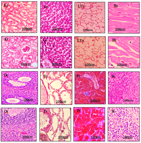
Figure 6: Photomicrographs of H & E stained (100x) section some vital organs of rats after 28 days subchronic toxicity study at 400 mg/kg b.w. A. absinthium extract. Kc, Kt: kidney control group, kidney treatment group, Lc, Lt: liver control group, liver treatment group, Luc, LUt: lung control group, lung treatment group, Hc, Ht: heart control group, heart treatment group, Oc, Ot: ovary control group, ovary treatment group, Tc, Tt: testes control group, testes treatment group, Pc, Pt: pancreas control group, pancreas treatment group, Sc, St: spleen control group, spleen treatment group.
Antiulcer Effects of A. absinthium on Wistar Rats
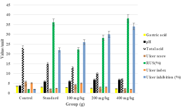
The results showed dose-dependent activities in all the experimental models. For example, mean ulcer scores decreased at a dose of 100 to 400 mg/kg in the animals while the percentage inhibition of ulceration by the extract increased to 80 % in the ethanol-induced ulcer model (Table 4). Similarly, significant (p < 0.05) reductions in ulcer areas and ulcer indices were noticed in the naproxen-induced ulcer model (Table 5). Moreso, results obtained from the pylorus ligation-induced model in rats after 20 days post-treatment showed dose-dependent activities such as gastric volume, pH, total acidity, ulcer indices and percentage inhibition of ulcerations (Figure 7). Our results, showed that antiulcer activity of the plant extract was strictly dose-dependent, and this was significant (p < 0.05) when compared to the control group.
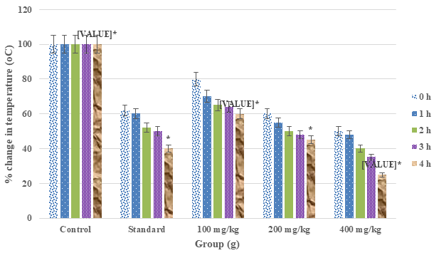
Antipyretic Effects of A. absinthium Leaf Extract
The results obtained showed a consistent decrease in rectal temperatures of the rats from 0 h to 4 h at 100 to 400 mg/kg b.w. extract doses. These decreases were significant (p < 0.05) when compared to the control group (Figure 8).
Antinociceptive Effects of A. absinthium Leaf Extract
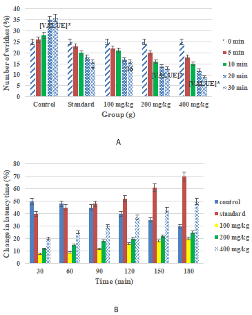
From the results obtained in both acetic acid and hot plate-induced pains, there were dose-dependent reductions in the contraction of abdominal muscles and response times in the withdrawal of limbs as well as changes in latency times (Figure 9). There were significant reductions at 400 mg/kg b.w. extract dose in several writhing and changes in latency time within 30 and 180 min respectively in both experiments.
Anti-Inflammatory Effect of A. absinthium Extract
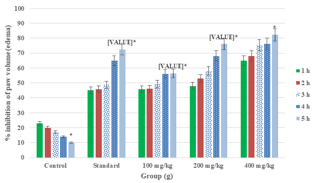
The result of the anti-inflammatory effect of A. absinthium leaf extract in carrageenan-induced rat paw oedema (Figure 10) showed that the extract displayed a significant (p < 0.05) decrease in paw diameter and increased percentage inhibition of inflammation in the animals within 5 h. From the results obtained, at a dose of 400 mg/kg b.w. extract the extract showed the highest inhibition of inflammation (86.12 %) in hind limb paws (oedema) of rats, and maintained for 5 h. The result obtained was not different significantly from the standard (positive control) anti-inflammatory drug aspirin (72.42 %).
Discussion
The study of pharmacognosy has contributed immensely towards the quality assurance of herbal drugs in recent years. This aspect of natural science has evolved from economic botany and extended into more advanced fields like ethnobotany, analytical chemistry, zoo pharmacognosy, medicinal chemistry, nanotechnology, molecular biology, phytochemistry, natural medicine, and ethnopharmacology [39]. It has been established that drugs of plant origin have formed a significant part of our healthcare system for the treatment of various diseases, which has increased their usage in many parts of the world. Most of these plants are readily available, low cost, and low toxicity, and they are components of novel pharmacophores on which drug development is based because of the presence of many primary and secondary metabolites [40].
In the current study, phytochemical analysis of the methanol leaf of A. absinthium L. extract revealed the presence of many constituents which have various therapeutic actions in humans. For instance, some secondary metabolites have been reported to possess arrays of biological activities such as antiulcer, antimalarial, anti-inflammatory, anticancer, antioxidant and antimicrobial [41]. Flavonoids, saponins and polyphenols possess immunomodulatory potentials by altering T1 and T2-helper cell balance, thereby inhibiting inflammation of the macrophages [37]. The presence of these metabolites (Table 1) in the current study must played anti-inflammatory functions in the animals as seen from the study. Moreover, thujone a monoterpene ketone isolated from the plant in this current study and analyzed by GC- MS and LC/MS is reported to be present in many medicinal plants and has been used in many herbal formulations as an anti-inflammatory agent. The mechanism of action of thujone was the inhibition of histamine, serotonin, bradykinins, and prostaglandins which are inflammatory regulators [42]. In this study, thujones and sabinyl acetate isolated from the plant (Table 2, Figure 1) must have played a similar role in the rats.
It was reported that at high doses, thujone a monoterpene ketone isolated from A. absinthium was neurotoxic in rats [42]. However, our study has shown that no mortality and histopathology effects were noticed in fourteen days and twenty-eight days of acute and subchronic toxicity studies respectively in rats. This is because, the animals did not show significant reduction or increase in body and organ weights (Figures 2 & 3), and parameters such as blood, lipids and liver function were within the acceptable limits (Table 3, Figures 4 & 5). Histopathology examinations of the kidney, liver, heart, lung, ovary, testes, pancreas and spleen showed clear blood vessels, no vascular congestion, necrosis, and extended lumen in the organs (Figure 6). However, slight distribution of spermatozoa seen in the testes was suggestive of spermatogenesis, which implied that the extract might boast sperm production in the animals. The study further showed the presence of other metabolites in the extract such as flavonoids and tannins might have neutralized the toxic effects of thujone and its derivatives at higher doses [21].
From the current study, in ethanol-induced ulcers, the A. absinthium extracts significantly decrease ulcer score and ulcer index with an increased reduction in ulcer score and inhibition percentage in a dose-dependent manner (Table 4). Ethanol is a solvent of preferred choice used to induce gastric ulcers in rats to evaluate the antiulcer effect of bioactive compounds. This is because, ethanol can cause various degrees of gastrointestinal abnormalities such as damage to mucosa linings, dehydration, cell membrane injury, cytotoxicity, and exfoliation as well as triggering pro- inflammation mediators [29]. Our study has shown that the extract was able to counter these side effects of ethanol in animals due to the presence of certain bioactive compounds such as flavonoids, saponins, and tannins. For example, flavonoids have been reported to elevate prostaglandin contents in the mucosa, reduce secretion of histamine by mast cells through the inhibition of growth of Helicobacter pylori (Family: Helicobacteraceae) histidine decarboxylase, H+/K+-ATPase as well as act as scavenging of free radicals, while saponins and tannins activate protective factors of mucous membrane and impermeability of mucosa outer layer to certain chemicals respectively [43]. Even though the aetiology of peptic ulcer is not well understood, the general belief was that it resulted from a lack of balance between causative agents and the integrity of mucosal linings via an endogenous defence system. Currently, various drugs have been used to boost the mucosa defence mechanism by elevating the secretion of mucosa, stabilization of epithelial cell surface or blockage of the synthesis of prostaglandins [43]. In the current study, various doses of the extract at 100, 200 and 400 mg/kg b.w. showed dose-dependent activity in naproxen-induced ulcers with decreased ulcer area and ulcer index while percentage inhibition of ulceration elevated at 400 mg/kg b.w. extract dose (Table 5). Similarly, the gastric volume, pH, total acidity, and ulcer score showed dose-dependent activities from the pylorus ligation-induced ulcer in rats (Figure 7). The antiulcer activity shown by the plant extract was better than the standard drugs which were significant (p < 0.05) when compared to the negative control. The results showed potential inhibitory and antisecretory effects of the plant extract on ulcer parameters such as volume of gastric acids, pH, ulcer area and ulcer index.
Our present study also showed that the methanol leaf extract of A. absinthium greatly displayed antipyretic potentials in Wistar rats, as seen by the progressive reductions in rectal temperatures against brewer’s yeast- induced pyrexia or fever (Figure 8). The extract showed its antipyretic activity in dose-dependent fashion because at a dose of 400 mg/kg b.w. extract, a significant (p < 0.05) reduction in rectal temperatures within 4 hours was achieved in the animals when compared to the standard drug. The results from our study further suggest that the extract must have inhibited the synthesis of prostaglandin and other promoters of inflammation that are often associated with fevers thereby lowering the rectal temperature of the rats. The antipyretic activity of the extract obtained from the present study was due to the presence of metabolites like phenols, tannins and beta-pinene revealed by GC-MS (Table 2), which exhibit various degrees of antipyretic effects in animals [13, 44].
Finally, the results obtained from antinociceptive and anti-inflammatory studies (Figure 9), the extracts also showed dose-dependent activities in the reduction of abdominal writhing caused by acetic acid or delayed responses to pain caused by hot plate and paw oedema volumes induced by carrageenan respectively at doses of 100 to 4000 mg/kg b.w. administered orally to the rats. These results showed that A. absinthium possessed antinociceptive and anti-inflammatory effects which were probably a result of the inhibition of some peripheral pain and inflammation promoters like serotonin, histamine, nitric oxide (NO), and bradykinin, thereby, preventing the release of prostaglandins. The presence of some bioactive compounds such as phenols and flavonoids in the plant has been reported to inhibit pro- analgesic and inflammatory endogenous mediators thereby activating antinociceptive and inflammatory inhibiting materials like non-steroidal anti-inflammatory compounds [45]. The study showed no involvement of opioid receptors in antinociceptive activity of the extract, but peripheral mechanism of pain reduction.
On a final note, the anti-ulcer, antipyretic, antinociceptive and anti-inflammatory activities of A. absinthium leaf extract observed in the current study were due to the presence of many secondary metabolites which inhibited various factors responsible for ulcers, fever, pain and inflammation in Wistar rats.
Conclusion
Our study revealed that the methanol leaf extract of A. absinthium possessed potential antiulcer, antipyretic, antinociceptive, and anti-inflammatory activities in in vivo rat models. These current findings further justified the use of the plant in Nigeria’s traditional medicine for the treatment of ulcers, fever, pains and inflammations. The presence of certain bioactive compounds in the leaf extract was responsible for the observed activities of the plant. It is suggested that the mechanism of actions of the extract for the observed activities should be evaluated further in animal models.
Conflict of Interest
We have none.
Acknowledgements
We are thankful to Mr. Sumaila Mejida of University of Lagos Central Research Laboratory, Lagos Nigeria for his immense technical assistant.
Funding
No funding.
Author Contribution
- Cletus Anes Ukwubile: Designed the research, Conducted experiment, performed data analysis
- and wrote the manuscript.
- Nnamdi David Menkiti: Carried out literature search, Conducted experiment and participated in
- data analysis.
- Shedrach Yakubu: Conducted experiment and performed data analysis. All authors read and
- approved the final manuscript before submission.
References
-
Sofowora A, Ogunbodede E, Onayade A (2013) The role and place of medicinal plants in the strategies for disease prevention. Afr J Tradit Complement Altern Me 10(5): 210-229.
-
Oliveira RDG, Mahon CPAN, Ascêncio PGM, Ascêncio SD, Balogun SO, et al. (2014) Evaluation of anti-inflammatory activity of hydroethanolic extract of Dilodendron bipinnatum Radlk. J Ethnopharmacol 155(1): 387-395.
-
Onasanwo SA, Fabiyi TD, Oluwole FS, Olaleye SB (2012) Analgesic and anti-inflammatory properties of the leaf extracts of Anacardium occidentalis in the laboratory rodents. Niger J Physiol Sci 27(1): 65-71.
-
Ukwubile C, Ikpefan E, Malgwi T, Umeokoli B (2020) Pharmacognostic study and physiochemical assessment on the leaves of Melastomastrum capitatum Fern. anticancer plant in Nigeria. Am J Physiol Biochem Pharmacol 10(1): 8-15.
-
Komansilan A, Abadi AL, Yanuwiadi B, Kaligis DA (2012) Isolation and Identification of Biolarvicide from Soursop ( Annona muricata Linn ) Seeds to Mosquito ( Aedes aegypti ) Larvae. Int J Eng Technol 12(3): 28-32.
-
Prachi P (2010) In Vitro Antimicrobial Activity and Phytochemical Analysis of the Leaves of Annona Muricata. Int J Pharma Res Dev 2(5): 1-6.
-
Adinortey MB, Ansah C, Galyuon I, Nyarko A (2013) In Vivo Models Used for Evaluation of Potential Antigastroduodenal Ulcer Agents. Ulcers 2013: 1-12.
-
Bessède E, Staedel C, Amador LAA, Nguyen PH, Chambonnier L, et al. (2014) Helicobacter pylori generates cells with cancer stem cell properties via epithelial-mesenchymal transition-like changes. Oncogene 33(32): 4123-4131.
-
Mworia JK, Kibiti CM, Ngugi MP, Ngeranwa JN (2019) Antipyretic potential of dichloromethane leaf extract of Eucalyptus globulus (Labill) and Senna didymobotrya (Fresenius) in rats models. Heliyon 5(12): e02924.
-
Yemitan OK, Adeyemi OO (2017) Mechanistic assessment of the analgesic, anti-inflammatory and antipyretic actions of Dalbergia saxatilis in animal models. Pharm Biol 55(1): 898-905.
-
Olorunniyi OF, Morenikeji OA (2014) In vivo antimalarial activity of crude aqueous leaf extract of Pyrenacantha staudtii against Plasmodium berghei (NK65) in infected mice. African J Pharm Pharmacol 8(12): 342-345.
-
Dawaki S, Al-Mekhlafi HM, Ithoi I, Ibrahim J, Atroosh WM, et al. (2016) Is Nigeria winning the battle against malaria? Prevalence, risk factors and KAP assessment among Hausa communities in Kano State. Malar J 15(1): 1-14.
-
Sharma VC, Kaushik A, Dey YN, Srivastava B, Wanjari M, et al. (2020) Analgesic, anti-inflammatory and antipyretic activities of ethanolic extract of stem bark of Anogeissus latifolia Roxb,” Clin. Phytoscience 6(1): 22.
-
Lee YM, Son E, Kim SH, Kim OS, Kim DS, et al. (2019) Anti- inflammatory and anti-osteoarthritis effect of Mollugo pentaphylla extract. Pharm Biol 57(1): 74-81.
-
Bezerra JJL, Oliveira JSR, Lima LMV, Silva MV, Araújo DRC, et al. (2022) Evaluation of the anti-inflammatory, antipyretic and antinociceptive activities of the hydroalcoholic extract of Rhynchospora nervosa (Vahl) Boeckeler (Cyperaceae). J Ethnopharmacol 284: 114811.
-
Alamgir ANM (2017) Introduction. Alamgir ANM, et al. (Eds.), Therapeutic Use of Medicinal Plants and Their Extracts: Volume 1. Springer International Publishing, Cham, pp: 1-17.
-
Shafi N, Khan GA, Ghauri EG (2004) Antiulcer effect of Artemisia absinthium L. In rats. Pak J Sci Ind Res 47(2): 130-134.
-
El-Ghani MMA (2016) Traditional medicinal plants of Nigeria: an overview. Agriculture and Biology J North Am 7(5): 220-247.
-
Ukwubile CA (2019) Acute and subchronic toxicity profiles of Melastomastrum capitatum (Vahl) Fern. (Melastomataceae) root aqueous extract in Swiss albino mice. Prog Chem Biochem Res 2(2): 74-83.
-
Kim GS, Zeng L, Alali F, Rogers LL, Wu FE, et al. (1998) Two new mono-tetrahydrofuran ring acetogenins, annomuricin E and muricapentocin, from the leaves of Annona muricata. J Nat Prod 61(4): 432-436.
-
Keskes H, Belhadj S, Jlail L, El Feki A, Damak M,et al. (2017) LC-MS-MS and GC-MS analyses of biologically active extracts and fractions from tunisian juniperus phoenice leaves. Pharm Biol vol 55(1): 88-95.
-
OECD (2022) Test Guideline No. 425 Acute Oral Toxicity: Up-and-Down Procedure, pp: 27.
-
Khalifa MA, Mohammad AEH, Ahmed BM (2022) Acute and Subacute Toxicity Studies of Cyperus Papyrus Ash on Wistar Albino Rats. Pharmaceutical and Biomedical Research 8(4): 279-290.
-
Kpemissi M, Metowogo K, Melila M, Veerapur VP, Negru M, et al. (2020) Acute and subchronic oral toxicity assessments of Combretum micranthum (Combretaceae) in Wistar rats. Toxicol Reports 7: 162-168.
-
Panda SK, Sarkar G, Acharjya M, Panda PK (2017) Antiulcer Activity of Amaranthus Spinosus Leaf Extract and Its Comparision With Famotidine in Shay Rats. J Drug Deliv Ther 7(2): 96-98.
-
Mekonnen AN, Atnafie SA, Atta MAW (2020) Evaluation of Antiulcer Activity of 80% Methanol Extract and Solvent Fractions of the Root of Croton macrostachyus Hocsht: Ex Del. (Euphorbiaceae) in Rodents. Evidence- based Complement Altern Me 2020: 2809270.
-
Brito SA, Barbosa IS, Almeida CLF, Medeiros JW, Silva Neto JC, et al. (2018) Evaluation of gastroprotective and ulcer healing activities of yellow mombin juice from spondias mombin l. PLoS One 13(11): 1-16.
-
Al-Batran R, Al-Bayaty F, Al-Obaidi MMJ, Abdualkader AM, Hadi HA, et al. (2013) In Vivo Antioxidant and Antiulcer Activity of Parkia speciosa Ethanolic Leaf Extract against Ethanol-Induced Gastric Ulcer in Rats. PLoS One 8(5): 2-12.
-
Andargie Y, Sisay W, Molla M, Norahun A, Singh P, et al. (2022) Evaluation of the Antiulcer Activity of Methanolic Extract and Solvent Fractions of the Leaves of Calpurnia aurea (Ait.) Benth. (Fabaceae) in Rats. Evidence-based Complement Altern Med 2022: 4199284.
-
Tripathi A, Singh S, Mukerjee A (2021) Antiulcer activity of ethanolic leaf extract of Capparis zeylanica against chemically induced ulcers. Futur. J Pharm Sci 7(1): 211.
-
Abebaw M, Mishra B, Asmelashe DG (2017) Evaluation of anti-ulcer activity of the leaf extract of Osyris quadripartita Decne ( Santalaceae ) in rats. J Exp Pharmacol 9: 1-11.
-
Muhammad N, Saeed M, Khan H (2012) Antipyretic, analgesic and anti-inflammatory activity of Viola betonicifolia whole plant. BMC Complement Altern Med 12: 59.
-
Sulaiman MR, Zakaria ZA, Chiong HS, Lai SK, Israf DA, et al. (2009) Antinociceptive and anti-inflammatory effects of stachytarpheta jamaicensis (L.) Vahl (Verbenaceae) in experimental animal models. Med Princ Pract 18(4): 272-279.
-
Yimer T, Birru EM, Adugna M, Geta M, Emiru YK, et al. (2020) Evaluation of analgesic and anti-inflammatory activities of 80% methanol root extract of echinops kebericho m. (asteraceae). J Inflamm Res 13: 647-658.
-
Hijazi MA, El-Mallah A, Aboul-Ela M, Ellakany A (2017) Evaluation of Analgesic Activity of Papaver libanoticum Extract in Mice: Involvement of Opioids Receptors. Evidence-based Complement Altern Med 2017: 8935085.
-
Ayertey F, Ofori-Attah E, Antwi S, Amoa-Bosompem M, Djameh G, et al. (2021) Anti-inflammatory activity and mechanism of action of ethanolic leaf extract of Morinda lucida Benth. J Tradit Complement Med 11(3): 249-258.
-
Borquaye LS, Laryea MK, Gasu EN, Boateng MA, Baffour PK, et al. (2020) Anti-inflammatory and antioxidant activities of extracts of Reissantia indica, Cissus cornifolia and Grosseria vignei. Cogent Biol 6(1): 1785755.
-
Fyad K, Belboukhari N, Hadj-Khelil AO, Sekkoum K (2020) Analgesic and anti-inflammatory activity of aqueous extract of Bubonium graveolens. Biomed Res Ther 7(9): 4002-4009.
-
Jones PW, Chin YW, Kinghorn DA (2006) The Role of Pharmacognosy in Modern Medicine and Pharmacy. Current Drug Targets 7(3): 247-264.
-
Badal S, Delgoda R (2017) Pharmacognosy: Fundamentals, Applications and Strategies 1st(Edn.), Elsevier, India, pp: 738.
-
Baeshen NA, Almulaiky YQ, Afifi M, Al-Farga A, Ali HA, et al. (2023) GC-MS Analysis of Bioactive Compounds Extracted from Plant Rhazya stricta Using Various Solvents. Plants Basel 12(4): 960.
-
Batiha GE, Olatunde A, El-Mleeh A, Hetta HF, Al-Rejaie S, et al. (2020) Bioactive compounds, pharmacological actions and pharmacokinetics of wormwood (Artemisia absinthium) Antibiot 9(6): 353.
-
Li Q, Hu X, Xuan Y, Ying J, Fei Y, et al. (2018) Kaempferol protects ethanol-induced gastric ulcers in mice via pro- inflammatory cytokines and NO. Acta Biochim Biophys Sin Shanghai 50(3): 246-253.
-
Ukwubile CA, Malgwi TS, Ikpefan EO, Modu B, Umeano VA, et al. (2023) Evaluation of physicochemical parameters, acute and subchronic toxicities, and anti-diabetic activity of Spondias venulosa (Engl.) Mart. ex Engl. leaf extract on alloxan-induced diabetic rats. J Ethnopharmacol 306: 116169.
-
Nakalembe L, Kasolo JN, Nyatia E, Lubega A, Bbosa GS, et al. (2019) Analgesic and Anti-Inflammatory Activity of Total Crude Leaf Extract of Phytolacca dodecandra in Wistar Albino Rats. Neurosci Med 10(3): 259-271.
- Potential Medicinal Herbs and Secondary Metabolites in Combating Corona Virus
- Monkeypox and its Clinical Implications in Pregnancy
- Pharmacognostical Analysis of the Leaves of Important Nervine Medicinal Plant: Strychnos Nux-Vomica L
- Assessment of Antibacterial Activity of Various Solvent Extracts of Dictyota Dichotoma Against Multidrug Resistant Bacterial Strain
- An Insights of Bioactive Elements on Malignancy: Mechanistic Avenues
- Advances and Future Directions in Pharmacognosy and Chinese Medicine