Missed Early Diagnosis of Rare Case Thoraco-Omphalopagus Conjoined Twins: Case Report
Introduction: Conjoined twins are rare congenital anomaly with mortality and morbidity. It is result of a faulty division of an embryo at 13-15 days of conception. Thoraco-omphalopagus that was undiagnosed early and cancelling on the prognosis of the outcome and termination of pregnancy would have been done early leading to avoidance of cesarean section and neonatal morbidity and mortality. Case Report: A 28-year-old para 3+0 G4 with twin gestation at 35/40 by a third trimester scan no other scan had been done. Before she was Unsure of her dates, also reported family history of twinning. Admitted to Kenyatta National Hospital as a referral from Nyahururu county hospital with a diagnosis of conjoined twins for specialized care. Obstetric ultrasound done revealed monochromic, monoamniotic viable Conjoined twins both flexed in breech. Twin A (34+4)/40 2289 grams, B 36/40 2368grams. BPP 8/8. Differential diagnosis of Thoraco-omphalopagus with shared and communicating heart and liver. Placenta fundo-posterior not low lying, with minimal infarctive areas. Conclusion: The conjoined twins are rare and complex, majority of cases are stillbirth or die soon after birth but when there is early ultrasound an essential requirement for safe medical termination of pregnancy to avoid more complication and knowing that the rate of survival is very low. We need to understand that early ultrasound mostly in first trimester is very important. Attempt at surgery depends on the type of conjoined twins, the degree and type of organs shared by the twins, as in our case both they shared heart and liver.
Introduction
The conjoined twins are the result of a faulty division of an embryo at 13-15 days of conception. Different pathogenic mechanism and theories have been postulated which include incomplete separation of the developing embryo [1]. The incidence is very rare and reported to be between 1:33000 to 1: 165000 pregnancies [2]. The conjoined twinning rise when the even occurs at about the primitive streak stage of development, at about 13-14 days after fertilization in the human and is exclusively associated with the monoamniotic monochorionic type of placentation [3]. The conjoined twins are twins whose bodies are anatomically joined in utero. The level of which the twins are attached can range from simple, involving skin and cartilage, to complex, including fusion of the skull, brains, or other vital organs [4]. The common types of conjoint twins include thoracopagus and omphalopagus is the least common with incidence of 0.5% [5]. The other less common forms of conjoint twins include thoraco- omphalopagus (joined at chest and abdomen), pyopagus (joined at buttocks), ischiopagus (joined at the ischium) and craniopagus (joined at the head) [5, 6]. Incidence of thoraco- omphalopagus is 28% [7]. Around 40% of the conjoint twins are stillbirth and around 35% die within 24 hours of delivery [8]. The female fetuses are more commonly affected with the ratio of male to female being 1:3, particularly in thoracopagus [5]. Ultrasonography is the most accurate technique, which can be used for its diagnosis.
Case Presentation
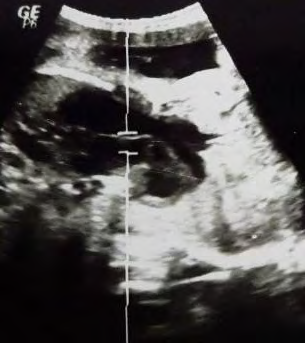
A 28-year-old para 3+0 G4 with twin gestation at 35/40 by a third trimester scan, Unsure of her dates. There was family history of twins. She had attended antenatal clinic once first contact at 28 weeks where her antenatal profile was unremarkable. Admitted to Kenyatta National Hospital as a referral from Nyahururu county hospital with a diagnosis of conjoined twins for specialized Care. Obstetric ultrasound done revealed monochromic, monoamniotic viable Conjoined twins both flexed in breech. Twin A (34+4)/40 2289 grams, B 36/40 2368grams. BPP 8/8. Differential diagnosis of Thoraco-omphalopagus with shared and communicating heart and liver. Placenta fund posterior not low lying, with minimal infarction areas. Amniotic fluid normal for gestation.
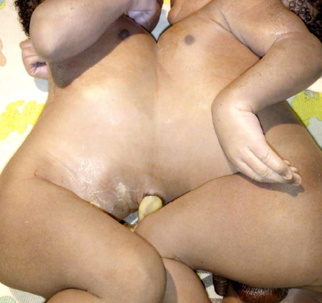
While in the ward the pediatric and surgical team reviewed patient and plan was to carry pregnancy to term 38/40 weeks, and plan for electives cesarean section where pediatrician, pediatric surgeon and pediatric cardiothoracic team would be present. 19 days later while in the ward patient progressed into labour and was prepared for emergency cesarean section. One cord and one placenta noted and expelled successfully. Outcome was two male twins conjoined at thorax and abdomen combined weight of 5420 grams, combined APGAR score of 4 /7/ 8. Both had well-formed individual upper and lower limbs, 5 digits each. Surgery completed successfully with estimated blood loss of 700mls.
Babies were admitted to NBU where the team of neurosurgeon and pediatric surgeon wanted to separate them but they faced challenge the babies were sharing the vital organs liver and heart. They unfortunately succumbed 2 days later.
Discussion
Conjoint twins are the result of a faulty division of an embryo at 13-15 days of conception. Different pathogenic mechanism and theories have been postulated which include incomplete separation of the developing embryo [1]. Conjoint twins on his classification depends upon the site of fusion and they are classified into thoraco-omphalopagus (chest, abdomen, or both as in our case), Pyopagus (Buttocks), Ischiopagus (ischium) and Craniopagus (head. The analysis of survived and aborted fetuses showed that most of the conjoint twins are female with a M: F ratio of 1:3 in our case we are reporting both were male. The survival rates reported range between 20-25% [9].
This type of conjoined twins may undergo complex surgery for separation if they are found not to be sharing vital organs. Early detection of this condition is of paramount importance so as to provide the parents with an option of safe termination of pregnancy [10]. The most common varieties encountered were thoraco amphalopagus (28%) thoracopagus (18,5%), omphalopagus (10%), parasitic twins (10%) and cranionpagus (6%). Of these, about40% were stillborn, and 60% live born, although only about 25% of those that survived to birth lived long enough to be candidates for surgery [11]. Looking on low rate of survival of this case mostly when they are sharing the vital organs it is very important to discover this type of malformation early for possible counseling to the couple and termination of the pregnancy. The ultrasound on first trimester has its importance in pregnancy. In this case the discovery was done in late prematurity.
Conclusion
Conjoined Twins are a complex occurrence and rare, it is associated with high perinatal mortality but when there is early ultrasound an essential requirement for safe medical termination of pregnancy to avoid more complication and knowing that the rate of survival is very low. We need to understand that early ultrasound mostly in first trimester is very important. This is gold standard investigation for identification of all defects and malformation.
References
-
Kilby MD, Bricker L (2016) Management of monochorionic twin pregnancy. BJOG124: e1-e45.
-
Tannuri ACA, Batatinha JAP, Velhote MCP, Tannuri U (2013) Conjoined twins- twenty years’ experience at a reference center in Brazil. Clinics (Sao Paulo) 68(3): 371-377.
-
Kaufman MH (2004) The embryology of conjoined twins. Childs Nerv Syst 20(8-9): 508-525.
-
DeRuilter C (2011) Conjoined Twins. The embryo project encyclopedia, Arizona state University, United States.
-
Mishra N, Rohilla M (2015) Thoraco-omphalopagus conjoint twin: A Case Report and Literature Review. Gynecology & Obstetrics Case report 1(1): 3.
-
Seo JW, Lee YS, Chi JG (1988) Cross-sectional illustration on major of conjoined twins. J Korean Med Sci 3(1): 19- 25.
-
Graham GM, Gaddipati S (2005) Diagnosis and management of obstetrical complications unique to multiple gestations. Semin Perinatol 29(5): 282-295.
-
Sakala EP (1986) Obstetric management of conjoined twins. Obstet Gynecol 67 (3 Suppl): 215-255.
-
Chiu CT, Hou SH, Lai HS, Lee PH, Lin FY, et al. (1994) Separation of thoracopagus conjoined twins. A case report. J Cardiovasc Surg (Torino) 35(5): 459-462.
-
Asmita S, Mohammed S, Prasanna M, Dahiphale DB, Prashant L (2017) Thoraco-omphalopagus conjoined twins: Imaging and Antenatal Diagnosis. International Journal of contemporary Medicine surgery and radiology 2(4): 124-126.
-
Hanson JW (1975) Letter: Incidence of conjoined twinning (letter). Lancet 2(7947): 1257.
- Pattern of Breast Lesions in Ovu Inland, Delta State, South Southern Nigeria
- Morphometric Analysis of the Human Femur: Exploring Platymetric and Robusticity Indices Among the Nigerian Population
- Anatomical Variation of Arteria Lusoria: Clinical Implications for Dysphagia Lusoria and Surgical Risk
- Morphometric Study of the Vertebral Body and Pedicle of Typical Cervical Vertebrae Using Radiological Image
- Epigenetic Mechanisms Driving Human Evolutionary Changes
- Neuroprotective Effects of Ginkgo Biloba Extract on Bilateral Common Carotid Artery Ischaemic Stroke Induced in Wistar Rat