Anatomy Image Series: Persistent Metopic Sutures in a Series of Dry Skulls of Hellenic Origin
The persistence of a complete metopic suture in adulthood is called metopism. Numerous studies report wide variations concerning the prevalence of metopic sutures in adult crania. Metopic suture lies to the midline of the forehead and extends from the frontal bone to the root of the nose. However various types of incomplete sutures do exist. A series of 126 dry skulls is examined. A total of 15 metopic sutures were revealed, 2 complete (1 combined) and 13 incompletes. The localization and suture types are important for clinical approaches, as during the evaluation of head traumas in the emergency department.
Introduction
The metopic, or inter frontal, or median frontal suture forms from the anterior fontanelle and proceeds toward the nasion area, closing form the masion towards bregma. It appears during the second month of the fetal life and closes in infancy or early childhood. However, it has been observed in individuals as old as 8 years of age [1].
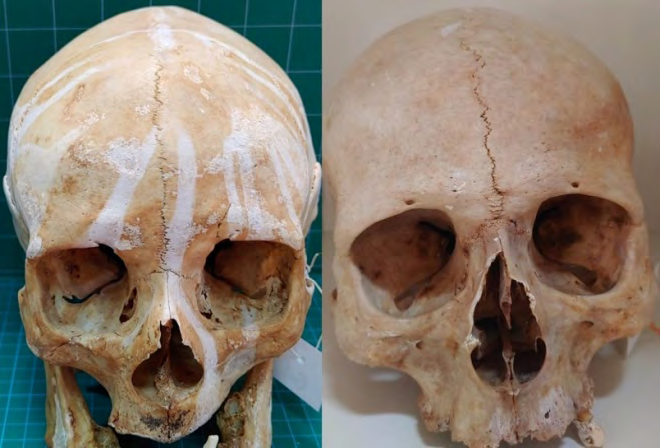
The reasons why such a suture persists into adulthood are to be determined. The metopic suture may appear as a complete or an incomplete variant [1]. The condition when in an adult cranium a complete suture is encountered is called metopism (Figure 1) [2], while the crania have been referred to as crania metopica, or crania bifida [3]. On the other hand, incomplete suture cases are divided into categories, remmant of the metopic suture, supranasal and supranasal combined with metopic, prominent supranasal rectangular, supranasal V-shaped double suture and supranasal triangle with lateral extensions [4].
The prevalence of metopism greatly varies among different geographic populations and between sexes, being reported for 0, 12% up to 23, and 6% [1]. Although the Image Article incomplete type research lacks thorough studies, it has been noted that its incidence vary from 0, 30% up to 0, 93%, while combined types about 1, 75% [2].
Case Series
The materials for this case series consisted of 126 adult human dry skulls of Caucasian (Hellenic) origin and unknown sex that were collected from the Ossuary of the Anatomy Laboratory of the Democritus University of Thrace, School of Medicine in Alexandroupolis, Greece. The criteria for the crania to be included were i) no signs of prior cranial surgery, bony malformation, or trauma and ii) adulthood defined by tooth eruption of the skulls. The authors retained approval for the present research’s protocol from the ethics committee of the abovementioned institution.
Results
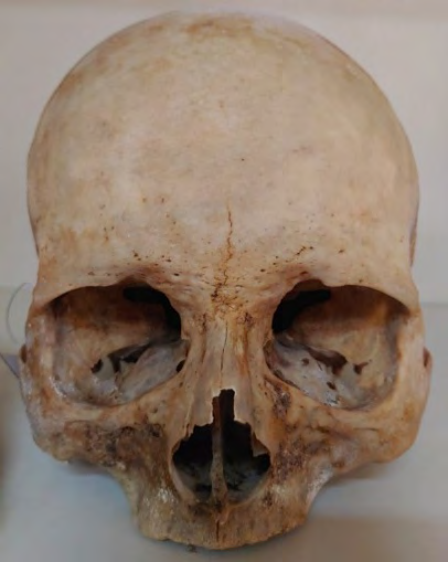
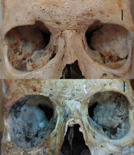
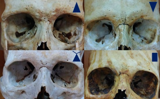
Among the 126 crania studied, 2 (1, 6%) were identified to acquire a complete metopic suture, and 13 (10, 3%) with an incomplete. As far as the metopism 1 of the 2 bony cranial specimens was of a combined type, complete linear figure (Figure 1). The types for the incomplete were 1 remnant (Figure 2) and 12 supranasal. The 12 supranasal were categorized as 2 triangle shaped, 2 triangle with lateral extensions, 2 V-shaped double, 3 double linear (Figure 3) and 3 linear (Figure 4).
Discussion
Small number of complete suture (metopism) alongside with increased findings of incomplete types, mismatching various studies [2] may be attributed to the sample itself. A cluster of genes is involved in sutures development. Thus genetics paly also its part for their appearance [5]. Metopic suture is related to several craniofacial defects including frontal sinus anomalies, cleft lip, and cleft palate. Most studies have reported that metopism is more prevalent in females than in males. Knowledge of the anatomy of the inter frontal sutures is crucial as they may mimic a vertical traumatic cranial fracture extending in the midline of the head in trauma cases [1]. Formed by bands of fibrous connective tissue which prevent premature bone separation, are initially flexible joints between the calvarian bones which develop to accommodate grown cranial and intracranial organs and structures. Taking into consideration the fact that sutures are shaping cranial morphology, they present a significant role among historical findings, anatomy, embryology and development, surgery, anthropology and cranial morphometrics [5].
Epilogue
Gross anatomy of the crania displaying metopic sutures differs significantly from the non-metopic ones. Although persistent inter frontal sutures are rather rare, they present a separate series and should be separately examined, even if they are part of a large and homogenous cranial sample. Nature’s diversity may masquerade a frontal bone fracture, triggering physicians’ alertness for topographic anatomy.
References
-
Zdilla MJ, Russell ML, Koons AW, Bliss KN, Mangus KR (2018) Metopism: a Study of the Persistent Metopic Suture. J Craniofac Surg 29(1): 204-208.Aksu F, Cirpan S, Mas NG, Karabekir S, Magden AO (2014) Anatomic features of metopic suture in adult dry skulls. J Craniofac Surg 25(3): 1044-1046.
-
Marciniak R, Nizankowski C (1959) Metopism and its correlation with the development of the frontal sinuses. Acta Radiol 51(5): 343-352.
-
Mann RW, Hunt DR, Lozanoff S (2016) Photographic Atlas Anatomical Variants of the Human Skeleton. Springfield 30(5): 744
-
Di Ieva A, Bruner E, Davidson J, Pisano P, Haider T, et al. (2013) Cranial sutures: a multidisciplinary review. Childs Nerv Syst 29(6): 893-905.
- Pattern of Breast Lesions in Ovu Inland, Delta State, South Southern Nigeria
- Morphometric Analysis of the Human Femur: Exploring Platymetric and Robusticity Indices Among the Nigerian Population
- Anatomical Variation of Arteria Lusoria: Clinical Implications for Dysphagia Lusoria and Surgical Risk
- Morphometric Study of the Vertebral Body and Pedicle of Typical Cervical Vertebrae Using Radiological Image
- Epigenetic Mechanisms Driving Human Evolutionary Changes
- Neuroprotective Effects of Ginkgo Biloba Extract on Bilateral Common Carotid Artery Ischaemic Stroke Induced in Wistar Rat